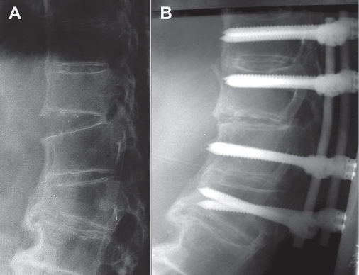
16 Thoracolumbar distraction-extension injuries are uncommon. The mechanism of injury is the opposite of the more common flexion-distraction injuries and involves hyperextension. The extension moment causes tensile failure (distraction) of the vertebral body as well as tensile or compressive failure of the posterior elements. This injury pattern is most common in patients with stiff or kyphotic thoracolumbar spines; for example, those with ankylosing spondylitis (AS) and diffuse idiopathic skeletal hyperostosis (DISH).1–5 These injures are most commonly seen at the thoracolumbar junction, although injuries in the mid-thoracic and lumbar spine are occasionally seen. Most occur as a result of low-energy trauma, such as a fall, although injuries from high-energy trauma can result in significant translation of the spine. The risk of neurologic injury varies but neurologic injury is much more likely to occur in the presence of spinal translation. Distraction-extension injures are specifically described within the context of a classification system by Ferguson and Allen.3 The authors reported one such injury in their series of 54 patients with thoracolumbar injuries. This particular mechanism of injury is not mentioned in the original Denis (or subsequent modifications) thoracolumbar injury classification system.6 Distraction-extension injuries represent a B3 distraction injury in the Magerl (AO Group) thoracolumbar classification system, where only three such injuries out of 1445 (0.21%) were identified.7 From a practical standpoint, it is simplest to think of this uncommon mechanism of injury as a reverse flexion-distraction injury, which can be morphologically sub-classified as follows: (1) osseous (injury to bone only), (2) soft-tissue (purely soft-tissue injury, such as transdiskal and ligamentous injuries), and (3) combination (osseous and soft-tissue injuries). Furthermore, the presence or absence of translation (subluxation/dislocation = grossly unstable) must be noted. Because of the rarity of these injuries, the diagnosis may be easily overlooked. In addition, they are often due to minor trauma, where suspicion for a significant spinal injury is typically low. Consequently, a high index of suspicion for this injury should be maintained in kyphotic elderly patients complaining of back pain following minor or major thoracolumbar trauma. The presence of anterior vertebral column widening (i.e., distraction) on plain radiographs, computed tomography (CT), or magnetic resonance imaging (MRI) confirms the diagnosis of an extension-distraction mechanism of injury (Fig. 16.1). However, the transverse nature of this injury, concomitant degenerative changes, osteoporosis, and the difficulties visualizing the posterior elements on plain radiographs, as well as the propensity for the more stable injuries to reduce in flexion, may render this injury undetectable on plain radiographs. Consequently, CT with sagittal and coronal reformats (a transverse plane of fracture is easy to miss on axial slices) and MRI are the imaging modalities of choice. Fig. 16.1 (A) Lateral plain radiograph of the midthoracic spine, showing unstable fracture through ankylosed intervertebral elements without neurologic compromise. (B) Posterior fusion with pedicle screw instrumentation provides stable fixation.
Thoracolumbar Distraction-Extension Injuries
![]() Classification
Classification
![]() Workup
Workup
History
Spinal Imaging

Stay updated, free articles. Join our Telegram channel

Full access? Get Clinical Tree


