Therapeutic Physical Agents


Therapeutic Physical Agents


Jeffrey R. Basford

G. David Baxter


This chapter reviews the physical agents with an emphasis on their clinical use, scientific basis, and effectiveness. Although the properties of many agents overlap, discussion will begin with superficial heat and cold and then progress to hydrotherapy, the diathermies, and electrical therapies. The chapter will conclude with an analysis of some of the less established modalities, such as lasers, monochromatic light, vibration, low-intensity electrical stimulation, and extracorporeal shock wave therapy.


HEAT AND COLD

Heat and cold have potent effects on tissue. For example, temperatures above 42°C may be painful, and prolonged exposure to those above 45°C may cause injury (1). Temperatures below 13°C are also uncomfortable, and systemic temperatures below 28°C can cause death (1). In addition, metabolic and enzymatic processes are temperature dependent: an increase of 3°C increases collagenase activity severalfold (2). Heating the hands to 45°C reduces metacarpophalangeal joint stiffness 20%, whereas cooling them to 18°C increases stiffness by a similar amount (3). Temperature changes of a few degrees affect nerve conduction velocities, and changes of 5°C to 7°C alter blood flow (4, 5, 6) and collagen extensibility (7). In practice, most clinical treatments attempt to warm tissues to between 40°C and 45°C. As a point of reference, a gradual 20-minute immersion of the body in 22°C cold or 42°C warm baths result in core temperature changes of 0.3°C to 0.4°C (8). Cold therapy can produce intense local effects; ice treatment over an inflamed knee joint reduces skin temperatures by 16°C and intra-articular temperatures by 5°C or 6°C (9). Conversely, hot paraffin is reported to increase local skin temperature and knee intra-articular temperature by 7.5°C and 1.7°C, respectively (9). Although the heating modalities differ, most gain their effects by producing analgesia, hyperemia, changes in local or systemic temperatures, and reduced muscle tone. As a result, they share many of the indications (Table 63-1) and contraindications (Table 63-2) of heat in general (10). Cold’s main effects are analgesia, reduced perfusion, and muscle tone reduction. As a result, cold has indications (Table 63-3) and contraindications (Table 63-4) that are often surprisingly similar to heat.


Superficial Heat

Tissue can be heated or cooled by conduction, convection, or conversion. The first of these, conduction, requires the physical contact of two or more objects at different temperatures. A hot pack in contact with a patient’s back exemplifies conductive heating. The second, convection, also involves the transfer of energy between objects at different temperatures. In this case, however, one object, the medium, is moving relative to the other. Whirlpool baths are the most common convective heating modality. (Note that the medium in contact with the patient is in motion. As a result, the heating and cooling of the immersed body is more intense than would occur in a conductive situation with a stationary medium.) The last approach, conversion, involves the transformation of one form of energy to another. Heat lamps and ultrasound devices heat conversively, since they rely on transformation of radiant or sound energy to heat.

The physical properties of superficial heating and cooling modalities differ, but none is able to overcome the combination of skin tolerance, tissue thermal conductivity, and the body’s responses to produce temperature changes of more than a few degrees at depths of a few centimeters. The following sections review the characteristics of the most common superficial agents.


Hot Packs

Hot packs (e.g., hydrocollator packs) typically consist of segmented cloth bags filled with a silicon dioxide that, when exposed to moisture, absorbs many times its own weight of water. These packs are available in various sizes and are stored in water bath reservoirs at temperatures of 70°C to 80°C. When needed, the packs are removed from the reservoirs and excess water is allowed to drain off. The packs are then wrapped in an insulating cover or toweling and placed on the patient (Fig. 63-1).

Hot packs are one of the most commonly used heat modalities (11) due to their advantages of low cost, minimal maintenance, long life (i.e., packs last as long as 5 years; reservoirs, up to 30 years), patient acceptance, and ease of use. A variety of home-use packs are available and many can be heated in a microwave oven.

Hot packs have few risks that are not outlined in Table 63-2. However, scalding is a possibility, and it is important that excess water is drained off before use, that the insulating pad/toweling is not wet, and that the pack is placed over, not under, the patient to avoid the body’s weight expressing hot water out of the pack and wetting the insulation.

We often think of hot packs as the source of a relatively stable temperature. This is not necessarily true. Thus, although
hot packs maintain therapeutic temperatures throughout a treatment session, they cool significantly over the 20- to 30-minute period. At one time, this cooling tendency was used therapeutically. Kenny packs, for example, were thin wool cloths that were soaked in 60°C water and spun dry before use. After spinning, they contained little water and cooled rapidly. Consequently, they were replaced at 5- to 10-minute intervals and produced a cyclic heating pattern that was considered particularly effective for muscle pain and spasms of poliomyelitis.


TABLE 63.1 General Indications for Therapeutic Heat


Pain


Muscle spasm


Contracture


Tension myalgia


Production of hyperemia


Acceleration of metabolic processes


Hematoma resolution


Bursitis


Tenosynovitis


Fibrositis


Fibromyalgia


Superficial thrombophlebitis


Induction of reflex vasodilation


Collagen-vascular diseases


Electric heating pads, hot water bottles, and circulating-water heating pads are alternatives to hot packs. Many of these do not cool spontaneously and not all have reliable timers or thermostats. Burns are possible and exposure may need to be limited in the elderly, people with diminished sensation, or those who may fall asleep during use.


Heat Lamps

Radiant heat, although less frequently used than in the past, is a versatile and easy way to warm superficial tissue. Specialized infrared (IR) sources may be used due to their convenience and durability. However, incandescent lights release most of their energy as heat, and clamp lamps using these bulbs can both be used as an inexpensive alternative.


TABLE 63.2 General Contraindications and Precautions for Therapeutic Heat


Acute inflammation, trauma, or hemorrhage


Bleeding disorders


Cutaneous insensitivity


Inability to communicate or respond to pain


Poor thermal regulation (e.g., from neuroleptics)


Malignancy


Edema


Ischemia


Atrophic skin


Scar tissue


Unstable angina or blood pressure


Decompensated heart failure within 6-8 weeks of a myocardial infarction (31)


TABLE 63.3 General Indications for Therapeutic Cold


Acute musculoskeletal trauma


Edema


Hemorrhage


Analgesia


Pain


Spasticity


Adjunct in muscle reeducation


Reduction of local and systemic metabolic activity


Skin temperatures are controlled by adjusting the distance between the heat source and the patient. Point sources such as light bulbs heat according to the “inverse square law”; that is, energy density decreases inversely in proportion with the square of the distance (1/r2). Elongated sources tend to follow a slower inverse distance (1/r) relationship. Heating is most intense when a source is perpendicular to the patient and decreases in proportion to the cosine of the angle between the beam and the perpendicular. In practice, heat sources are typically placed directly above, and about 40 to 50 cm from, the patient.


Choice

The physiologic effects of dry heat from a heat lamp differ little from those of moist heat of a hot pack (12). As a result, the choice depends on ease of use and preference. If the patient is in bed or cannot tolerate pressure, radiant heat may be a better option. Similarly, if a patient prefers moisture, the choice is also easy.


Safety

The precautions in Table 63-2 apply to the superficial heating agents (10). In addition, these modalities can burn the patient and, with chronic use, may produce a permanent brown skin discoloration (erythema ab igne).


TABLE 63.4 General Precautions and Contraindications for Therapeutic Cold


Ischemia


Cold intolerance


Raynaud’s phenomenon or disease


Severe cold pressor responses


Cold allergy


Inability to communicate or respond to pain


Poor thermal regulation


Cutaneous insensitivity


FIGURE 63-1. Hot pack treatment of the low back. The pack is covered with an insulated wrapper and separated from the patient with several layers of toweling. Note that the patient must be positioned carefully and be able to tolerate the weight of the pack.


Effectiveness

As noted above, superficial heat and cold have clear and established effects of a variety of physiological, metabolic, and tissue effects. As such, their main use has been for the amelioration of discomfort and, to a lesser extent, the control of swelling and edema. Given their limited penetration, usage is usually restricted to the musculoskeletal system where there is support of benefits for its use as an adjunct to the treatment of a variety of musculoskeletal conditions such as low back pain (13,14), contractures (15), and rheumatoid arthritis (16).


Hydrotherapy

Hydrotherapy uses fluid to transfer thermal energy and mechanical forces to tissue. Whirlpool baths (Fig. 63-2) and Hubbard tanks (Fig. 63-3) use agitated water to produce convective heating, cooling, massage, and gentle debridement. Agitation, however, is not essential, and sitz baths, paraffin baths, and contrast baths all use a stationary medium. Immersion itself has effects. Subjects placed in water up to their chins will have transient elevations of their serum atrial natriuretic protein that are independent of temperature and result from increased venous return or right atrial loading (17). This section discusses hydrotherapy as well as other alternatives, such as balneotherapy and fluidotherapy.


FIGURE 63-2. Whirlpool treatment of the lower extremity. Water temperatures may range from 11°C to 43°C, depending on the patient’s condition and the amount of surface area treated. Positioning as well as entering and leaving the bath are facilitated with a hydraulic chair.


FIGURE 63-3. Hubbard tank treatment. These tanks are large, are expensive to operate, and occupy large amounts of floor space. Nevertheless, they are necessary for the cleaning of large wounds and helpful in treating patients with conditions involving multiple joints.


Whirlpool Baths and Hubbard Tanks

Tanks vary in size from small portable whirlpools designed to treat a single extremity to Hubbard tanks containing thousands of liters. Pools and tanks are not always essential; hand-held shower heads and small water jets are often used for local treatment such as the irrigation and debridement of deep wounds and burns.

Temperature choice depends on the amount of immersion, treatment goals, and the patient’s medical condition. Neutral temperatures of 33°C to 36°C are usually well tolerated on limited portions of the body, although for a healthy patient, temperatures of 42°C to as much as 45°C or 46°C are possible. Full-body immersion for 20 minutes can increase systemic temperatures by 0.3°C (8); Hubbard tank temperatures are usually limited to 39°C. Temperature selection should take into account the fact that for any given temperature, turbulent water heats and cools more vigorously than stationary water.

Whirlpools and Hubbard tanks are well suited for wound and burn treatment in which gentle agitation, heat, and solvent action are needed. Neutral temperatures (to somewhat warmer, depending on comfort) are chosen. Once the patient is immersed, agitation is increased to provide gentle debridement and aid in dressing removal.


“Sterile” tanks should be specified for burns and wounds. Although true sterility is not possible, a sufficient approximation is possible to make disease transmission unlikely. If wounds are large or if there is a significant exposure of internal tissue, sodium chloride may be added to the water (5 kg or more for a Hubbard tank) to improve comfort and lessen the risks of hemolysis and electrolyte imbalance. Additional agents such as potassium permanganate and gentle detergents may be added as desired.

Hydrotherapy is a common adjunct to the treatment of rheumatoid arthritis, “muscle spasm,” and joint stiffness after cast removal. Immobilized patients and those with wounds often find the concept frightening, and treatment should be reviewed before the first session with an emphasis on its comfort, transfers, and any use of plinths and hoists.

Hydrotherapy is resource intensive, expensive, and consumes large amounts of hot water and floor space. As a result, therapy clinics are tending to discard their larger units and to use the smallest baths possible.


Contrast Baths

Contrast baths use two reservoirs, one is typically at 38°C to 40°C and the other significantly cooler at about 13°C to 16°C. The theory of their use is that the alternating exposure to heat and cold results in a reflex hyperemia and neurological desensitization. Treatment typically begins with the distal extremity placed in the warm bath for about 10 minutes and then proceeds to four cycles of alternating 1- to 4-minute cold and 4- to 6-minute warm soaks. If edema is an issue, a case can be made for ending with a cool, rather than a warm, soak.

Contrast baths may be most commonly used in treatment programs for rheumatoid arthritis and complex regional pain syndrome I (CRPS I). People with rheumatoid arthritis often benefit but may find simple warm-water soaks as effective and less difficult. Patients with CRPS I may prefer to begin with less extreme bath temperatures and also seem to benefit, but this improvement is difficult to separate from the effects of other desensitization and activity measures. There is limited evidence to suggest that contrast baths may also improve autonomic regulation and blood pressure in people with hypertension (18); research findings in other popular areas of application such as sports medicine are limited (19).


Sitz Baths

Warm sitz baths are enshrined in the treatment of hemorrhoids, anorectal fistulas, and postpartum pain. While there has been criticism about the limitations of our knowledge of sitz bath effectiveness (20), the available research provides support for this practice. For example, sitting in water between 40°C and 50°C (with warmer temperatures perhaps more effective) lessens sphincter activity and anal pressures in normal subjects as well as those with hemorrhoids and anorectal fistulas (21). A recent trial following anal surgery found those receiving sitz baths obtained pain relief equivalent to that produced by oral analgesics (22,23). While research provides limited reports of side effects, there is, as with any heat and water modality, a risk of scalding and perineal burn at higher temperatures (24).


Edema

The ancient Greeks and 18th-century physicians treated edema with water immersion, a practice mimicked today with compressive sleeves. Research supports this intuitively reasonable treatment; immersion increases natriuretic proteins (17) and renal water and salt loss in normal subjects as well as those with the nephrotic syndrome and cirrhosis (25). Because warmth produces a reactive vasodilation, neutral temperatures appear to be the most effective.


Water-Based Exercise

Water-based exercise and therapeutic pools are a popular way to provide gentle, progressive exercise in a setting that permits limited weight bearing. The approach has shown benefits (26), but its value over land-based programs has been more difficult to establish. Although patients with rheumatoid and osteoarthritis often like the warmth and support of water-based exercise, benefits may not be as clear as they might seem. For example, a number of studies involving land- and water-based programs have found that water-based programs provide little or no benefits over their land-based counterparts for patients with osteoarthritis, rheumatoid arthritis, juvenile inflammatory arthritis, and anterior cruciate rehabilitation (27, 28, 29). Although many believe exercise is beneficial for patients with fibromyalgia, the benefits of water- over land-based approaches remain unclear (30, 31, 32, 33, 34).


Balneotherapy

Balneotherapy (spa) therapy is now little more than a curiosity in North America. In Europe, however, acceptance remains strong and visits to spas are supported by some governments and insurance programs (35). Treatments involve a combination of physical therapy, water baths, mud treatments, mineral water consumption, and education that may take place in a resortlike atmosphere for periods as long as 3 weeks. The fundamental tenet of the approach is the belief that water containing dissolved gases (such as nitrogen and carbon dioxide), elements (e.g., calcium, magnesium, zinc, and cobalt), and compounds (e.g., hydrogen sulfide) has therapeutic effects.

Balneotherapy research has traditionally been hampered by poor quality and an environment in which a patient is undergoing multiple changes in his or her lifestyle and exercise patterns. Research quality is improving, however, and higher quality controlled studies are becoming available. Furthermore, although intact skin is relatively impervious, we know that blood concentrations of substances such as bromine and rubidium may be increased after bathing in patients with impaired skin barriers (36,37).

Although most U.S. physicians are skeptical, some controlled investigations support the use of balneotherapy for the inflammatory and degenerative arthritides. For example, although the differences are often small, patients with rheumatoid arthritis, ankylosing spondylitis, and osteoarthritis appear
to respond similarly to or somewhat better than they do to alternative treatments (35,36,38, 39, 40, 41, 42, 43, 44, 45, 46). There are many studies, but there is limited evidence that patients with nonradiating low back pain may do better following balneotherapy than a control treatment (47). In all, balneotherapy studies are often limited by poor size, incomplete blinding, and short follow-up. Nevertheless, the findings are intriguing and warrant honest consideration.


Fluidotherapy

Hydrotherapy usually uses water as the heat-exchanging medium, but substances such as pulverized corncobs and small beads “fluidized” by hot air jets may be substituted. Although these devices have been available since the 1970s, the benefits of this high-temperature, low-heat capacity approach over other approaches for improving joint mobility and sensory desensitization remain controversial (48).


Safety

The general precautions of heat (Table 63-2) apply to hydrotherapy. Drowning, cardiac disease, systemic hyperthermia, and disease transmission are also concerns, but they may be overemphasized. Hot water-associated seizures are rare but are known to occur (49).

Cardiac disease may not be an absolute contraindication to hyperthermia as was once thought. For example, even though sauna baths (80°C to 100°C) elevate body temperatures by 1°C or 2°C (50,51), research indicates that they may improve the function and quality of life of individuals with severe congestive heart failure (CHF) (52). Furthermore, Finnish heart attack survivors return to sauna bathing without apparent increased risk, and 15-minute soaks in 40°C hot tubs do not create ischemic electrocardiogram changes or alter systolic and diastolic blood pressures more than cardiac rehabilitation-as-sociated stationary bicycle exercises (50,51,53).

Hydrotherapy-associated infections seem to be rare. However, there may be some reproductive consequences: neural tube defects may be increased (relative risk, 2.6 to 2.9) in the children of women who take sauna baths during early pregnancy (54), and sperm counts may be lowered after isolated or repeated sauna sessions (55).


Paraffin Baths

Paraffin baths are thermostatically controlled reservoirs filled with a 1:7 mixture of mineral oil and paraffin. Bath temperatures (45°C to 54°C) are higher than in most hydrotherapy but are well tolerated because of the low heat capacity of the mixture and the tendency for an insulating layer of wax to build up on the surface of the treated area.

Two paraffin treatment approaches predominate. Dipping is the most common and consists of the patient submerging the treated extremity in the bath ten times, with pauses between dips to permit a layer of paraffin to solidify. The treated area is then covered with a plastic sheet and placed in an insulating cover for about 20 minutes (Figs. 63-4 and 63-5). The paraffin is then stripped off and returned to the container.


FIGURE 63-4. Paraffin bath treatment. Two approaches are common. In the dipping technique, the extremity is immersed in the bath, removed briefly to allow the wax to solidify, and redipped for a total of ten repetitions. After the dipping, the extremity is wrapped in an insulating cover for about 20 minutes before the wax is removed. The immersion technique provides a more vigorous heating and is similar to the dipping approach, except that after a number of dips, the extremity is kept immersed in the paraffin.

Dipping initially increases skin temperatures to about 47°C, but by the end of a 30-minute session, skin temperatures fall to within a few degrees of baseline. Deeper tissues respond to a lesser extent; subcutaneous temperatures may increase by 3°C, and intramuscular/intra-articular temperatures may increase by 1°C (56).


FIGURE 63-5. Paraffin-dipping technique. After completion of the dipping, the extremity is placed in a plastic-coated, insulated bag to slow cooling. As an alternative, the hand may be wrapped in a plastic sheet and toweling.


Continuous immersion is an alternative approach in which the treated extremity is dipped six to ten times in the paraffin and then kept immersed for 20 to 30 minutes. Heating is more intense with this method but is still well tolerated because a layer of insulating solidified paraffin forms on the skin. Immersion produces the same initial maximum skin temperature as dipping; however, temperatures decrease less rapidly. At the end of a session, skin temperatures are about 41.5°C with subcutaneous and superficial intramuscular temperature increases (about 5°C and 3°C, respectively), which are higher and more persistent than what occurs with dipping (56). The dip-and-wrap method in conjunction with limb elevation is preferable if the patient is predisposed to edema.

Paraffin baths are often used to treat hand contractures associated with rheumatoid arthritis, scleroderma, burns, and injury. Dipping or immersion is the most common technique, but, at times, paraffin may be brushed on difficult-to-treat areas. Reports in the literature are scant, but these messy treatments seem frequently to be helpful and capable of producing significant temperature changes and improvements in joint mobility (9,48,57).


Safety

A thermometer should be kept in the reservoir, and paraffin temperature should be checked (typical values are about 48°C) before use to ensure that the paraffin is at the correct temperature and to avoid burns. (A film of solidified wax around the margins of a reservoir is a sign that the temperature is not dangerously elevated.) Small commercial units are available for home use. If the feet have poor circulation, a few insulating layers of paraffin may be brushed on before they are dipped or immersed.

Most clinicians do not heat acutely inflamed joints and tissues vigorously because even a few degrees of temperature elevation (as may occur with paraffin baths or hot soaks) increases intra-articular enzymatic activity (2). Although there has been controversy about its appropriateness, warmth clearly improves comfort, and most clinicians use superficial heat in the subacute situation.


Diathermy

There are three diathermy (literally “through heating”) agents: ultrasound, shortwave diathermy, and microwave diathermy. Ultrasound is the most commonly used of the three, but short-wave diathermy, despite a gradual decline, is still in use as is pulsed shortwave (which is claimed to be athermic). Microwave diathermy no longer has a place in routine therapy practice but continues to have specialized medical applications. All three will be reviewed but with a declining emphasis commensurate with their current use.


Ultrasound

Ultrasound is sound that occurs at frequencies above the 17,000- to 20,000-Hz limit of human hearing. As such, it shares the characteristics of sound in general: its waves consist of alternating compressions and rarefactions; it requires

a medium for transmission; it transmits energy; and it can be focused, refracted, and reflected. Although arguments are made for a variety of frequencies, most therapeutic ultrasound treatment occurs between 0.8 and 3 MHz due to the practical considerations of focusing, penetration, and standardization.


Biophysics

Ultrasound has both thermal and nonthermal effects. Heat production, with its goals of hyperemia, enhanced soft-tissue extensibility, and lessened pain and muscle tone, is the best known. Nonthermal processes—which include cavitation, streaming, standing waves, mechanical deformation, and shock waves—may also be sought because of their ability to alter cell membrane permeability and function (58). The first of these nonthermal processes, cavitation, occurs when high-intensity ultrasound passes through a liquid and produces small bubbles that may either rhythmically oscillate in size (stable cavitation) or grow and abruptly collapse (unstable cavitation). In either case, large temperature and pressure changes may occur (59) and produce localized tissue distortion and injury. Pressure asymmetries produced by the presence of an ultrasonic beam can generate large shear forces that in turn may lead to media movement (streaming, microstreaming), tissue damage, or accelerated metabolic processes (58,60). Standing waves are generated from the superposition of sound waves and produce fixed regions of high and low pressure at half-wavelength intervals (which for 1 MHz ultrasound [tissue velocity 1,500 m/s] is about 0.75 mm) (61). Graphic effects are possible; ultrasound exposure produces repetitive bands of red blood cells in chick embryo vessels (62).

Ultrasound penetration into tissue depends on a number of factors. Frequency is particularly important as penetration decreases by a factor of 6 as the frequency increases from 0.3 to 3.3 MHz (63). Orientation is also critical. For example, about 50% of a 0.87-MHz ultrasound beam penetrates 7 cm in a direction parallel to muscle fibers, but the same beam penetrates only 2 cm in a transverse direction (63). Tissue type is also significant. Fifty percent of an ultrasound beam penetrates several centimeters in muscle, only a few tenths of a millimeter in bone, and 7 to 8 cm in fat (63,64). In practice, 3-MHz ultrasound is used for superficial tissues such as those of the hand and temporomandibular joint, as most of its energy is absorbed within 1 to 2 cm of the skin’s surface. Lower frequency beams are used when deeper penetration is desired.

Ultrasound treatments are frequently delivered to anisotropic tissue, and it should be remembered that localized areas of temperature elevations of 5°C or more may occur at sound absorption discontinuities such as those that occur at bone-soft-tissue interfaces (63, 64, 65). Heat is lost from tissue as the result of conduction and cooling effects of the local blood flow.


Equipment

Ultrasound machines typically use ferromagnetic lead zirconate titanate (PZT) ceramics to convert electrical energy into sound. Machines are increasingly computerized, and in addition to
indicating treatment time, wavelength and waveform are now often provided with predesigned treatment programs. Additional capabilities such as waveform modulation and concurrent electrical stimulation are also common.

Ultrasound frequencies are relatively stable and usually remain within 5% of the manufacturer’s specifications (66,67). Machines, however, should be routinely calibrated as output powers and intensities (power output divided by the active area of the applicator) may vary by 20% or more during a session and as a unit ages (66,67).


Technique

There are two philosophies of ultrasound therapy. The most widely held is that ultrasound’s benefits are due to heating. This approach typically uses an unmodulated, continuouswave (CW), or high-intensity pulsed beam with intensities of 0.5 to 2.5 W/cm2. The second approach emphasizes ultrasound’s nonthermal properties. In this case, the beam is modulated to deliver brief pulses of high-intensity ultrasound separated by longer pauses of no power and as a result lower average energy intensity. Thus, heating is minimized and ultrasound’s nonthermal effects are emphasized.

Ultrasound is usually delivered by moving the applicator (Fig. 63-6) over the treated area in slow (1 to 2 cm/s), overlapping strokes. Treatments cover areas of about 100 cm
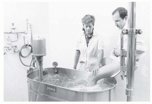
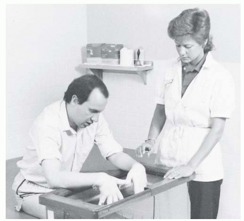
2 and last 5 to 10 minutes. Indirect ultrasound, while less common, is used to treat irregular surfaces such as the foot and ankle where it is difficult to keep the applicator in contact with the skin. In these situations, the body part is placed in a container filled with degassed water. The applicator is held a short distance away (0.5 to 3.0 cm) and moved without touching the skin. Power intensities may need to be higher due to transmission losses.

Coupling between the applicator and the skin is not a trivial issue. The treatment area should be cleansed before treatment, and a coupling agent is necessary. Degassed water (water that has been allowed to sit for several hours) is used for indirect ultrasound because the dissolved gases in water fresh from the tap form bubbles during treatment and attenuate the beam. Little practical difference exists between commercial gels and mineral oil for direct applications, with transmissivity similar to that for degassed water (68). However, mineral oil becomes watery and most people use the commercial products because of the convenience. Coupling agents should not be salt based (e.g., those used for EMG or ECG), as the salt may damage the applicator.


FIGURE 63-6. Direct-contact ultrasound treatment of the elbow. Note the use of a folded towel to support the patient comfortably. Although not shown in the picture, a coupling agent is needed to acoustically couple the applicator and the skin.

Phonophoresis is a variant of ultrasound in which biologically active substances are combined with the coupling medium in the hope that the ultrasound will force the active material into tissue. Although this technique has been in use since the 1960s, neither its effectiveness, penetration, optimal frequency, appropriate coupling mediums/active materials nor amount of material lost to the subcutaneous circulation is well established. Although claims of increased cortisol concentrations at depths of several centimeters after corticosteroid phonophoresis have been made, our own research as well as that of others (69,70) finds limited evidence for deep penetration. Clinical reports are mixed. Thus, although some clinical studies report phonophoresis with a variety of agents successful in terms of improved shoulder range of motion and pain following the treatment (71) as well as in the treatment of keloids and sarcoid nodules (72), other work involving an assortment of musculoskeletal conditions may find the approach no more effective than ultrasound alone (73).


Indications

Musculoskeletal Conditions. The research relating to musculoskeletal condition is surprisingly inconclusive (74). For example, a study of 63 patients with calcific shoulder tendonitis treated with pulsed ultrasound (0.89 MHz, 2.5 W/cm2) found that the treated patients had significantly larger improvements in their pain and decrease in calcium deposits relative to their sham controls at the end of an intensive 6-week treatment program. However, this difference had disappeared at follow-up 9 months later (75). Although ultrasound may be more beneficial than corticosteroid injection in the treatment of shoulder pain, other studies and reviews find it no more effective than placebo or nonsteroidal anti-inflammatory medications for a variety of conditions ranging from subacromial bursitis or lateral epicondylitis (76) to heel pain (77). More tellingly, perhaps, an evidence-based practice guideline panel came to the conclusion that although therapeutic ultrasound was effective in the treatment of calcific tendonitis of the shoulder, there was no compelling evidence of its clinical benefits for other sources of musculoskeletal pain (78). Although these studies raise legitimate issues about its effectiveness in such conditions, many physicians and therapists remain convinced that ultrasound is useful for the treatment of at least some musculoskeletal pain. In support of this, several reviews have found evidence of some benefits in the form of increased motion and lessened stiffness from the cautious use of ultrasound in
patients with rheumatoid arthritis (79, 80, 81). A recent small scale study of ultrasound treatment of myofascial trigger points found significant pain relief from conventional relative to low-intensity ultrasound stimulation (82).

Contractures. Ultrasound is effective in increasing the range of motion of the heel cords, periarthritic shoulders, and contracted hips (83,84). In fact, due to its penetration and focusing capabilities, it is the only agent that can significantly heat (by 8°C to 10°C) the hip joint (65). Hand and Dupuytren contractures also may benefit from ultrasound (85), although a small study of burns did not find treatment beneficial (86). Collagen and tendon extensibility increases as temperatures increase and decreases as tissue cools. As a result, stretching should begin during heating and should continue as the tissue cools and “sets.”

Soft-Tissue Wounds and Inflammation. Ultrasound treatment of wounds and inflammation is based on the belief that either its heat (by increasing blood flow, metabolic, or enzymatic activity) or nonthermal effects (perhaps by changing cell wall permeability) accelerate healing. Laboratory studies offer some support (87). Human studies, however, provide a more mixed picture with a recent review of decubitus ulcers concluding that the evidence was too weak to assess (88). Some feel that inflammation and swelling are indications for ultrasound. Others believe that the heating and membrane permeability changes associated with treatment prohibit its use in acutely inflamed conditions such as in recent trauma and active rheumatoid arthritis.

Trauma. Although ultrasound may aggravate tissue damage and swelling if used too soon after an injury, subacute hematomas (89) and postpartum perineal pain (90) may improve more rapidly with treatment. Ankle sprains are a common indication for ultrasound treatment. Even here, benefits are unclear with a systematic review involving more than 570 patients concluding that current evidence, at best, supported the presence of limited benefit (74). A number of clinical and animal studies find evidence that ultrasound at intensities on the order of 1 W/cm2 may provide at least short-term benefits in the treatment of symptomatic carpal tunnel syndrome (91,92). Although these reports are intriguing, benefits are not always evident and remain controversial (93).

Fractures. Low-intensity ultrasound improves the repair of bony injuries (94). For example, 30 mW/cm2 pulsed 1.5-MHz ultrasound accelerates the healing of closed and open grade 1 fractures (95,96). Although ultrasound is not typically used for this purpose, its use has been approved by the Food and Drug Administration (FDA) for the treatment of some fractures. In view of the fact that 5% to 10% of fractures heal slowly (97), there may be a wider applicability for this treatment in the future.

Other Indications. Postherpetic neuralgia is often resistant to conventional treatment. Treatment with both pulsed and continuous 1- to 1.5-MHz ultrasound has been evaluated. Unfortunately, the studies have again often been poorly controlled, and the results are unclear; some investigators have found improvement (98,99), whereas others have not (100). As ultrasound has thermal, and possibly nonthermal, effects on nerve conduction (101), further evaluation seems appropriate. Plantar warts, keloids, scars, and even chronic sinusitis are additional refractory conditions for which pulsed and conventional ultrasound has inconclusive benefits (102, 103, 104).


Precautions and Contraindications

Ultrasound produces intense heating and, under some conditions, potentially destructive nonthermal effects, such as shockwaves, cavitation, and media motion. The precautions in Table 63-2

Only gold members can continue reading. Log In or Register to continue

Stay updated, free articles. Join our Telegram channel

May 25, 2016 | Posted by in PHYSICAL MEDICINE & REHABILITATION | Comments Off on Therapeutic Physical Agents

Full access? Get Clinical Tree

Get Clinical Tree app for offline access