Fig. 45.1
The adjacent structures of the backside of the left hip joint after lifting up the gluteus maximus are seen. P piriformis, SG superior gemellus, OI obturator internus, IG inferior gemellus, QF quadratus femoris
A vertical incision is made above greater trochanter to approach the hip joint laterally. Thigh fascia (fascia lata) is observed, and then the skin and the superficial fascia are passed. In this region, the fascia thickens to form the iliotibial tract, which lies toward the leg. Gluteus medius, which holds on to the greater trochanter superiorly, and the tendinous attachment of vastus lateralis, which lies in the lateral part of the thigh, can be exposed when the fascia is incised. It is important to recognize the formation of muscles in this area. The tendons of these muscles can be detached from greater trochanter with a “V” shaped incision, with the opening of “V” facing the anterior side in order to protect the muscle fibers [10]. The profound branches of the superior gluteal vessels and nerves passing through the upper part of the greater sciatic foramen (suprapiriform foramen) and lying between gluteus medius and gluteus minimus have to be preserved. These neurovascular structures are found approximately 3–5 cm above the greater trochanter. It should be noted that the distance decreases as we progress toward the front [11]. The terminal branch of the superior gluteal nerve goes to the tensor fascia latae. This muscle is significant, as it is the only muscle innervated posteriorly and flexes the thigh.
For the anterior or anterolateral approaches of the hip joint, skin incision is started from the anterior superior iliac spine to between the tensor fascia latae and sartorius. Lateral femoral cutaneous nerve is the most important structure which must be protected during this incision. This nerve passes under the inguinal ligament and usually over the proximal attachment of the sartorius and enters the fascia of thigh, approximately 2.5 cm below the anterior superior iliac spine [12]. Incisions are performed on the thigh fascia, and deeper levels are reached. The nerve and sartorius must be protected by retracting medially. The incision is deepened in the internervous plane between the sartorius innervated by the femoral nerve and the tensor fascia latae innervated by the superior gluteal nerve. In order to reach the hip joint, we pass between gluteus medius and biceps femoris on the same intravenous plane. Meanwhile, the ascending branch of the lateral femoral circumflex artery lying along the anterior side of the hip joint can cause bleedings [10] (Fig. 45.2). Anterior part of the hip joint capsule can be reached with the removal of the blood vessels and connective tissue.
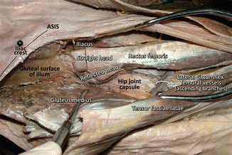
Fig. 45.2
The anterior side of the right hip joint was approached passing between first sartorius and tensor fascia latae, and second gluteus medius and rectus femoris. During this approach, attention must be paid to bleeding from the ascending branch of the lateral femoral circumflex artery. ASIS anterior superior iliac spine
Femoral vessels and nerves in the femoral triangle can be injured during retraction if these are not closely noticed. Femoral triangle is the most important part which is found in the anterior superior part of the thigh (Fig. 45.3). The base of the femoral triangle is upward; its apex is downward and constituted superiorly by inguinal ligament, medially by adductor longus, and laterally by sartorius. Fascia lata which covers the muscles is observed in this area when the skin and subcutaneous tissue are removed. Saphenous opening where the great saphenous vein enters deeper plane is located on the fascia lata covering femoral triangle. Hence, femoral hernias protrude from this area to underneath the skin. After passing beneath the inguinal ligament, external iliac artery goes into the femoral triangle, and it is named as femoral artery. Deep artery of the thigh (profunda femoris artery), which is supplying the posterior side of the thigh is found 2–5 cm beneath the inguinal ligament, and it originates from the external posterior part of the femoral artery. Femoral artery travels through adductor hiatus, and it is named as popliteal artery in popliteal fossa; it ends at the lower border of the popliteus, where it is separated into the two terminal branches: anterior and posterior tibial arteries [8, 10].
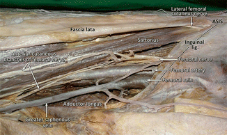
Fig. 45.3
Fascia lata covering the right femoral triangle was removed, and the main neurovascular structures located in this region were made visible. Attention must be paid to the structures located from the medial to lateral, under the inguinal ligament. ASIS anterior superior iliac spine
Femoral vein, artery, and nerve (VAN) can be observed from medial to lateral in the femoral triangle, just beneath the inguinal ligament, when the deep fascia is passed [9] (Fig. 45.3). Profunda femoris artery can also be observed coming out from the external posterior part of the femoral artery. Lateral circumflex femoral artery, a branch of profunda femoris artery, is divided into branches slightly below the neck of the femur, and its ascending branch arises through the front of the hip joint [10]. Medial circumflex femoral artery, which is the other branch of profunda femoris artery, passes between the first pectineus and iliopsoas, and then the obturator externus and the adductor brevis muscles. It is separated into branches beneath the quadratus femoris. The branches given to the joint constitute the most important blood supply of femoral head in the adults. Head of femur is supplied by the branches of lateral and medial circumflex femoral artery and acetabular branch of obturator artery during growth. Acetabular branch of obturator artery passes through the ligament of head of femur (ligamentum teres) and reaches the head of femur [8]. This artery loses its significance after 4 years of age.
The adductor canal (canalis adductorius or Hunter’s canal) is a tunnel in the middle third of the femur and is found between vastus medialis, adductor longus, adductor magnus, and anteromedial intermuscular septum (subsartorial fascia, vastoadductor intermuscular septum). The starting point is the apex of the femoral triangle, and the exit is the adductor hiatus in the tendon of adductor magnus. Adductor canal is also known as subsartorial canal since it is found beneath the sartorius muscle. Femoral vessels pass through the adductor canal and go into the fossa poplitea and travel in here as popliteal vessels. Saphenus nerve is found in the adductor canal with femoral vessels and does not come out of the lower opening of this canal. Slightly above, it pierces the subsartorial fascia to exit the canal and reaches beneath sartorius. It becomes superficial between the sartorius and gracilis muscles and travels along with the great saphenous vein later [13, 14].
There are many sensory fibers found in the nerve of vastus medialis. Therefore, it is the thickest branch of femoral nerve that travels to the muscles. During the flexion of knee, lateral movement is prevented by the fibers of vastus medialis attached to the patella, which pull the patella to the medial origin to sustain the stability [15, 16]. Hence, any damage to this nerve will affect the stability of the knee.
Aponeurosis of tensor fasciae latae joins with the fascia lata, and a thick band-shaped aponeurotic structure, which is gaining strength is formed from the ilium to tibia on the lateral side of the thigh. This band-shaped structure is called iliotibial tract [8, 9].
Knee and Leg Anatomy
Popliteal fossa is a rhomboid-shaped area limited by ischiocrural muscles [hamstring muscles; lateral hamstring = biceps femoris; medial hamstring = semitendinosus + semimembranosus) superiorly and gastrocnemius muscle with its lateral and medial heads inferiorly. Plantaris joins these limits in the inferior lateral area. Small saphenous vein and sural nerve, which are lying between the heads of the gastrocnemius muscle can be observed when the superficial fascia of the lower half of the popliteal fossa is dissected gently. Separation of the branches of sciatic nerve (tibial and common fibular nerves) can be seen when the hamstring muscles in the upper half of popliteal fossa are parted away. This separation can also occur rather above in the gluteal area. Tibial nerve stays at the posterolateral surface of the popliteal vessels at the back of the knee joint in the inferior section; however, fibular nerve travels parallel with the medial side of the tendon of biceps femoris muscle. Popliteal vessels (popliteal artery and vein) are covered with a sheath and pass through a deeper plane, compared to the tibial nerve. Popliteal artery is found in the closest plane to the knee joint capsule. This vicinity must be noticed for both arthroscopic and open surgical procedures of the knee joint [17].
Fibular nerve which follows the medial edge of biceps femoris winds around the head of fibula and goes between soleus and peroneus longus muscles [18]. Herein, the fibular nerve travels superficially right behind the skin. Therefore, fibular neck fractures and the surgical procedures to this area must be performed carefully [19, 20].
The skin branches separating from the tibial and fibular nerves (medial and lateral sural cutaneous nerves, respectively) generally merge inside the superficial fascia at the posterior face of the gastrocnemius muscle to form the sural nerve. Sural nerve goes along with the small saphenous vein and travels back to the lateral malleolus. At the lateral face of the calcaneus, it spreads to the most lateral part of the dorsal face of the foot [21].
Knee is defined as the structures and layers found in the medial and lateral area. There are differences between sources; however, these structures can be defined as below.
There are three layers found in the medial part of the knee [22–26] (Fig. 45.4):
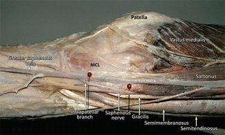
Fig. 45.4
The structures on the medial side of right knee. Attention must be paid to the saphenous nerve arising between the sartorius and gracilis tendons and the infrapatellar branch of this nerve
I.
Structures found in the first layer: (1) sartorius muscle and its fascia; (2) medial patellar retinaculum; (3) saphenous nerve (courses between the first and second layer)
II.
Structures found in the second layer: (1) gracilis; (2) semitendinosus; (3) superficial layer of the medial collateral ligament; (4) medial patellofemoral ligament; (5) semimembranosus
III.
Structures found in the third layer: (1) deeper layer of the medial collateral ligament; (2) joint capsule (coronary ligament)
There are three layers found in the lateral part of the knee [23, 27–30] (Fig. 45.5):
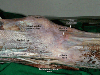
Fig. 45.5
The superficial structures on the lateral side of the right knee. Common fibular (peroneal) nerve was located just behind biceps femoris tendon, and quite superficially
I.
Structures found in the first layer: (1) iliotibial tract; (2) tendon of biceps femoris muscle; (3) common fibular (peroneal) nerve
II.
Structures found in the second layer: (1) lateral patellar retinaculum; (2) lateral patellofemoral ligament
III.
Structures found in the third layer: superficial: (1) lateral collateral ligament; (2) fabellofibular ligament; (3) lateral geniculate artery (runs between deep and superficial layer)
Deep: (1) arcuate ligament; (2) popliteus tendon; (3) popliteofibular ligament; (4) joint capsule (coronary ligament)
There are two arterial networks around the knee joint, deep, and superficial. The one which is superficial (patellar network – rete patellare) forms between the skin and deep fascia. On the other hand, deep plexus (genicular anastomosis – rete articulare genus) is found close to the joint surfaces of tibia and femur. The branches separating from this plexus supply the fibrous and synovial membranes of the knee joint. The arteries constituting this network are as follows: descending genicular artery which is the branch of femoral artery, descending branch of the lateral circumflex femoral artery, recurrent branch of the anterior tibial artery, superior lateral and medial genicular arteries with inferior lateral and medial genicular arteries, which are the branches of the popliteal artery, and circumflex fibular artery, which is the branch of the posterior tibial artery [8].
< div class='tao-gold-member'>
Only gold members can continue reading. Log In or Register to continue
Related posts:

Stay updated, free articles. Join our Telegram channel

Full access? Get Clinical Tree


