Fig. 48.1
Spinal column consists of cervical, thoracic, lumbar, and sacrococcygeal regions. It has curvatures in the sagittal plane, while it is totally straight in the coronal plane
Every anatomic region demonstrates unique vertebral morphology and all vertebrae are separated by intervertebral discs (Fig. 48.2). The unit including two adjacent vertebrae and one disc in between is called as a “functional spine unit.” Summation of all movements raised from these functional units gives spinal column a whole remarkable degree of movement. The following movements are possible in the spinal column: flexion, extension, lateral flexion, rotation, and circumduction. The type and range of movements differ in every region, depending on the thickness of the disc, orientation of facet joints, and tethering of paraspinal structures such as ribs.
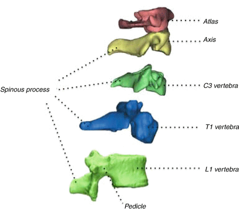
Fig. 48.2
Different vertebral morphologies of all regions. Cervical region has three unique types of vertebra
Basic Anatomy
Cervical Region
All cervical vertebrae are similar, other than the atlas (C1) and axis (C2), which are unique in shape. Atlas differs from the others in that it does not have a body. However, it consists of two lateral masses, which are connected together with an anterior and a posterior arch (Fig. 48.3). It also does not have a spinous process that is represented by a tubercle located at the apex of the posterior arch. On the superior surface of each lateral mass is a concave facet which articulates with the corresponding occipital condyle. There is also a groove at the posterior side of each lateral mass. This groove contains a vertebral artery and first spinal nerve. Vertebral artery injuries may occur in this location during the C1–2 transarticular screw fixation procedure. Especially in the presence of high-riding vertebral artery, this potential risk becomes higher. It is suggested to take three-dimensional angiographic tomography preoperatively for visualization of possible variations of vertebral artery before the C1–2 transarticular fixation procedure [25].
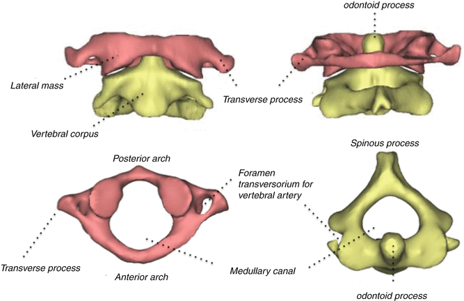
Fig. 48.3
Atlantoaxial complex is seen in first row. Axial views of atlas and axis are shown in second row
Axis is another unique vertebra within the spinal column. It has a vertical bony protuberance which projects upward from the corpus called dens axis or odontoid process. Odontoid process acts as a pivot around where the atlas and head rotate (Fig. 48.3). It has many ligamentous attachments that are very important for atlantoaxial stability. Fractures of the odontoid process may result in nonunion, if, especially, it is broken from its base (Type-II). Histomorphometric analysis showed that the base of the odontoid process has reduced trabecular bone volume, poorer trabecular interconnection, and thinner cortex (cortical thickness is one-third of the other parts) [1]. Another important morphologic feature of axis is its bifid spinous process. This large bony structure provides a prominent landmark for palpation and powerful leverage for the attachment of suboccipital muscles. Due to this important function, surgeons should protect the spinous process of C2 vertebra in order to prevent possible instability.
Subaxial (third to seventh) cervical vertebrae are similar in structure. These have smallest vertebral corpus but largest vertebral foramina of the vertebral column. The superior surface of the vertebral corpus is concave at the transverse plane and convex at the sagittal plane. There are bony elevations called uncinate process in each side. This process has a separate articulation with the uncinate process of the adjacent vertebra known as uncovertebral joint or joint of Luschka. Transverse processes arise from two parts that are from the vertebral corpus anteriorly and from the articular process posteriorly. Transverse processes contain foramen transversariums wherein lie the vertebral artery, vein, and sympathetic plexus bilaterally (Fig. 48.4). Only C7 does not contain vertebral artery.
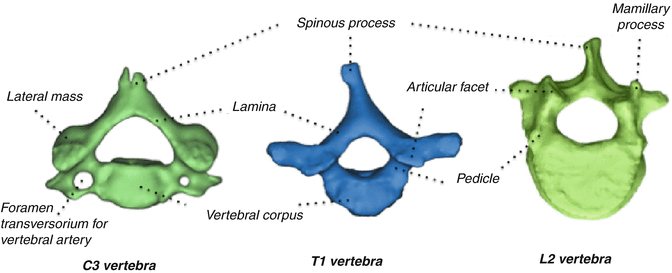
Fig. 48.4
Subaxial cervical, thoracic, and lumbar vertebra samples are shown in the axial plane. It is to be noted that they are all similar but have slight differences
There are lateral masses adjacent to the facet joints bilaterally. These thick bony structures can be used as fixation points during posterior instrumentation procedures. Screw placement to these structures is relatively safe when compared with the cervical pedicle screw placement. Average depth of lateral mass on the sagittal plane is 12.83 ± 1.28 mm, while the average width on the coronal plane is 11.92 ± 0.96 mm. Screw trajectory must be divergent around 20° on the transverse plane and directed caudally 30° on the sagittal plane to prevent vertebral artery and root injuries [17].
Thoracic Region
There are 12 thoracic vertebrae in this region. Major morphologic differences rise from the components of the thoracic cage. Ribs connect to the spine by costotransversal and costovertebral joints and limit the movements of the spinal column. Other reasons for limited motion in thoracic region are orientation of facet joints, sternum, strong intercostal fascia, and the radiate ligament that attaches the head of rib and adjacent vertebra.
Thoracic vertebral body has a cylindrical shape with almost equal anteroposterior and mediolateral dimensions. Vertebral bodies decrease in size from T1 to T3 and then increase to T12, gradually. The primary function of thoracic vertebrae is to bear the weight of the trunk, since the sagittal vertical axis crosses just anterior to the vertebral bodies in the midthoracic region [11] (Fig. 48.5). There are bilateral demifacets on the lateral surfaces of the body, which articulate with the rib heads.
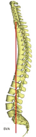
Fig. 48.5
Sagittal vertical axis (SVA) is a relative line that is drawn from C7, perpendicular to the ground. It should be passed ±2 cm around sacral promontorium in normal subjects
Vertebral foramen is near round shape and small. Medullary canal is narrowest at T6. However, this narrowness extends between T4 and T9. Thus, the spinal cord becomes vulnerable in this part of the medullary canal due to any kind of space occupying lesion or foreign body such as pedicle screw. Pedicles are short bony connections between the body and laminae. Pedicle morphology is especially important for inserting pedicle screws during any posterior instrumentation procedure. Pedicle diameter in transverse plane is the most critical variable for thoracic pedicle screw insertion. Usually, the smallest pedicles are T4 and T6, with the transverse diameter ranging between 4.7 and 6.1 mm. On the other hand, the largest pedicle is T12, with the diameter varying from 6.3 to 7.8 mm. Pedicles progressively become larger between T4 and T1 in the cephalad direction [9].
If the screw diameter exceeds 80 % of outer pedicular diameter there will be three possibility: Pedicle will adapt by expansion, pedicle will be cut out by screw threats or pedicle will break. Pedicle fracture is seen mostly in the lateral wall, because the medial cortex of the pedicle is two to three times thicker than the lateral cortex [10, 16].
Angle of the pedicles on transverse plane also differs from level to level. It decreases gradually from 30° convergent at T1 to neutral at T12. The transverse pedicle angle decreases caudally down to around 14° in the fourth thoracic vertebra. It remains around 14° between T4 and T9 and decreases sequentially to neutral in the lower thoracic spine [22, 26].
Thoracic facets are oriented in the coronal plane. Thus, they allow primarily lateral bending and axial rotation. The superior articular facet of the caudal vertebrae forms the roof of the neural foramen. Transverse processes of thoracic vertebra join the pedicles and the laminae at their bases. They extend laterally and posteriorly to make room for the ribs to pass anterior to them. Thoracic vertebrae typically have long and slender spinous processes that point downward and overlap the vertebral arches of the vertebra below. Thus, thoracic laminectomy requires removal of the inferior portion of the cephalad–spinous process (Fig. 48.4).
Lumbar Region and Sacrum
Lumbar segments are quite larger than the cervical and thoracic segments. There are five lumbar vertebrae, and the bodies of these are oval-shaped on the axial plane. Mediolateral diameter is larger than the anteroposterior diameter. Sagittal vertical axis bisects most of the lumbar vertebrae. Thus, lumbar spine bears the whole weight of the trunk, together with the head and upper extremities. Body of the lumbar vertebrae consists of very well designed trabecular system for this function.
Pedicles of lumbar vertebrae are quite large. Pedicle screw, even as large as 8 mm in diameter, can be placed within the pedicles of the lumbar spine. In fact, first pedicle screw was inserted within the lumbar pedicle by Boucher [2].
Laminae are the bony plates projecting each of the pedicles toward the midline. The shape of the lamina in the lumbar region is similar with the thoracic and cervical spine. However, part of the lamina between the superior and inferior articular processes of the lumbar vertebra, called as pars interarticularis or isthmus, has clinical importance. The biomechanical significance of isthmus comes from its location in a transitional zone of forces. It lies at the junction of vertically oriented lamina and horizontally projecting pedicle. Because of this morphological feature, isthmus exposes serious shear forces. Developmental fusion defect of growth centers or fatigue fracture of isthmus is described as spondylolysis. Spondylolysis is usually seen at the L5 level. Moreover, L5 may slip anteriorly over the lower S1 segment due to spondylolysis, if these vertebrae have dysplastic background. This pathology is defined as spondylolisthesis.
Spinous process, which is formed by the connection of laminae in the midline, can be easily palpated under the skin. Most surgeons use this examination while determining the surgical levels. The spinous process intersected by the line that is drawn between the contralateral iliac crests is generally L4.
Extending laterally from the junction of the pedicle and the lamina on each side, there are transversely oriented flat bony bars. These are called as transverse processes. Transverse processes are excellent graft beds beneath well-vascularized paraspinal muscles for posterolateral fusion. Surgeons use this graft bed by decortication of tranverse processes on each side within fusion levels and placement of grafts between them. Another excellent structure for fusion procedure is the facet joint. Articular facets that are covered by hyaline cartilage and facet capsule support the movement function of the spinal column. They also contribute to the stability of the functional anatomic unit with their special orientation.
At the base of the transverse process and lateral to the superior articular facet, there is a bony prominence called mammillary process, which is a very good reference point for inserting the lumbar pedicle screw (Fig. 48.4). But, every lumbar vertebrae may not have a mammillary process. Another point to place the lumbar pedicle screw is the transection of two perpendicular lines: (1) Vertical line drawn from the lateral facet. (2) Horizontal line bisects the transverse process.
The sacrum is a large, irregular, and triangular bone at the base of the spinal column. Its broad upper part joins the lumbar spine by S1 vertebra, while the lower part joins the coccyx. It also connects both os coxae laterally, and they form a pelvic ring together. S1 pedicles are broad but short structures. Inserting a pedicle screw to the S1 pedicle is technically different from that to the lumbar spine. In order to place a longer pedicle screw, its trajectory should be more divergent, and it should penetrate the anterior cortex with a couple of threads for better purchase. Biomechanically strong screw purchase will be accomplished by this technique [14]. There are bony wings on each side at the top of the sacrum called sacral ala. Basically, these structures are identical to the transverse process and also appropriate for screw insertion. But, the last is technically different than the standard pedicle screw fixation and needs convergent trajectory.
Ligaments of Spine
Ligamentous structures are crucial for proper stability of the spinal column. The main ligamentous structures of the spinal column are as follows: ligamentum nuchae (LN), anterior longitudinal ligament (ALL), posterior longitudinal ligament (PLL), ligamentum flavum (LF), facet capsule, interspinous ligament (IL), and supraspinous ligament (SL).
< div class='tao-gold-member'>
Only gold members can continue reading. Log In or Register to continue
Related posts:

Stay updated, free articles. Join our Telegram channel

Full access? Get Clinical Tree


