Fig. 22.1
Illustrates standing full-length radiographs. This allows for assessment of varus and determination of mechanical axis
Fig. 22.2
Demonstrates the difference between primary, double, and triple varus
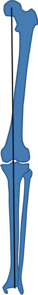
Fig. 22.3
Mechanical axis shown in this figure is drawn from the center of the femoral head to the center of the ankle joint (not plafond)
Dugdale et al. used this film to identify the amount of correction needed [17]. First, a line is drawn from the center of the femoral head to the point along the tibial plateau through which the desired mechanical axis will pass. Next, a line from the center of the tibiotalar joint to this point is draw [17]. The angle created by the intersection of these lines is the amount of correction needed with HTO to achieve the desired mechanical axis (Fig. 22.4). Dugdale et al. also stated that every 1° of varus is proportional to a 1-mm increase in lateral joint line widening [17]. In addition, the A-P view of the knee can be very helpful to observe Segond and arcuate avulsion fractures of the lateral capsule and fibular head, respectively.
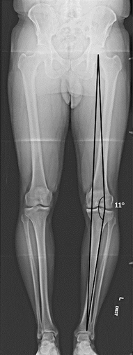
Fig. 22.4
Demonstrates how to determine mechanical axis correction. The point through which the desired mechanical axis will pass is first identified. The angle created by the intersection of the femoral and tibial mechanical axis and the desired point is the amount of correction needed
Supine Full-Length Bilateral A-P Radiograph
Supine radiographs are important to obtain because they eliminate the added varus brought about by insufficiency to the lateral and posterolateral soft-tissue structures. This allows the surgeon to measure the true amount of correction needed.
Lateral Radiograph
A lateral X-ray is essential to measure the posterior tibial slope. Dating back to the 1970s, a number of methods have been suggested for calculating the posterior tibial slope [20, 21]. Brazier et al. compared the tibial slope measurement methods and concluded that the proximal tibial anatomic axis (PTAA) and posterior tibial cortex (PTC) methods are the most reliable [21]. In the PTAA method, a line is drawn along the proximal tibial anatomical axis and another is drawn along the tibial plateau. The angle between these two lines is the tibial slope [20]. In the PTC method, a line is drawn along the PTC and another is drawn along the medial tibial plateau. The angle between these two lines is the tibial slope [21].
Merchant’s View
This radiograph is obtained by having the patient lie in the supine position with the knees at 45° of flexion over the end of the table. The knees are held to maintain the femora parallel to the floor. The beam is directed proximal to distal, forming a 30° angle with the table. The film cassette is placed about 30 cm below the knees, perpendicular to the tibial shaft. This view is helpful for looking at the patellofemoral joint. Specifically, in this patient group, Merchant’s view helps assess patellofemoral degenerative changes, patellar tilt, and maltracking.
Rosenberg’s View
The patient stands on both legs with thumbs pointing ahead and the patellae touching the film cassette. The knees are at 45° of flexion (25° between the femora and the cassette, and 20° between the tibiae and the cassette), and the X-ray beam is directed posterior to anterior, 10° caudal, so the posterior and the anterior margins of the tibial plateau are superimposed. This view is ideal for assessing the lateral compartment.
Stress View
A number of methods have been described for obtaining a stress view of the knee. Jacobsen first described a method for obtaining a lateral stress radiograph of the knee in 1976 [22]. This description calls for the patient to lie in the lateral decubitus position with the knee flexed to 90°. The heel is fixed to a stand, and the arm of the Telos GA II (Telos, Weterstadt, Germany) applies a posterior force to the tibia [22]. A lateral X-ray is then taken in this position. A radiograph is then taken with the knee in 25° of flexion. This method is very important in chronic PCL -deficient knees to evaluate both anterior and posterior tibial translation with regard to the femur, and it is useful to detect fixed posterior tibial subluxation. The lateral stress view using the kneeling method asks the patient to kneel on a flat, elevated surface with the knee flexed to 90° while a lateral X-ray is taken [23]. Meanwhile, the hamstring contraction method has the patient lie in the lateral decubitus position or in a seated position with the knee at 90° of flexion and the heel fixed to a stand. A lateral X-ray is taken while the patient contracts his/her hamstring [24]. The gravity method asks the patient to lie supine with the hip and knee flexed to 90°, supported by an assistant, with the leg in neutral rotation. In this position, a lateral X-ray is taken [25]. Lastly, Puddu et al. described an axial stress test in 2000 [26]. The patient lies in supine position with both knees at 70° of flexion, feet in moderate plantar flexion, and the tibia in neutral rotation. The X-ray beam is directed parallel to the longitudinal patellar axis, from distal to proximal, and the distance between the anterior tibial profile and the center of the femoral groove is measured [26]. The side-to-side difference is the amount of posterior instability.
Jung et al. compared all five of these methods, focusing on posterior translation, side-to-side difference, condyle rotation, time to perform the test, and pain during the test. Considering all these factors, they stated that the most effective methods are Telos view at 90° of knee flexion and the kneeling method, even if they are painful and time-consuming [24]. Despite being the most expensive method, the Telos method is the most reliable in detecting posterior tibial subluxation [24].
MRI
Magnetic resonance imaging (MRI ) is useful in evaluating the PCL -deficient knee, especially associated soft-tissue injuries and subchondral bony edema. Gross et al. introduced a classification system for PCL tears and noted 100 % sensitivity and specificity for diagnosing a PCL tear using T1-weighted MRI [27]. Bellelli et al. incorporated T2-weighted spin echo (SE) and short-tau inversion recovery (STIR) scans to augment the classification system established by Gross et al [28]. In the new classification scheme, the two bundles of the PCL were treated independently. Type I lesions involve high-intensity signal within the ligament with low intensity along the borders of the ligament. Type II lesions are partial and involve the dorsal edge of the ligament (posteromedial bundle). Type III lesions are partial and involve the ventral edge of the ligament (anterolateral bundle). Type IV lesions represent a complete tear with both the dorsal and ventral edges involved [28].
MRI is also helpful for evaluating the PLC. LaPrade et al. recommend using at least a 1.5-T scanner with dedicated PLC sectioning using thin-sliced (2 mm) proton density coronal oblique images, which include the entire fibular head and styloid [5]. Doing so affords evaluation of the iliotibial band, long head of the biceps femoris, short head of the biceps femoris, fibular collateral ligament, popliteus complex, and the fabellofibular ligament.
Indications
In general, indications for HTO in the setting of PCL deficiency include the following:
Young, healthy patients in whom arthroplasty would fail due to excessive wear
Good vascular status
Pain and/or disability interfering with daily living
Compliant patient who will abide by postoperative protocols
Contraindications
Contraindications to HTO include the following:
Severe osteoporosis
Inflammatory disease
Infection
Severe tricompartmental OA
Prior lateral meniscectomy
Knee flexion < 90°
Relative contraindications include the following:
Morbid obesity
Age greater than 65 years
Patellofemoral OA
Tobacco use
Preoperative Planning
Careful preoperative planning is essential prior to performing an HTO. PCL and PLC lesions are often associated with malalignment of the knee, and they should be addressed in a staged fashion 6–8 months after HTO if the knee is still unstable. Poor results are common after soft-tissue procedures alone in the setting of the malaligned knee. This is due to the forces on these structures, which do not decrease if the underlying malalignment is not corrected, as the bony deformity overstresses them. Instead, HTO reduces these forces and improves the stability and biomechanics of the knee [5, 13, 19, 29–31].
If medial compartment OA with joint-space narrowing is present, then the new weight bearing axis should be positioned to intersect at 62–66 % of the tibial plateau (where 0 % indicates the medial margin of the tibial plateau and 100 % the lateral margin), as this alignment increases the pressure on the lateral compartment of the knee [17, 19]. This slight overcorrection has been shown to prevent progression of medial compartment OA and early recurrence of varus [19]. If degenerative narrowing of the medial compartment is not present, the new mechanical axis should split the tibial plateau in half [19]. Medial opening wedge HTO improves symptoms of patellofemoral OA because the anterior translation of the tibia reduces the tension on the patellar tendon, thereby reducing stress on the lateral facet [3]. Because of this, patellofemoral pain is not a contraindication to HTO.
The type of osteotomy performed to correct the deformity alters the posterior tibial slope in a fairly predictable manner. Lateral closing wedge and dome HTOs have been shown to decrease posterior tibial slope [32]. Although decreasing slope may be beneficial in the anterior cruciate ligament (ACL)-deficient knee, it may exacerbate instability in a PCL -deficient knee. Conversely, medial opening HTO tends to increase posterior tibial slope [33]. Noyes et al. determined that the height of the anterior osteotomy gap at the tibial tubercle must be one half the height of the posteromedial gap to maintain normal sagittal alignment [34]. Furthermore, the authors showed that a gap error of a single millimeter will result in approximately 2° of change in the posterior tibial slope [34].
If meniscal lesions are associated, they should be addressed at the same time as. HTO. A preoperative rehabilitation protocol consisting of strengthening and gait training has been suggested in the literature as a measure to avoid recurrence of hyperextension varus thrust gait after surgery [19]. In the patient with a chronic PCL -deficient knee associated with double or triple varus, HTO should be performed before soft-tissue procedures. The patient should be evaluated 6–8 months later, and soft-tissue reconstruction can subsequently be undertaken if the knee remains unstable.
Operative Technique
The patient is placed on the operating room table in a supine position. A thigh tourniquet can be used on the operative leg. However, this may occlude possible vascular injury that may need to be addressed after the tourniquet is let down. Some authors prefer to use iliac crest bone autograft harvested from the ipsilateral side. In this case, the iliac crest is also prepped and draped along with the surgical limb.
The initial incision is medially based over the pes anserine vertically oriented halfway between the tibial tubercle and the PTC. The sartorius fascia is sharply incised parallel to the underlying hamstring tendons. Care should be taken to preserve the sartorius fascia so that it can be repaired upon closure of the wound. In the case of concurrent ligament reconstruction of the ACL or PCL, the hamstring may be harvested at this time. The superficial medial collateral ligament (MCL) is then exposed by retracting the hamstrings (if still in place) medially. The superior portion of the MCL attachment is released with an elevator to expose the posteromedial border of the tibia. On the anterior aspect of the tibia, the fascia is dissected at the level of the patellar tendon insertion. Visualization and exposure are key and blunt retractors are placed under the MCL and patellar tendon.
Related posts:
 Results of Treatment of Posterior Cruciate Ligament Surgery
 Anatomy and Biomechanics of the Posterior Cruciate Ligament and Their Surgical Implications
 Combined Posterior Cruciate Ligament and Posteromedial Reconstruction
 All-Arthroscopic Tibial Inlay Double-Bundle Posterior Cruciate Ligament Reconstruction
 Clinical and Arthroscopic Evaluation of the Posterior-Cruciate-Ligament- Injured Knee
 Combined PCL, Posteromedial Corner, and Posterolateral Corner Reconstruction
Stay updated, free articles. Join our Telegram channel

Full access? Get Clinical Tree


