Fig. 11.1
Chondrosarcoma located in the right femur. Coronal images with T1-weighted (a), fat saturated (FS) T2-weighted (b), and contrast enhanced FS T1-weighted images (c). Note the associated soft tissue mass in the medial aspect of the femoral neck
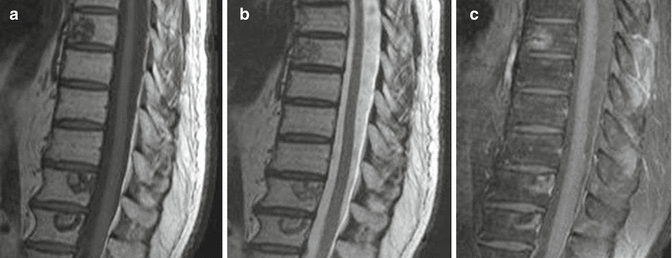
Fig. 11.2
Dorsal and proximal lumbar spine obtained in the sagittal plane with T1-weighted (a), T2-weighted (b) and contrast enhanced FS T1-weighted images (c). Multiple metastatic lesions in the vertebrae in a case with known prostate cancer
While sagittal and coronal images may provide a broad perspective pertaining to the extension of the tumoral lesion with its full length in the bone marrow, axial images may display a better visualization of its neurovascular involvement.
Vascularity and permeability of the tumoral lesion may be assessed by using the paramagnetic contrast agents. Edema and cystic lesions show high signal intensity on T2-weighted images. Though dependent on their pathology, in general, musculoskeletal tumors are hypointense on T1-weighted images and hyperintense on T2-weighted images. In this content, a fibrous tumor with a high collagen content may display hypointense signal on T2-weighted images.
Some tumoral lesions are observed with their specific findings on MR images. For example, the cortex and medullary cavity of an osteochondroma is continuous with that of the involved bone. The thickness and morphology of the cartilage cap as well as an accompanying bursa formation can be defined on fluid-sensitive sequences (Fig. 11.3). Fluid-fluid (blood/serum) levels of an aneurysmal bone cyst (differential diagnoses should also include some other lesions as well as telangiectatic osteosarcoma), the periosteal reaction, and sclerosis around the nidus of an osteoid osteoma and its reactive edema are called “Aunt Minnie lesions” diagnosed on MR images.
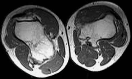
Fig. 11.3
Transverse T1-weighted MR image of both thigh. The cortical and marrow continuity of the lesions with the underlying bone is well seen. Note the compression of the popliteal vascular structures by these lesions
Dynamic contrast-enhanced MR imaging can be referred for (1) determining the most appropriate route for biopsy sampling by evaluating the viable tumor tissue before intervention, (2) evaluating the change in tumoral size and necrosis as a means of treatment response after chemotherapy, and (3) assessing the residual and recurrent tumoral masses after the surgery.
Bone marrow involvement and the soft tissue component around the tumors can be shown to a better extent with MR imaging than with CT, owing to the inherited high soft tissue resolution of the previous technique. However, calcification in the tumoral tissue, cortical bone destruction, and periosteal reaction are perceived better with CT. In the complex anatomic regions such as the spine, tumor extension into the canal, accompanying soft tissue and bony invasion, can be evaluated on MR images.
Spinal Column Imaging
Magnetic resonance imaging is a highly referred modality in evaluating the discal pathologies of the vertebral column. Degeneration of an intervertebral disc and its other pathologies such as bulging and protrusion and their relation with that of thecal sac and the nerves can be readily seen on MR images [23] (Fig. 11.4). While the sensitivity of MR imaging is higher than that of CT for discal pathologies, it is comparable in the evaluation of spinal stenosis [24]. In addition, granulation tissue in the postoperative setting can be easily distinguished from recurrent/residual disc herniation on fat-suppressed T1-weighted images after contrast media administration. Granulation tissue shows contrast enhancement in contrast to recurrent/residual disc herniation [25].
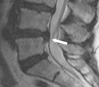
Fig. 11.4
Sagittal T2-weighted image of L4–5 disc extrusion (arrow)
Magnetic resonance myelography is a noninvasive means in demonstrating spinal canal narrowing by using coronal T2-weighted sequences, without any interventional procedure and necessitating the use of intrathecal contrast agents.
Spinal cord tumors, spondylitis – spondylodiscitis – and accompanying any abscess formation can be evaluated in detail with MR imaging, especially on post-contrast images [26] (Fig. 11.5). Spinal involvement secondary to specific infectious agents such as Brucella and tuberculosis, which are frequently seen in our country, can be evaluated on MR imaging.
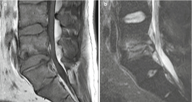
Fig. 11.5
Distal lumbar spine on sagittal plane with T1-weighted (a) and FS T2-weighted (b) images. There are irregularities at the end- plates of L4 and L5 vertebrae with hyperintense signal intensities both on the end-plates and the intervertebral disc (b), corresponding to vertebral osteomyelitis and discitis
Compression fractures of the vertebrae particularly seen in osteoporotic patients, the extension of the fractured bony fragments into the spinal canal, and the differentiation from malignant vertebral compression fractures can be made with high accuracy with MR imaging. In these patients, diffusion-weighted images added to the conventional sequences may be of help in making a distinction between these two processes, which in malignities, diffusion restriction can be observed.
Imaging in Trauma
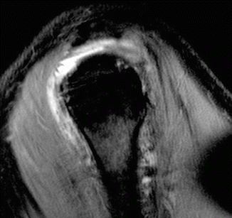
Radiography for occult fractures of the spine CT is the preferred imaging modalities. However, MR imaging is particularly needed for the evaluation of trauma effects on soft tissues, such as tendons, ligaments, and muscle compartments. Bone marrow edema like intensity changes, i.e., high signal intensity on fluid-sensitive sequences and low intensity on T1-weighted images, is observed after trauma, secondary to the contusion of the trabeculae and hemorrhage. Bone marrow edema associated with occult fractures can readily be demonstrated on fat-saturated sequences. Fracture lines can easily be perceived on T1-weighted images with their linear low signal intensities [27]. Especially for the early diagnosis of scaphoid and femoral neck fractures, which are not seen on radiograms, MR imaging helps to identify the fracture lines and bone marrow edema with the high soft tissue resolution that this method yields (Fig. 11.6) [10].
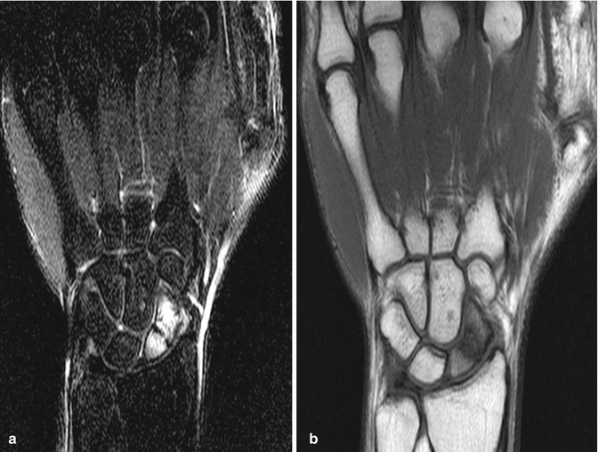
Fig. 11.6
Scaphoid fracture and accompanying edema, FS T2-weighted images (a) and T1-weighted images (b)
Imaging Particular Joints
Temporomandibular Joint Imaging
The temporomandibular joint (TMJ) has a fibrous disc that resembles the meniscus of the knee, both in appearance and tissue composition. The disc looks like a bow tie on sagittal images with the triangles of the bow called “anterior or posterior band.” The posterior band of the disc is much greater than the anterior band. A thin intermediate zone lies in between these two bands. The joint is evaluated while the patient is of open and closed mouth positions. The fibrocartilaginous disc is seen with low signal intensity on both sagittal T1- and T2-weighted images. The disc dislocations can be assessed at an open mouth position by evaluating the mandibular condyle-articular eminence relationship with the disc. Coronal images may show articular surfaces and so can reveal degenerative changes, if there is any [28].
Knee Imaging
The internal derangements of the joint that necessitates MR imaging may involve the menisci, ligaments (cruciate, collateral, patellar ligament), cartilage surfaces, and bone.
The normal meniscus is of low intensity on both T1- and T2-weighted sequences owing to its fibrocartilaginous tissue composition. Degeneration of the meniscus shows high signal intensity that does not extend to the articular surface on fluid-sensitive sequences. If this high signal intensity reaches to the articular surface, we should describe this as a tear that may be evaluated in more detail on coronal and sagittal proton-weighted images (Fig. 11.7). The two most important criteria for meniscal tears are an abnormal shape of the meniscus and high signal intensity unequivocally contacting the surface on proton-weighted images. Meniscal tears can be described roughly as longitudinal and vertical according to the orientation of the collagen bundles of the meniscus itself. A combination of these types can be reported as complex tears.
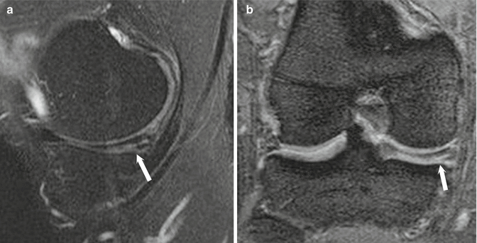
Fig. 11.7
Sagittal (a) and coronal (b) FS T2-weighted image. Medial meniscus tear of the dorsal horn extending lower articular surface (arrow) is observed
The anterior and posterior cruciate ligaments are revealed with their low signal intensity and in the sagittal images throughout their length. When completely torn, the ligaments are not observed with their full continuity (Fig. 11.8). Gradient echo sequences are helpful in evaluating the cartilage. Subchondral bone marrow edema with cartilage defects can be strikingly observed on fluid-sensitive sequences [29]. The patellar and quadriceps tendons show low signal intensity like other tendons, and the degeneration and surrounding inflammation are revealed with the high intensity on T2-weighted images.
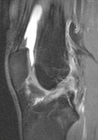
Fig. 11.8
Anterior cruciate ligament tear. Ligament is not visualized with its normal signal intensity and contour
Shoulder Imaging
Shoulder joint is the third most common site of musculoskeletal pain occurring in 7–25 % of general population [30]. The shoulder MR imaging is often referred for evaluating the rotator cuff lesions. With MR imaging, the rotator cuff, long head of the biceps tendon, glenoid labrum, cartilage, acromioclavicular joint, adjacent muscle, and bone structures are evaluated. Rotator cuff pathologies are commonly encountered in patients with shoulder impingement syndrome. Especially examining simultaneously the oblique, sagittal, and coronal fat-suppressed fluid-sensitive sequences reveals the partial- or full-thickness tendon tears in their high signal intensity (Fig. 11.9). Rotator cuff tears can be observed as:
 < div class='tao-gold-member'>


Only gold members can continue reading. Log In or Register to continue
Related posts:

Stay updated, free articles. Join our Telegram channel
Full access? Get Clinical Tree
