Tendon Sheaths and Bursae
Shahan K. Sarrafian
Armen S. Kelikian
SYNOVIAL TENDON SHEATHS
The tendons of the leg at the level of the ankle and the plantar aspect of the foot engage in fibrous, fibro-osseous tunnels acting as pulleys or as retention systems. Within these tunnels, the tendons are surrounded by synovial sheaths to facilitate their gliding.
Structurally, a synovial tendon sheath has a parietal layer lining the deep surface of the fibrous or fibro-osseous canal, a visceral layer covering the tendon, and a mesotenon connecting the latter to the parietal lining. The synovial sheath forms a cavity dosed at both ends, and proximally, synovial folds permit the play of the tendon through the sheath.
Tibialis Anterior
The synovial tendon sheath of the tibialis anterior (Figs. 6.1 and 6.2) extends from above the proximal arm of the inferior extensor retinaculum to the level of the talonavicular interline. Hartman mentions that the synovial sac extends 5.75 cm proximal to a line joining the center of the malleoli.1 The length of this synovial sheath is given as 6 to 8 cm or 8 to 10 cm.2, 3 Hartman reports that the distal end of the synovial sac reaches the talonavicular joint in 60%, is shorter in 22%, and is longer in 18%.1 There is a complete mesotenon to the tibialis anterior tendon throughout the length of the sheath, located on the deep surface of the tendon.
Extensor Hallucis Longus
The synovial tendon sheath of the extensor hallucis longus (see Figs. 6.1 and 6.2) starts lower than the synovial sac of the tibialis anterior tendon but extends more distally. It originates slightly above the upper arm of the inferior extensor retinaculum, at 1.75 cm proximal to the intermalleolar interline, just above the talotibial articular interline.1, 4 The lower limit of this synovial sac is more difficult to delineate; it usually reaches the level of the first cuneometatarsal joint. Less frequently, this distal end of the synovial sheath extends distal to the cuneometatarsal joint interline and advances more or less on the dorsum of the first metatarsal bone.2 Hartman mentions that the synovial sac terminates at the level of the cuneo1-metatarsal1 joint in 34%, proximal to it in 34%, and distal to it in 32%, and that it may reach the lower third of the first metatarsal in 24%.
Extensor Digitorum Longus
The synovial tendon sheath of the extensor digitorum longus (see Figs. 6.1 and 6.2) is a large but short synovial sac that enlarges as it extends distally and also covers the tendon of the peroneus tertius. It starts 2 to 3 cm above the ankle joint, about 1 cm above the upper limit of the extensor hallucis longus tendon sheath.3 The synovial sac passes under the undivided segment of the inferior extensor retinaculum, enlarges to cover the diverging tendons, and ends at the level of the cuneonavicular joint in a large blind sac that is subdivided into small saclike projections that are 0.5 cm in length and are located on the superficial aspect of the tendons.3 There is no prolongation over the tendon of the peroneus tertius.
Extensor Digitorum Brevis
Lovell and Tanner describe synovial sheaths to the tendons of the extensor digitorum brevis (see Fig. 6.1) when the tendons are well developed.3 The synovial sheath of the extensor digitorum brevis to the big toe is the longest, measuring 5.5 cm; it originates under the fascial band extending from metatarsal 1 to 2 and terminates at the proximal end of the proximal phalanx.
The synovial sheaths of toes 2, 3, and 4 are 3 to 4 cm in length; they start from close to where the tendons originate, pass under the digital extensor aponeurosis, and terminate at the level of the proximal phalanges.3
Peroneus Longus and Peroneus Brevis
The synovial sheath of the peroneus longus and peroneus brevis is complex (Figs. 6.3 and 6.4). It is common to both tendons behind the lateral malleolus and bifurcates for a short distance above and for a longer distance below the malleolus. The common synovial sheath extends 2.5 to 3.5 cm above the tip of the lateral malleolus.3 The proximal bifurcation extends 2.5 cm on the peroneus longus and 1.5 cm on the peroneus brevis.3 Inferiorly, the synovial sac bifurcates at the level of the peroneal tubercle of the calcaneus, which separates the two tendons. The bifurcation extension of the peroneus brevis is located above the tubercle and extends up to within 2 cm of the insertion of the tendon on the base of the fifth metatarsal or to the level of the calcaneocuboid joint.
The synovial sac of the peroneus longus terminates in the region of the groove of the cuboid. On the superficial surface of the tendon, the sac extends for about 1 cm distal to the peroneal tubercle, whereas on the osseous surface it extends into the groove of the cuboid entering the planta pedis.3
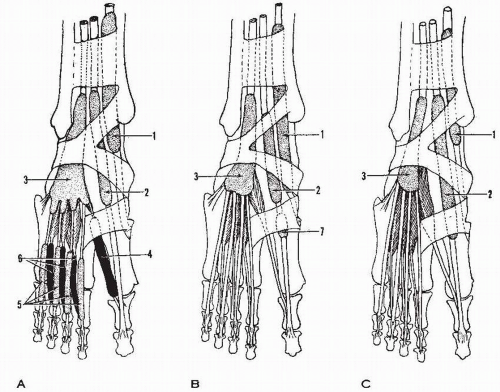 Figure 6.1 Extensor tendon sheaths. (A) According to Lovell and Tanner.3 (B) According to most French anatomists. (C) According to most German anatomists. (1, tendon sheath of tibialis anterior; 2, tendon sheath of extensor hallucis longus; 3, tendon sheath of extensor digitorum longus; 4, tendon sheath of extensor hallucis brevis; 5, tendon sheath of extensor digitorum longus, digital portion; 6, tendon sheath of extensor digitorum brevis; 7, distal segment of extensor hallucis longus tendon sheath.) (From Jones FW. Structure and Function as Seen in the Foot. 2nd ed. Longon: Baillière, Tindall, & Cox; 1949:229-245.) |
In the sole of the foot, the peroneus longus tendon receives a second synovial sheath, which extends from the groove of the cuboid to the insertion of the tendon on the base of the first metatarsal. At the level of the cuboid, this synovial sac extends laterally less on the superficial surface of the tendon and more on the deep surface; this brings the two sacs close to one another on the osseous surface of the tendon. At this level, the two synovial sacs of the peroneus longus tendon are separated only by a thin, cellular, transparent lamella, or they may even communicate (about one-third of cases).2
Behind the lateral malleolus, the common synovial sheath of the peronei tendons is in close contact with the synovium of the ankle joint above and below the posterior talofibular ligament. The peronei tendon sheath and the calcaneofibular ligament cross each other in an “X” and are in intimate contact. The possible communication of the common synovial sheath of the peronei with the ankle joint is of importance in the arthrographic assessment of ligamentous injuries of the ankle joint.
According to Brostrom and Prins and based on their arthrographic assessment of injured ankles in healthy individuals, there is no communication between the synovial sheath of the peronei and the synovial cavity of the ankle joint.5, 6 The data provided by investigators is as follows: postmortem—in 18 ankles, 0 communication; in 40 ankles, 0 communication; in vivo—in 45 ankles, 0 communication; in 68 ankles, 0 communication; in 17 ankles, 14% communication; in 95 ankles, 26% communication.5, 7
Tibialis Posterior
The synovial sheath of the tibialis posterior tendon (Fig. 6.5; see Fig. 6.7) is 7 to 9 cm in length and starts 6 cm proximal to the tip of the medial malleolus. It descends along the tendon in the retromalleolar groove and terminates close to the tuberosity of the navicular, extending more on the osseous surface and less on the superficial surface. The tibialis posterior tendon has no mesotenon.
The synovial sheath sometimes communicates with that of the flexor digitorum longus or with the synovial cavity of the ankle joint (in 5.8% of cases).3, 6
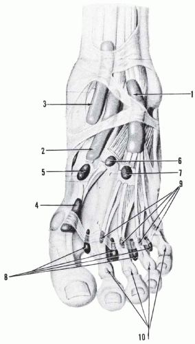 Figure 6.2 Tendon sheaths and bursae of the dorsum of the foot. (1, tendon sheath of extensor digitorum longus and peroneus tertius; 2, tendon sheath of extensor hallucis longus; 3, tendon sheath of tibialis anterior; 4, distal synovial space of extensor hallucis longus [may be tendon sheath or bursa]; 5, bursa between metatarso1-cuneiform1 joint and extensor hallucis longus tendon; 6, 7, bursae between tarsometatarsal joint and extensor digitorum brevis, extensor hallucis brevis; 8, intermetatarsophalangeal bursae seen in upper and anterior parts; 9, bursae between extensor digitorum longus tendons and dorsal extensor aponeurosis at level of metatarsophalangeal joint; 10, subcutaneous bursae over proximal interphalangeal joints.) (From Hartman H. Die Sehnenscheiden und synovialsäcke des fusses. Morphoi Arbeit. 1896;5:214.) |
Flexor Digitorum Longus
The synovial sheath of the flexor digitorum longus (see Figs. 6.5, 6.6, and 6.7) has two components: malleolar and digital.
The malleolar synovial sac starts about 5 cm above the tip of the medial malleolus, slightly lower than the sac of the tibialis posterior. The synovial sac accompanies the tendon of the flexor digitorum longus in the calcaneal canal and terminates in the sole of the foot where the flexor digitorum longus tendon crosses that of the flexor hallucis longus. On the superficial aspect of the tendon, the sheath falls 1 cm short of this crossing point, and a mesotenon is present on its entire length.3 The synovial sheath of the flexor digitorum longus may communicate with those of the tibialis posterior and flexor hallucis longus tendons.
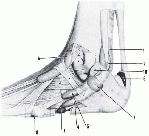 Figure 6.3 Tendon sheaths and bursae of the lateral aspect of the ankle and foot. The combined tendon sheath of the peronei tendons splits into two: peroneus longus and peroneus brevis above and peroneous longus and peroneus brevis below. (1, peroneus longus above; 2, peroneus brevis above; 3, peronei tendons; 4, peroneus longus below; 5, peroneus brevis below; 6, tendon sheath of extensor digitorum longus and peroneus tertius; 7, bursa between peroneus longus tendon and abductor digiti quinti; 8, bursa between metatarsal 5 and abductor digiti quinti; 9, pre-Achilles bursa; 10, lateral malleolar subcutaneous bursa.) (From Hartman H. Die Sehnenscheiden und synovialsäcke des fusses. Morphoi Arbeit. 1896;5:214.) |
Related posts:


Stay updated, free articles. Join our Telegram channel

Full access? Get Clinical Tree


