Osteology


Osteology


Shahan K. Sarrafian

Armen S. Kelikian


LOWER ENDS OF THE FIBULA AND TIBIA

The lower ends of the fibula and tibia form an anatomic and functional unit providing the osseoligamentous retention system to the talus and contributing to the ligamentous stabilization of the calcaneus at the subtalar joint. The bimalleolar retaining fork is rigid medially and movable laterally.


Lower End of the Fibula

The lower end of the fibula is divided into the distal fibular shaft and the lateral malleolus.


Distal Fibular Shaft

The distal one-fourth of the fibular shaft terminates at the level of the tibial plafond. It has two surfaces, lateral and medial, separated by an anterior and a posterior border (Fig. 2.1).


Medial Surface

The interosseous crest divides the upper segment of the medial surface into an anterior narrow segment giving attachment to the peroneus tertius and a broader posterior part, flat or markedly convex, for the lower fibers of the flexor hallucis longus muscle. Where the anterior border and the interosseous crest merge, an oblique line originates and is directed downward and posteriorly, delineating a triangular area with a distal base and an anterosuperior apex. This triangular surface is covered by rugosities and gives insertion to the tibiofibular interosseous ligament, which is in continuity at the apex with the interosseous membrane. Distal to the insertion of the interosseous membrane is a triangular smooth surface with an anterior base and a posterior apex. The broader anterior part, which has an average height of 1 cm, corresponds to the tibioperoneal recess lined by periosteal synovium. The narrow posterior part gives insertion to a fatty synovial fringe that descends in the ankle joint (Fig. 2.2).


Lateral Surface

At the level of the distal fibula, the anterior border divides into two branches, anterior and posterior. The anterior branch remains anterior, merges with the interosseous crest, and continues into the anterior border of the lateral malleolus. The posterior branch or oblique crest is directed downward and posteriorly and continues with the posterior border of the lateral malleolus, forming the lateral border of the peroneal sulcus. This oblique crest delineates two surfaces: anteroinferior and posterosuperior. The anteroinferior segment is limited by the anterior and posterior divisions of the anterior border. The surface is laterally oriented, flat, and subcutaneous. The posterosuperior segment is initially posterolateral in orientation. Further down, through a twist, it becomes posterior and is in continuity with the posterior aspect of the lateral malleolus. The lower fibers of the peroneus brevis muscle originate from the upper segment of this surface. Both peronei muscle tendons, initially in a lateral position, follow the posterior shift of this segment. The anteroinferior and posterosuperior surfaces are slanted relative to each other along the oblique crest, and this must be taken into consideration during the application of a plate to a fractured fibular shaft at this level.

The anterior border was described previously. The posterior border is distinct proximally but loses its definition distally and dissipates toward the medial border of the peroneal groove.


Lateral Malleolus

The lateral malleolus is pyramidal in contour and presents three surfaces: lateral, medial, and posterior (Figs. 2.3 and 2.4). The lateral and medial surfaces converge toward the anterior border. The posterior surface is limited laterally by the oblique fibular crest and medially by the direct extension of the posterior border of the fibular shaft. The apex of the pyramid is inferoposterior. The lateral malleolus is projected outward and descends 1 cm further than the medial malleolus.


Lateral Surface

The lateral surface of the malleolus is smooth, convex, and subcutaneous and is in continuity with the anteroinferior segment of the fibular lateral surface.


Medial Surface

The medial surface is limited at a level corresponding to the incisura fibularis of the distal tibia. A large triangular articular

surface occupies the anterosuperior aspect. The base of the triangle is proximal and convex. The apex is anteroinferior, located on the anterior border of the malleolus. The anterior border is inclined backward, whereas the posterior border is directed anteroinferiorly. The surface is convex along its long axis and corresponds to the lateral articular surface of the talus. Behind the posterosuperior angle of the triangular articular surface is the round posterior fibular tubercle, which gives origin to the deep component of the posterior tibiofibular ligament. Below the tubercle and behind the triangular articular surface is the digital fossa. The upper segment of the fossa is cribriform, with multiple vascular foramina. The lower segment gives origin to the posterior talofibular ligament. The superficial components of the posterior tibiofibular ligament originate from the posterior border of the peroneal tubercle and digital fossa (see Fig. 2.2).


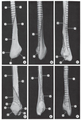
Figure 2.1 Distal fibula and lateral malleolus. (A) Lateral surface. (B) Posterolateral view. (C) Posteromedialview. (D) Medial surface. (E) Anterior surface. (F) Posterior surface. (1, surface of origin of peroneus brevis muscle—this surface becomes posterior distally and continues as posterior surface of lateral malleolus; 2, anterior border of fibular shaft for insertion of anterior peroneal septum; 3, posterior division branch of anterior border [2]—it forms a crest and continues as lateral border of posterior surface of lateral malleolus; 4, anterior division branch of anterior border; 5, subcutaneous surface; 6 posterior border of lateral malleolus; 7, tip of lateral malleolus; 8, anterior border of lateral malleolus; 9, sulcus of peronei tendons; 10, line of insertion of deep transverse fascia of leg; 11, line of insertion of posterior peroneal septum; 12, posterior tubercle of medial surface of lateral malleolus; 13, digital fossa; 14, surface corresponding to peroneotibial recess; 15, articular surface corresponding to lateral surface of talus; 16, insertion of tibiofibular interosseous ligament; 17, insertion line of the interosseous membrane.) The anterior division line (4) of the anterior fibular border and the line of the interosseous membrane (17) join distally.


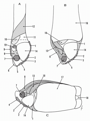
Figure 2.2 (A) Medial surface of distal fibula and lateral malleolus. (B) Same view as in (A). Distal tibia connected to fibula. (C) Inferior view of the distal tibiofibular complex. (1, anterior tibiofibular ligament; 2,main component of anterior talofibular ligament; 3, secondary band of anterior talofibular ligament; 4, calcaneofibular ligament; 5, tip of lateral malleolus, free of insertion; 6, gliding surface of peronei tendons; 7, posterior talofibular ligament; 8, cribriform fossa; 9, superficial component of posterior tibiofibular ligament; 10,synovial fringe; 11, peroneal surface corresponding to tibioperoneal recess; 12, insertion of tibiofibular interosseous ligament; 13, deep component of posterior tibiofibular ligament; 14, articular surface for the lateral surface of the talus; 15, posterosuperior tuberosity; 16, tibia; 17, tibia plafond; 18, medial malleolus.)

The lateral malleolus is in contact with the incisura fibularis of the tibia through a minute crescentic cartilage-coated surface that is in continuity with the triangular articular surface of the fibula.


Posterior Surface

The posterior surface is broad proximally and tapers distally. The tendons of the peroneus brevis and peroneus longus follow the twist of the fibular corpus and lie on the posterior surface. The tendon of the peroneus brevis is against the bone, with the tendon of the longus on top of it. Usually, a sulcus is present on this surface. Edwards, in a study of 178 dry fibulas, gave the following data in regard to the contour of the posterior surface: definite sulcus present, 82%; flat surface, 11%; convex surface, 7%.1The width

of the sulcus is given as the narrowest, 5 mm; the majority (62%), 6 to 7 mm; and the widest, 10 mm. The lateral border of the posterior surface may become prominent and form a lateral bony ridge. “It helps to form a flange against which the tendons of the peroneal muscles play, and it gives attachment to some of the fibers of the superior peroneal retinaculum.1 The occurrence of this lateral bony ridge, based on Edwards’ data, is as follows: well-developed lateral bony ridge, 22%; slightly developed lateral bony ridge, 48% absence of a developed lateral bony ridge, 30%.-1 The majority of the ridges are 2 mm high, but occasionally the ridges may reach an elevation of 4 mm. Cartilage covering may increase the ridge 1 to 2 mm, and often the ridge is formed by cartilage only1


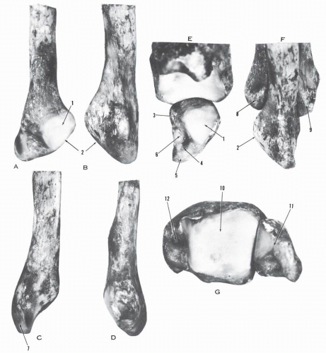
Figure 2.3 (A) Medial view of left fibula. (B) Lateral view of fibula. (C) Posterior view of fibula. (D) Anterior view of fibula. (E) Medial view of tibia-lateral malleolus. (F) Lateral view of distal fibula-tibia. (G) Inferior view of distal tibia-fibula. 1, articular surface; 2, anterior border; 3, posterosuperior tubercle; 4, insertion tubercle of posterior talofibular ligament; 5, tip of lateral malleolus; 6, digital fossa; 7, gliding surface for peronei tendon; 8, anterior tibial tubercle; 9, posterior tibial tubercle; 10, tibial plafond; 11, lateral malleolus; 12, medial malleolus.)


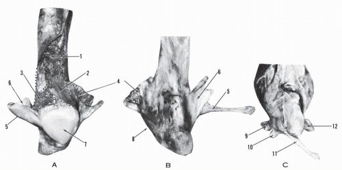
Figure 2.4 (A) Medial view of left distal fibula and lateral malleolus. (B,C) Lateral view of lateral malleolus. (1, insertion of tibiofibular interosseous ligament; 2, fibular component of tibiofibular recess; 3, insertion of synovial fringe; 4, anterior tibiofibular ligament; 5, 6, posterior tibiofibular ligament; 7, articular surface; 8, anterior border; 9, main component of anterior talofibular ligament; 10, secondary component of anterior talofibular ligament; 11, calcaneofibular ligament; 12, posterior talofibular ligament.)

Edwards further reports on the presence of a prominence on the medial border on the posterior surface that forms a medial ridge in about 50% or, in the remaining half, a rounded tubercle.1 In 4% of the fibulas, an intermediate low ridge is present between the lateral and medial ridges.

The peroneal sulcus, when present, is very shallow. Ozbag et al.2 investigated the morphometric features of the lateral malleolar groove in 93 specimens: 80 dry bones and 13 foot specimens. The malleolar groove was concave in 68% (63 specimens) and convex or flat in 32%. The mean groove width was 9.2 ±1.6 mm and the groove depth was 1.0 ± 0.5 mm.

The anterior border of the lateral malleolus is thin above and thick below. The contour is strongly convex anteriorly. A longitudinal tubercle extending from the level of the anterosuperior angle of the articular surface to the midsegment gives insertion to the anterior tibiofibular ligament. Below this level, the anterior border bears flat tubercles corresponding to the insertion of the two bands of the anterior talofibular ligament. Further distally but still anterior in location is the insertion of the calcaneofibular ligament. The apex of the lateral malleolus is free of insertion.

The posterolateral and posteromedial borders of the lateral malleolus are covered in the descriptions of the posterior and medial surfaces.


Lower End of the Tibia

The lower end of the tibia is formed by five surfaces: inferior, anterior, posterior, lateral, and medial (Fig. 2.5). The latter is prolonged distally by the medial malleolus.


Figure 2.5 (A) Anterior aspect of left distal tibia. (B) Posterior aspect of distal tibia. (C) Lateral aspect of distal tibia. (D) Medial aspect of distal tibia and medial malleolus. (E) Lateral aspect of medial malleolus. (F) Inferior view of distal tibia. (1, medial malleolus; 2, sulcus for tibialis posterior tendon; 3, anterior colliculus; 4, intercollicular groove; 5, posterior colliculus; 6, anterior tibial tubercle; 7, posterior tibial tubercle.)


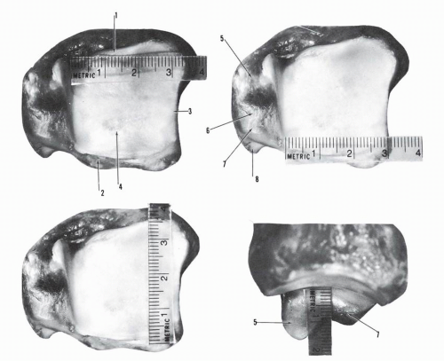
Figure 2.6 Distal tibia and medial malleolus. The anterior border of the distal tibia is longer than the posterior border, and the lateral border of the distal tibia is longer than the medial border. The anterior colliculus of the medial malleolus is 0.5 cm longer than the posterior colliculus. (1, anterior border of distal tibia; 2, posterior border of distal tibia; 3, lateral border of distal tibia—incisura tibialis; 4, tibial plafond; 5, anterior colliculus of medial malleolus; 6, intercollicular groove of medial malleolus; 7, posterior colliculus of medial malleolus; 8, groove for tibialis posterior tendon.)


Inferior Surface

The inferior surface is articular and corresponds to the dome of the talus. It is concave anteroposteriorly and slightly convex transversely because of the presence of a slightly elevated ridge dividing the surface into a wider lateral and a narrower inner segment.

The lateral border is larger than the medial and the anterior border is longer than the posterior (Fig. 2.6). Geometrically, this surface is a section of a frustum of a cone with an average medial conical angle of 22 degrees ± 4 degrees.3 This angle ranges from 0 to 35 degrees.3 An angle of 0 degrees corresponds to a cylindrical surface. The radius of this cylinder is an average of 2 cm, and the corresponding articular arc measures 60 degrees.4 In any position of the talus, the tibial plafond covers only two-thirds of the talar surface, and one-third remains uncovered (Fig. 2.7). With the long axis of the tibia, the tibial plafond makes an angle of 93.3 degrees ± 3.2 degrees, with a range of 88 to 100 degrees.3

The posterior border of the inferior articular surface is lower than the anterior. The direct implantation of the transverse component of the deep posterior tibiofibular ligament on the lateral half of this border forms a true labrum, thus increasing the depth of the containing surface (see Fig. 2.7).


Anterior Surface

The anterior surface is in continuity with the lateral surface of the tibial shaft. It is limited laterally by the interosseous border and medially by the anterior border. The surface is narrow proximally and enlarges distally, where it acquires a convexity in both the transverse and vertical directions.

A transverse ridge is present at 0.5 to 1 cm proximal to the anterior border and gives insertion to the anterior articular capsule. The transverse segment of the bone located between the articular border and the transverse ridge recedes posteriorly and

is an intra-articular segment. This surface may bear a small articular surface (squatting facet), usually lateral in location and very occasionally medial and lateral. The distribution of these facets is as shown in Table 2.1.


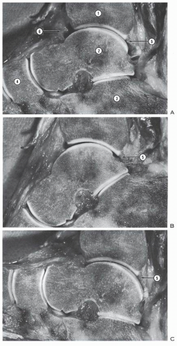
Figure 2.7 Sagittal cross-section of the ankle. (A) Ankle in neutral. (B) Ankle in plantar flexion. (C) Ankle in dorsiflexion. In any position, the articular surface of the distal tibia covers only twothirds of the corresponding talar articular surface. (1, tibia; 2, talus; 3, calcaneus; 4, navicular; 5, deep component of tibiofibular ligament forming a labrum; 6, anterior adipose body with large anterior joint cavity.)


TABLE 2.1 DISTRIBUTION OF SQUATTING FACETS


Author


Number of Tibias


Lateral Facet (%)


Medial Facet (%)


Wooda


118 European


17


1.7


236 Australians


80.5


2.1


Singhb


292 Indian


77.4


1.7


a Wood WQ. The tibia of the Australian aboriginie. J Anat. 1920;52:232.
b Singh I. Squatting facets on the talus and tibia in Indians. J Anat. 1959;93:540.


Posterior Surface

The posterior surface is in continuity with the posterior surface of the tibial shaft. The proximal segment is smooth and slightly convex. The distal segment bears an oblique groove medially, directed downward and inward, corresponding to the tendon of the tibialis posterior. This segment is in continuity with the posterior surface of the medial malleolus. A second, much less delineated groove may be recognized corresponding to the tendon of the flexor digitorum longus (see Fig. 2.5).


Lateral Surface

The lateral surface is triangular with an inferior base and a superior apex. It has the contour of a vertical gutter. The apex continues with the lateral border of the tibial shaft. The anterior and posterior borders are continued distally by soft crests that terminate in an anterior and a posterior tubercle.

The anterior tubercle, larger than the posterior, gives attachment to the anterior tibiofibular ligament, which extends its fibers into the anterior surface of the distal tibia. The posterior tubercle gives attachment to the deep component of the posterior tibiofibular ligament, which extends its insertion through the transverse band onto the posterior border of the tibia. The superficial component of the posterior tibiofibular ligament has a broad attachment on the posterior tubercle and the posterior surface of the distal tibia, reaching the lateral border of the groove for the tibialis posterior tendon. The anterior tubercle overlaps the fibula, and this relationship is given interpretation in the radiologic study of the tibiofibular syndesmosis.

The tibiofibular interosseous ligament inserts on the rugosities of the upper segment of the lateral surface. The inferior segment presents a smooth, small triangular surface (base anterior, apex posterior), corresponding to the tibiofibular recess described previously. This segment is limited inferiorly by a minute crescentic cartilage-coated surface corresponding to a similar surface on the fibula.


Medial Surface

The medial surface is smooth, directed obliquely downward and inward. It is larger proximally narrows progressively distally, and continues with the medial surface of the medial malleolus. It is limited by the anterior and posterior borders of the tibial shaft. This surface gives insertion to the upper arm of the inferior extensor retinaculum and to the flexor retinaculum.


Medial Malleolus

The medial malleolus (see Figs. 2.5 and 2.6) is a strong apophysis implanted at an obtuse angle into the medial aspect of the distal tibia. It is large at the base anteroposteriorly and flat and narrow transversely. It is formed by two segments or colliculi separated by the intercollicular groove. The anterior colliculus descends lower, usually 0.5 cm, than the posterior colliculus. The intercollicular groove is large and measures 0.5 to 1 cm in width. The deep talotibial component of the deltoid ligament inserts in the intercollicular groove, the anterior aspect of the posterior colliculus, and the posterior border of the anterior colliculus. The super-ficial deltoid ligament inserts on the medial surface and anterior border of the anterior colliculus and extends the attachment on the medial subcutaneous surface of the malleolus. The lateral surface of the malleolus is articular with the comma-shaped articular medial surface of the talus. The posterior border of the medial malleolus bears a groove and gives attachment to the fibrous tunnel of the tibialis posterior tendon.

The lateral malleolus, the tibial plafond, and the medial malleolus form a bony unit, the malleolar fork, covering and holding the talus on three sides. The long axis of the ankle mortise is directed posterolaterally in the transverse plane and makes an angle of 23 degrees with the transverse axis of the tibial plateaus (Fig. 2.8). This torsion locates anatomically the medial malleolus anteromedially and the lateral malleolus posterolaterally.


TALUS

The talus is an intercalated bone located between the ankle bimalleolar fork and the tarsus. It is moored with strong ligaments but has no tendinous attachments.


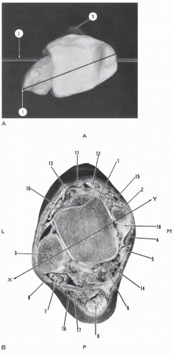
Figure 2.8 (A) Distal tibia-fibula, right ankle, inferior view. (1, bimalleolar axis; 2, transverse axis, transtibial plateau; 3, anterior tibial tubercle.) The bimalleolar axis is oriented posterolaterally. The lateral malleolus is posterior, and the medial malleolus is anterior. (B) Cross-section of left ankle, lower surface, passing 1 cm above the tip of the medial malleolus. (A, anterior; P, posterior; L, lateral; M, medial; 1, talus; 2, anterior colliculus of medial malleolus; 3, lateral malleolus; 4,tibialis posterior tendon and tunnel; 5, flexor digitorum longus tendon and tunnel; 6, flexor hallucis longus tendon-muscle; 7, peroneus longus tendon; 8, peroneus brevis, inverted U-shaped tendon; 9, Achilles tendon; 10, extensor digitorum longus tendon; 11, extensor hallucis longus tendon; 12, tibialis anterior tendon; 13, dorsalis pedis artery and veins; 14, posterior tibial neurovascular bundle; 15, greater saphenous vein; 16, lesser saphenous vein; 17, sural nerve; 18, deltoid ligament, deep talotibial component [note relationship of tibialis posterior tendon and deltoid ligament]; X-Y, bimalleolar axis oriented posterolaterally.)


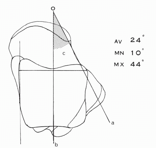
Figure 2.9 Declination angle (c) of the talar neck relative to the body, (a) Long axis of neck, (b) Long axis of body.

The talus is formed by three parts: the body (corpus tali), the neck (collum), and the head (caput). The body is defined as the part of the bone located posterior to an imaginary plane passing through the anterior border of the superior surface of the trochlea tali and the posterior calcaneal surface. The neck is the segment of bone anterior to this plane, located between the body and the head. The body and the neck are not coaxial. In the horizontal plane, the neck shifts medially and makes an angle of declination with the long axis of the trochlea tali. This angle is variable (Fig. 2.9), as indicated in Table 2.2.

In the sagittal plane, the neck is deviated downward relative to the talar body and makes an angle of inclination (Fig. 2.10; see Table 2.2).


TABLE 2.2 ANGLES OF DECLINATION AND INCLINATION IN TALUS


Author


Number of Tali


Declination Angle


Inclination Angle


Testuta


22 degrees


115 degrees


Paturetb


20 to 30 degrees


115 degrees


Sewell


1006 Egyptian


18 degrees average,
7 degrees minimum,
43 degrees maximum


112 degrees average, 98 degrees
minimum, 127 degrees
maximum


Present series


100


24 degrees average,
10 degrees minimum,
44 degrees maximum


114 degrees average (24-degree
plantar tilt), 95 degrees minimum
(5-degree plantar tilt), 140 degrees
maximum (50-degree plantar tilt)


a Testut L. Traité d’Anatomie Humaine, Vol. 1.7th ed. Paris: Doin; 1921:368.
b Paturet G. Traité d’Anatomie Humaine, Vol. 2. Paris: Masson; 1951:573.


The length and width of the bone measured on 100 dry tali are as follows (Fig. 2.11):

Length: average, 48 mm; maximum, 60 mm; minimum, 40 mm

Width: average, 37 mm; maximum, 45 mm; minimum, 30 mm.


Body

The body (corpus tali) has five surfaces (Fig. 2.12): superior, lateral, medial, posterior, and inferior.


Superior Surface

The superior surface of the talar body is pulley shaped (Fig. 2.13) and articulates with the distal surface of the tibia and the transverse component of the inferior and posterior tibiofibular ligament. The groove of the pulley runs near the medial border, which makes the lateral segment of the surface wider than the medial. The surface is markedly convex anteroposteriorly, with a sagittal radius of convexity of 20 mm (average). It presents a mild concavity transversely. This concavity may be shallow or deep and is the norm in 80% of the tali; in the remaining 20%, the transverse curvature is more complex, with a medial concavity and a lateral convexity. These talar curvatures have their interlocking counterparts in the distal tibias.

The medial border of the trochlear surface is straight, slightly lower than the lateral, and soft in contour. The lateral border is oblique, directed posteromedially, and beveled in its posterior segment, thus forming a triangular facet (the facies articularis intermedia corporis tali). The lateral border is sharper than the medial in the midsegment. Because of the obliquity of the lateral border, the trochlear surface is wedge-shaped and is narrower posteriorly.

The difference in width between the anterior and posterior transverse diameters, including the triangular facet, is shown in Table 2.3.


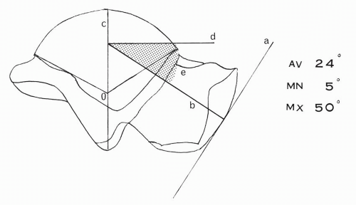
Figure 2.10 Inclination angle (e) of the talar neck relative to the body. The center O of the lateral trochlear arc is determined. The arc is bisected by the radius OC. A tangent is drawn at the apex of the navicular articular surface. A perpendicular line b is drawn at the tangential point. The line b gives the direction of the talar neck and intersects the radius OC of the talar trochlear arc. At this point of intersection, a perpendicular line d is traced, determining the inclination angle e.

The anterior border of the trochlear surface is variable in contour: it may be straight, slightly concave, convex in its entirety, or in the shape of an elongated “S.” Extension facets from the superior articular surface onto the neck are seen both medially and laterally (see Fig. 2.12). A medial extension facet is always accompanied by a forward prolongation of the medial malleolar articular surface of the talus; however, the reverse is not true. The frequency of occurrence of a medial extension facet from the trochlear surface is shown in Table 2.4.

A lateral extension surface is to be differentiated from a squatting facet. The criteria of differentiation are clearly stated by Singh.5 A lateral extension surface continues the convexity of the trochlear surface. During dorsiflexion, this facet establishes contact with the lower end of the distal tibia and not with its anterior border. In contradistinction, a squatting facet, in continuity with the trochlear surface, is concave anteroposteriorly and is directed upward and occasionally backward. During dorsiflexion of the foot, it establishes contact with the anterior margin of the distal tibia. The frequency of occurrence of these lateral prolongations is shown in Table 2.5.


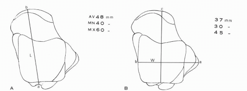
Figure 2.11 Measurements of talus. (A) Length of talus is (L) determined by a line joining the apex of the navicular articular surface to the flexor hallucis longus groove. (B) Width of talus (W) determined with caliper holding the talus at the tip of the lateral process and the middle of the medial trochlear line. The direction of the caliper is maintained perpendicular to the latter.


Lateral Surface

The lateral surface of the talar body is mostly occupied by a large trigonal articular surface, the facies malleolus lateralis. The curved base of this articular surface corresponds to the lateral border of the trochlear surface. The lateral profile of the base is almost an arc of a true circle measuring 106 degrees ± 13.3, 6 The surface is concave in the vertical direction and slightly convex transversely. The convexity is more pronounced in the

apical portion. Rarely, a concavity replaces the convexity in this location. The vertical concavity is determined by the outward projection of the lateral talar process. The angle of projection as measured in 100 tali is 32 degrees average, 55 degrees maximum, and 15 degrees minimum (Fig. 2.14).


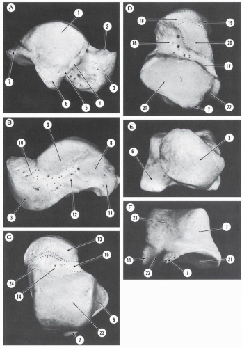
Figure 2.12 Talus. (A) Lateral aspect. (B) Medial aspect. (C) Superior aspect. (D) Inferior aspect. (E) Anterior aspect. (F) Posterior aspect. (1, articular surface—facies malleolus lateralis; 2, cervical collar; 3, articular surface—facies articularis navicularis; 4, 5, tubercles for insertions of anterior talofibular ligaments; 6, lateral process; 7, posterolateral tubercle; 8, oval surface for insertion of talotibial component of deltoid ligament; 9, articular surface—facies malleolaris medialis; 10, talar neck; 11, posteromedial tubercle; 12, tubercle of insertion of deltoid ligament; 13, segment of talar neck located within talonavicular joint; 14, segment of talar neck located within talotibial joint; 15, extra-articular segment of talar neck where a bursa may be found against which glides medial root of inferior extensor retinaculum; 16, sinus tarsi; 17, canalis tarsi; 18, anterior calcaneal articular surface of the talar head; 19, articular segment of talar head corresponding to superomedial and inferior calcaneonavicular ligaments; 20, middle calcaneal articular surface of talar neck; 21, posterior calcaneal articular surface of the talar body; 22, canal of the flexor hallucis longus tendon; 23, trochlear surface; 24, anteromedial extension of trochlear.


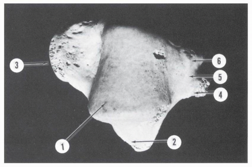
Figure 2.13 Superior aspect of talus. (1, talar pulley; 2, lateral process; 3, talar head; 4, posterolateral tubercle; 5, canal of flexor hallucis longus; 6, posteromedial tubercle.)

The lateral talocalcaneal ligament inserts on the apex of the lateral process.

Along the anterior border of the trigonal articular surface are two tubercles for the insertion of the anterior talofibular ligament, the lower tubercle being less pronounced. Sometimes the tubercles are replaced by a depression or notch. Occasionally, a small accessory articular surface is seen on the anterior segment of the lateral process. This surface, the facies externa accessoria corporis tali, is in continuity with the posterior calcaneal surface. When well developed, it is triangular with an inferior base and oriented anteroinferiorly (Fig. 2.15). In 100 Egyptian tali, this accessory surface was present in 10.15%.7 In the present series of 100 tali, a large accessory surface was present in 4%, and an accessory surface of variable size was present in 34%. Martus et al.8 surveyed 79 paired tali calcanei among 43 skeletons from individuals who had an average age of 13.4 years at the time of death. Skeletons of individuals less than 1 year had not been included. An accessory anterolateral facet of the talus was identified in 34% (27 specimens) and was large in 2.5% (2 specimens). The accessory anterolateral talar facet was significantly associated with dorsal talar beaking in 29% (Fig. 2.16).


TABLE 2.3 DIFFERENCE IN ANTERIOR AND POSTERIOR TRANSVERSE DIAMETERS


Author


Number of Tali


Difference


Testuta


5-6 mm


Poirier and Charpyb


4-5 mm


Inmanc


100


2.4 mm ± 1.3 mm average


Present series


100


4.2 mm average, 2 mm minimum, 6 mm maximum


a Testut L. Traité d’Anatomie Humaine, Vol. 1. 7th ed. Paris: Doin; 1921:632.
b Poirier P, Charpy A. Traitéd’Anatomie Humaine, Vol. 1. Paris: Masson; 1899:758.
c InmanVT. The Joints of the Ankle. Baltimore: Williams & Wilkins; 1976;2.


TABLE 2.4 FREQUENCY OF OCCURRENCE OF MEDIAL EXTENSION FACET


Author


Number of Tali


Occurrence(%)


Singha


300 Indian


55


Present series


100


36


Sewellb


1006 Egyptian


19


Barnettc


100 European


11


a Singh I. Squatting facets on the talus and tibia in Indians. J Anat. 1959;93:540.
b Sewell RBS. A study of astragalus. III. The collum tali. J Anat Physiol. 1906; 39:74.
c Barnett CH. Squatting facets on the European talus. J Anat Physiol. 1954; 88:509.


Along the posteroinferior border of the lateral malleolar surface there is a groove that gives attachment to the posterior talofibular ligament. This groove extends forward, usually up to the midsegment of the posteroinferior border, where it makes a notch. Rarely, it is continued forward to the apex of the facet.


Medial Surface

The medial surface is divided into two fields: superior and inferior. The superior segment is occupied by the facies malleolaris medialis or the auricular facet. This articular surface is comma-shaped, and the long axis is oriented anteroposteriorly. The anterior part is broad and circular; the tail is thin and posterior. The superior border of this surface forms the medial border of the trochlea. This border is convex
anteroposteriorly. The anterior third of this curve is part of a circle with a radius smaller than that of the lateral surface. The posterior two-thirds is an arc of a circle, the radius of which is larger than that of the lateral profile.6 Inman, contouring the trochlear surface in planes perpendicular to the functional axis of the ankle, found the medial side of the trochlea to be an arc of a circle in 80% of the tali and to deviate from it in the remaining 20%.3 The average arc on the medial side is 103 degrees ± 14.3


TABLE 2.5 FREQUENCY OF OCCURRENCE OF LATERAL EXTENSION AND SQUATTING FACETS


Occurrence (%)


Author


Tali


Later al Extension Facet


Squatting Facet


Total


Singha


300 Indian


54.6


26.6


81.2


Present series


100


36


33


69


Barnettb


100 European


17


2


19


a Singh I. Squatting facets on the talus and tibia in Indians. J Anat. 1959;93:540.
b Barnett CH. Squatting facets on the European talus. J Anat Physiol. 1954; 88:509.


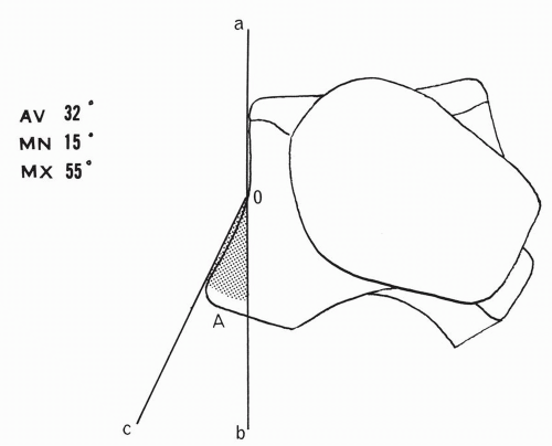
Figure 2.14 Angle of lateral projection (A) of talar lateral process. aob, tangential line to lateral surface; co, tangential line to lateral process.


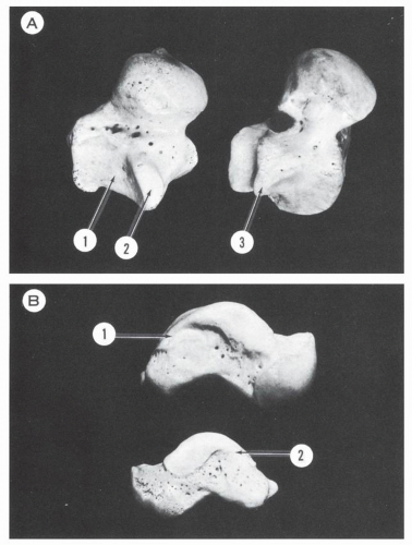
Figure 2.15 (A) Inferolateral view of tali. (1, posterior calcaneal articular surface with 2, facies externa accessoria; 3, absent accessory facet.) (B) Medial view of tali. (1, 2, posterior extension of medial articular facet.)


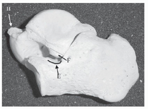
Figure 2.16 Lateral view of the talus and calcaneus, showing an accessory anterolateral talar facet (F), beaking of the dorsal aspect of the talar neck (B), and a calcaneal neck anterior extension facet (E). (From Martus JE, Fermino JE, Caird MS, et al. Accessory anterolateral facet of the pediatric talus. An anatomical study. J Bone Joint Surg Am. 2008;90:2453.)

The medial facet is often extended anteriorly over the medial aspect of the collum tali beyond the level of the anterior border of the trochlea. The frequency of occurrence of this extension was 96% in 300 Indian tali5; in the present series of 100 tali, it was 91% (55% isolated extension, 36% in association with anterior extension of trochlear surface).

When well developed the anterior extension of the medial articular surface projects medially and downward and articulates in strong dorsiflexion with the corresponding anterior aspect of the medial malleolus, which is then covered with articular cartilage. A posterior extension of the medial articular surface behind the area of attachment of the deep portion of the deltoid ligament is also seen (see Fig. 2.15).

The inferior segment is occupied in the anterior half by a depressed surface perforated by numerous vascular foramina. Under the tail of the articular surface, the posterior half is occupied by a large oval surface, flat or elevated, which gives insertion to the talotibial deep component of the deltoid ligament.


Posterior Surface

The posterior surface or processus posterior tali comprises the posterolateral and posteromedial tubercles flanking the sulcus for the flexor hallucis longus tendon.


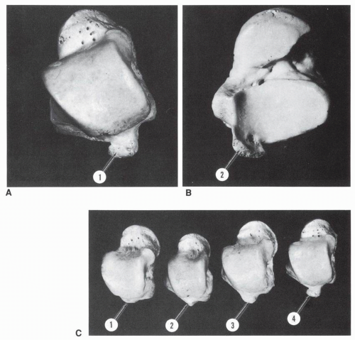
Figure 2.17 (A) Trigonal process, superior view. (B) Trigonal process, inferior view. Its articular surface is in continuity with that of the posterior calcaneal articular surface. (C) Variations in the size of the trigonal process. (1, absent; 2, moderate; 3, medium; 4, large.)

The posterolateral tubercle is large, more prominent than the medial tubercle. The size varies from a barely perceptible structure to a well-developed tubercle projecting posterolaterally from the talus (Fig. 2.17). This tubercle presents an inferior articular surface in continuity with the posterolateral corner of the posterior calcaneal surface of the talus. The superior surface is irregular and nonarticular and gives insertion on the lateral aspect to the posterior talofibular ligament and the talar component of the fibuloastragalocalcaneal ligament of Rouvière and Canela Lazaro.9 The deep layer of the flexor retinaculum inserts on the medial aspect, whereas the posterior talocalcaneal ligament attaches to its inferior border.

An accessory bone, os trigonum (Figs. 2.18 and 2.19), may be found in connection with the posterolateral tubercle. This ossicle has three surfaces: anterior, inferior, and posterior. The anterior surface articulates with the posterolateral tubercle or is attached to the latter with fibrous, fibrocartilaginous, or cartilaginous tissue. The inferior surface articulates with the os calcis. The posterior surface is nonarticular. The capsuloligamentous structures attaching on the posterolateral tubercle extend their insertions on this surface. The frequency of occurrence of the os trigonum in adults is shown in Table 2.6.

Sewell found a percentage of “separation” of 10.9% in 1006 tali.7 “Separation” includes the presence of a notch in the margin of the lateral process, a groove on the articular surface, a combination of both, or a frank separation. The last occurred in 24.1% of the separated group, which was 3% of the total group.

A fused os trigonum is called a trigonal process (see Fig. 2.17). The os trigonum is more often bilateral than unilateral. Very rarely, it is found in two equal or unequal parts. Distinct on one side, it may be present as a trigonal process on the other.

The medial tubercle is of variable size. It is in continuity with the medial talar surface and gives attachment to the deep and superficial layers of the talotibial components of the deltoid ligament, the medial talocalcaneal ligament, and the tunnel of the flexor hallucis longus tendon. Rarely, the tubercle may be very

large and may extend downward over the os calcis, contributing to a talocalcaneal coalition (Fig. 2.20).


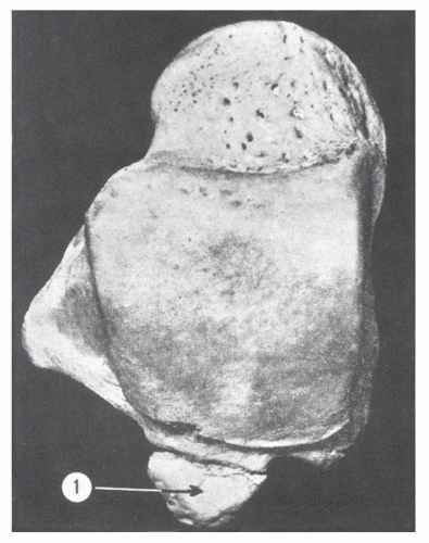
Figure 2.18 Os trigonum (1). (From Dwight T. Variations of the Bones of the Hands and Feet: A Clinical Atlas. Philadelphia: JB Lippincctt; 1907:14-23.)


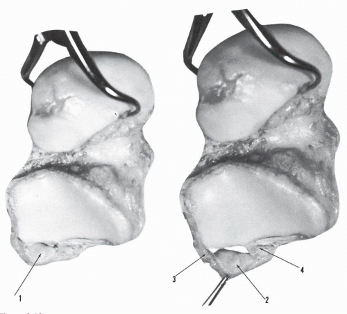
Figure 2.19 Os trigonum. (1, 2, inferior articular surface; 3, 4, ligaments of attachment on each side: thin anterior capsular structure has been removed.)


TABLE 2.6 FREQUENCY OF OCCURRENCE OF OS TRIGONUM IN ADULTS


Author


Number of Tali


Occurrence (%)


Thompsona


438


2.7


Stiedab


305


5.9


Pfitznerc


841


6.1


Grantd


558


7.7


a Report of Committee of Collection Investigation of the Anatomical Society of Great Britain and Ireland for the Year 1899-1890. J Anat Physiol. 1891;25;98.
b Stieda L. Der Talus und das Os Trigonum Bardelebens beim Menschen. Anat Anz. 1899;4:305-319, 336-351.
c Pfitzner W. Beiträge zur Kenntniss des Menschlichen Externitätenskelets: VI. Die Variationen in Aufbau des Fusskelets. In: Schwalbe, ed. Morphologische Arbeiten. Jena: Gustav Fischer; 1896:245-527.
d Grant JCB. Grant’s Atlas of Anatomy. 5th ed. Baltimore: Williams & Wilkins; 1962:356.


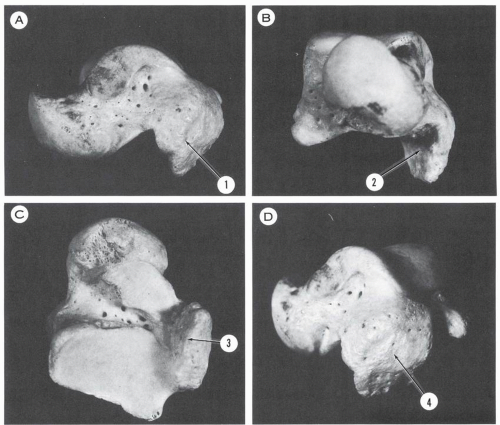
Figure 2.20 Talus. (A) Medial aspect. (B) Anterior aspect. (C) Inferior aspect. (D) Posteromedial aspect. Large posteromedial talar tubercle (1 to 4) probably forming a coalition with its corresponding calcaneus.

The sulcus of the flexor hallucis longus tendon is located between the posterolateral and the posteromedial tubercle. It is directed obliquely downward and inward and is curved anteriorly. The angle made by the long axis of the sulcus with the transverse trochlear axis in 100 tali is 68 degrees average, 85 degrees maximum, and 55 degrees minimum (Fig. 2.21A).


Inferior Surface

The inferior surface is occupied by the facies articularis calcanea posterior. The long axis of this articular surface is directed anterolaterally. The angle made by this axis with the anterior border of the trochlea in 100 tali is 37 degrees average, 50 degrees maximum, and 26 degrees minimum (Fig. 2.21B).

The articular surface is quadrilateral, rectangular medially, and more or less oval laterally. The surface is strongly concave in the long axis and usually flat or very minimally concave transversely. The anteromedial border is usually convex and forms the posterior border of the tarsal canal and the sinus tarsi. This border extends obliquely from the medial tubercle to the anterior surface of the lateral process of the talus. Occasionally, the border is straight or has a complex configuration (see Fig. 2.12). The posterolateral border is straight and parallel to the long axis. Of the two short sides, the medial is straight and directed posterolaterally and supports the posterior process of the talus; the lateral border is convex and supports the base of the lateral process of the talus.

Accessory articular surfaces may be present in continuity with the posterior calcaneal surface. Extending from the anterolateral corner is the facies externa accessoria corporis tali (see Fig. 2.15). A small facet may be present in the anteromedial corner, covering the undersurface of the medial tubercle. A trigonal process or a large lateral tubercle prolongs the articular surface posteriorly (see Fig. 2.17).

The posterior calcaneal surface may establish union with the facies articularis calcanea media, creating a single articulating surface running along the undersurface of the bone and closing the tarsal canal medially. In other instances, these two surfaces fuse through a direct anterior extension from the posterior calcaneal surface; without the medial detour, this extension completely obliterates the tarsal canal and a segment of the sinus tarsi (Fig. 2.22).


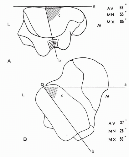
Figure 2.21 Angles of talus. (A) Posterior aspect of talus. Inclination angle (c) of the sulcus for the flexor hallucis longus tendon. (B) Inferior surface of talus. Angle (c) formed by the long axis ob of the posterior calcaneal surface with a line oa parallel to the anterior trochlear border. (L, lateral; M, medial.)


Neck

The neck (collum tali) is the segment of the talus located between the body posteriorly and the head anteriorly (see Fig. 2.12). Its average length is 17 mm, with a maximum of 23 mm and a minimum of 12 mm (Fig. 2.23).

The neck is projected anteromedially and downward, as described previously. The lateral border is slightly concave and well delineated, whereas the medial border is round and at times not discernible. The neck presents four surfaces: superior, lateral, inferior, and medial.


Superior Surface

The superior surface is limited anteriorly by the articular surface corresponding to the navicular and posteriorly by the anterior border of the trochlea. The lateral half of the surface is mostly occupied by a deep, concave cribriform fossa. The remaining anterior part of this surface is occupied by a bony prominence or a smooth, flat bony segment. The medial half of the superior surface is inclined medially because of the rotation of the talar head. In certain tali, a transverse cervical ridge or collar runs parallel to the articular surface of the head.10 The talotibial capsule inserts close to the malleolar facets laterally, medially, and distal to the cribriform fossa, which remains intra-articular. The talonavicular capsule inserts transversely along the articular surface of the head. On the lateral aspect of the neck, the capsules of the talotibial and talonavicular joints are separated by a bare extra-articular bony segment, over which glides the medial root of the inferior extensor retinaculum; a bursa may be found in this location. The superficial talotibial component of the deltoid ligament inserts on the medial aspect of the cervical surface. The articular facets extending from the trochlear surface onto the superior aspect of the neck, the facies articularis interna and externa collae tali, including the squatting facet located laterally, were discussed previously


Lateral Surface

The lateral surface of the neck is converted to a ridge extending from the anterolateral corner of the trochlea and talar body to the articular surface of the head. It is oriented anteromedially and presents a slight concavity. It may give insertion to the medial root of the inferior extensor retinaculum.


Inferior Surface

The inferior surface of the neck is formed by two nonarticulating segments, corresponding to the sinus tarsi and the tarsal canal, and an articular surface. The latter forms the facies articularis calcanea media, which occupies the anteromedial segment of the cervical surface. It has a variable contour and may be oval, elliptic, pyriform, or pentagonal. It is in continuity with the facies articularis calcanea anterior and the articular segment

corresponding to the inferior calcaneonavicular ligament (Figs. 2.22 and 2.24). A ridge may delineate these surfaces. Occasionally, a separation notch is seen between the surfaces; if the notch is deep enough, a near-complete separation occurs between the middle and anterior calcaneal surfaces. In rare instances, a complete separation is present.


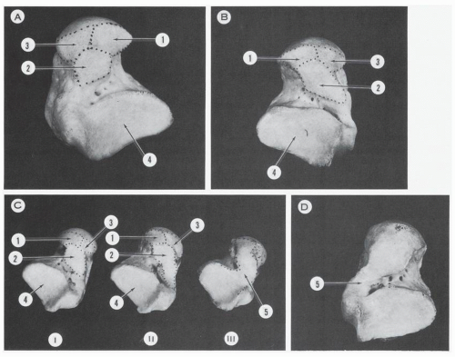
Figure 2.22 Variations in size and contour of the inferior articular surfaces of the talus. (A) Common configuration of the articular surfaces. (B) Posterior extension of the middle calcaneal surface. (C) (I) Moderate posterior extension of middle calcaneal surface. (II) Marked posterior extension of middle calcaneal surface. (III) Fusion (5) of all articular surfaces, obliterating the tarsal canal and a segment of the sinus tarsi. (D) Fusion (5) of the middle and posterior calcaneal surfaces on the medial aspect of the tarsal canal, which is still maintained. (1, anterior calcaneal articular surface of talar head; 2, middle calcaneal articular surface of talar neck; 3, articular segment of talar head corresponding to superomedial and inferior calcaneonavicular ligament; 4, posterior calcaneal articular surface of talar body.)


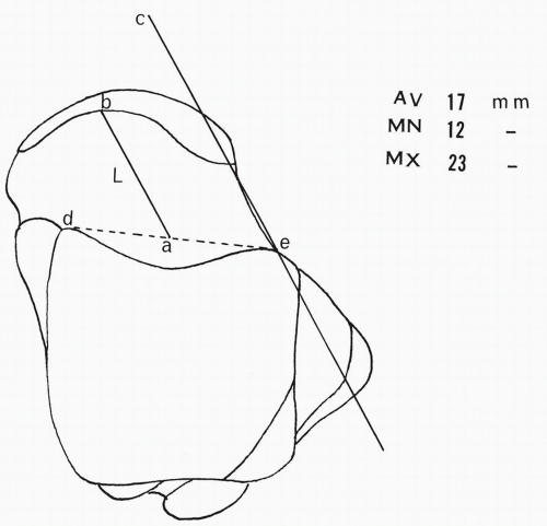
Figure 2.23 Length of talar neck L. An anterior trochlear line de is drawn as indicated. The midpoint a is determined. A line ec is drawn along the talar neck. From the point a a line ab is drawn, parallel to line ec. The segment ab is considered the length of the talar neck, and it terminates where the articular surface is encountered.


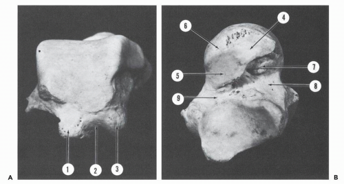
Figure 2.24 (A) Posterior aspect of talus. (B) Inferior aspect of talus. (1, posterolateral tubercle; 2, sulcus for flexor hallucis longus tendon; 3, posteromedial tubercle; 4, anterior calcaneal articular surface; 5, middle calcaneal articular surface; 6, articular segment of head corresponding to superomedial and inferomedial calcaneonavicular ligaments; 7, tubercle for cervical ligament; 8, sinus tarsi; 9, tarsal canal.)

The bony segment corresponding to the sinus tarsi occupies the lateral half of the cervical surface. It is triangular, with a lateral base and a posteromedial apex continuing in the tarsal canal. Anteriorly, it bears a tubercle (tuberculum cervicis tali) that gives insertion to the cervical ligament.11 In the present series of 100 tali, the tubercle was identified in 37%. Vascular foramina are distributed on the inner aspect of this surface. The lateral segment bears only a few vascular foramina.

The segment corresponding to the tarsal canal is located between the facies articularis calcanea media and the facies articularis calcanea posterior. It has a narrow, oblique surface oriented posteromedially. Laterally, it communicates with the sinus tarsi. Its medial opening is anterior to the talar posteromedial tubercle. Multiple vascular foramina are distributed along its longitudinal axis. A longitudinal crest may be present, giving insertion to the interosseous talocalcaneal ligament of the canalis tarsi and to the oblique calcaneotalar band of the inferior extensor retinaculum. As previously described, this sulcus interarticularis may be completely obliterated if a fusion occurs between the posterior and middle calcaneal articulating surfaces.


Medial Surface

The medial surface of the neck is higher than the lateral surface. It represents the forward extension of the nonarticular segment of the medial surface of the talar body and provides insertion to the talonavicular capsule and ligaments. Occasionally, a posterior extension of the articulating surface of the head or an anterior extension of the medial malleolar articulating surface considerably narrows the medial surface of the neck.


Head

The talar head (caput) articulates with the navicular, the calcaneus, and the calcaneonavicular ligaments. These articular fields are usually recognizable (see Fig. 2.22). The head is turned along a longitudinal axis relative to the talar body; the rotation is clockwise on the right and counterclockwise on the left. Because of this rotation, the navicular articular surfaces are higher laterally and lower medially, and its longitudinal axis is oriented upward and laterally. The longitudinal axis rotation relative to the transverse plane in 1006 Egyptian tali12 was 45 degrees average, 62 degrees maximum, and 25 degrees minimum; in the present series of 100 tali, it was 49 degrees average, 65 degrees maximum, and 30 degrees minimum (Figs. 2.25 and 2.26).


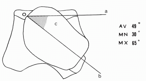
Figure 2.25 Angle (c) of lateral rotation of the talar head,(do, line parallel to trochlear surface; bo, long axis of head)


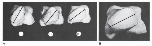
Figure 2.26 Variations in lateral rotation of talar head. (A) (I) Marked rotation. (II) Moderate rotation. (Ill) Minimal rotation. (B) Minimal rotation.

The facies articularis navicularis is the largest of the three surfaces. It is convex along its long and short axes. The superolateral and lateral borders are sharply defined from the neck surface; the superomedial border, less defined, may be beveled. The navicular articular field is in continuity inferiorly with the facies articularis calcanea anterior and the facet for the inferior calcaneonavicular ligament (see Fig. 2.22). A low ridge or a change of direction of the surface demarcates the division.

The medial, inferior segment of the elliptic articular surface corresponds to the deep surface of the superomedial calcaneonavicular ligament and may have a more or less flat contour.

The anterior calcaneal articular surface is nearly quadrilateral or oval, and its surface curvature is clearly different from that of the navicular surface. It is flat and is continuous anteriorly with the navicular surface and posteriorly with the middle articular calcaneal surface, from which it may be separated by a ridge or a notch of variable depth. It is adjacent medially to the articular segment corresponding to the inferior calcaneonavicular ligament. This last surface is wedged between the navicular surface anteriorly and the middle calcaneal articular surface posteriorly.


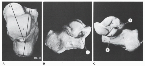
Figure 2.27 Talocalcaneal relationship. (A) Superior view—angle between long axis of calcaneus b and axis of talar neck a, 30 to 35 degrees. (B) Lateral view. (C) Medial view. (1, sinus tarsi; 2, medial opening of tarsal canal between posterior border of sustentaculum tali and anterior border of talar posteromedial tubercle; 3, sustentaculum tali.)


CALCANEUS

The calcaneus is the largest bone of the foot. The long axis is directed anteriorly, upward, and laterally. The upward tilt determines an angle of inclination relative to the horizontal plane— calcaneal pitch—and measures 10 to 30 degrees.13 The long axes of the calcaneus and of the talar neck normally make an angle of 30 to 35 degrees in the horizontal plane (Fig. 2.27).14

The length and width of the calcanei vary (Fig. 2.28): in 750 calcanei, the length was 94 mm maximum and 48 mm minimum;
the width was 53 mm maximum and 26 mm minimum.15 In the present series of 50 calcanei, the length was 75 mm average, 83 mm maximum, and 65 mm minimum; the width was 40 mm average, 46 mm maximum, and 35 mm minimum. The average breadth × 100/length index in the present series is 53 and may range between 50 and 60.15 The height of the os calcis is close to 50% of the length; in 50 calcanei, the average height was 40 mm, maximum 47 mm, and minimum 33.5 mm. The calcaneus is in the form of an irregular rectangle solid and presents six surfaces: superior, inferior, lateral, medial, posterior, and anterior.


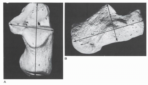
Figure 2.28 Dorsal (A) and lateral (B) views of calcaneus. (H, height; L, length; W, width.)


Superior Surface

The superior surface is divided into three parts: posterior, middle, and anterior (Fig. 2.29).


Posterior Third

The posterior third of the superior surface is nonarticular, narrow, transversely convex, and longitudinally concave. It is perforated by multiple vascular foramina, and the surface corresponds to the pre-Achilles corpus adiposum. Posterolaterally, it gives insertion to the calcaneal component of the ligament of Rouvière and Canela Lazaro.9 The anterior segment gives attachment to the posterior talocalcaneal ligament and to the deep crural aponeurosis.


Middle Third

The middle third of the superior surface supports the large facies articularis talaris posterior. This articular surface makes a sharp change in orientation relative to the posterior segment. It inclines anteriorly and creates a step contour. The angle of inclination in 50 calcanei was average 65.5 degrees, maximum 75 degrees, and minimum 55 degrees (Fig. 2.30, see Fig. 2.34).

Boehler has determined roentgenographically an angle expressing the height of the posterior talar surface.16 This “tuberjoint angle” (see Fig. 2.30) measures 30 to 35 degrees. This angle, measured anatomically, yields the following distribution in the present series of 50 calcanei: 17 to 20 degrees, 2 calcanei; 21 to 30 degrees, 25 calcanei; 31 to 40 degrees, 20 calcanei; 41 to 44 degrees, 3 calcanei; average 32 degrees.

The long axis of the posterior articular surface is directed forward, downward, and outward. The surface is convex along the longitudinal axis and represents a segment of a cone. The apex of the cone is directed toward the sustentaculum tali, and the axis of the cone—the axis of revolution of the surface or the axis of motion along this surface—points anteromedially, intersecting the sustentaculum tali on the inner side at a nearly right angle in the adult.4 The radius of the curvature along the greatest diameter (at the base of the cone) averages 30 mm, with a minimum of 12 mm and a maximum of over 40 mm.15

Manter considers the posterior talar articular surface as an oblique helicoid or screw-shaped surface inasmuch as sections made perpendicular to the joint surface axis reveal “spiral rather than circular arcs.”17 Inman, contouring the posterior talar articular surface with a dial indicator, demonstrated a screwlike behavior of the surface in only 58% of 42 specimens and concluded that “the remarkable variation is the important factor” in considering the geometry of this surface.3


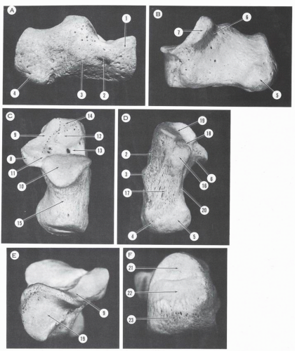
Figure 2.29 Calcaneus. (A) Lateral surface. (B) Medial surface. (C) Superior surface. (D) Inferior surface. (E) Anterior surface. (F) Posterior surface. (1, great apophysis; 2, trochlear process; 3, eminentia retrotrochlearis; 4, lateral tuberosity; 5, medial tuberosity; 6, canal for flexor hallucis longus tendon; 7, medial surface of sustentaculum tali; 8, posterior border of sustentaculum tali; 9, fused anterior and middle talar articular surfaces; 10, posterior talar articular surface; 11, canalis tarsi; 12, sinus tarsi—bony eminence; 13, sinus tarsi—fossa calcanei; 14, sinus tarsi—insertion surface of bifurcate ligament; 15, posterior third of superior surface; 16, anterior tuberosity of inferior surface; 17, longitudinally striated inferior surface; 18, coronoid fossa; 19, cuboidal articular surface; 20, medial calcaneal canal; 21, upper third of posterior surface, corresponding to pre-Achilles bursa; 22, 23, middle and lower thirds of posterior surface, corresponding to insertion of Achilles tendon.)


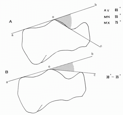
Figure 2.30 (A) Angle of inclination boc of the posterior talar articular surface. (B) Boehler tuberjoint angle boc.

Three accessory or extension facets may be present relative to the facies articularis talaris posterior: anterior, posterior, and medial. The anterior facet is seen in the anterolateral corner, extending onto the floor of the calcaneal fossa in a tonguelike projection. The counterpart to this surface on the talar side is the facies externa accessoria. The posterior facet is a trianglelike projection over the posterior third of the superior surface, corresponding to the presence of a trigonal process or to an os trigonum. The medial extension is directed toward the facies articularis talaris media and at times may succeed in establishing a union, thus obliterating the posterior end of the sulcus calcanei. The frequency of occurrence of these accessory facets is as shown in Table 2.7.


Anterior Third

The surface of the anterior third is formed by the sinus tarsi, the sulcus calcanei, and the facies articularis talaris anterior and media (see Figs. 2.27 and 2.29). The long axes of these last two articular surfaces and of the facies articularis talaris posterior make a diverging angle open anterolaterally

The facies articularis talaris media and anterior form a continuous supportive surface located on the medial aspect of the sinus tarsi and sinus canal. The long axis of the surface is directed forward and laterally. These two surfaces form a concavity along the long axis, corresponding to the convexity of the talar head. The anterior surface is supported by the beak of the os calcis, and the middle surface is supported by the sustentaculum tali. Variations are present in the contour and the degree of separation of these two surfaces. Bunning and Barnett classify the calcanei into three types: A, B, and C.18 In type A, the anterior and middle surfaces are separate, and in type B they are confluent. In type C, the anterior, middle, and
posterior facets are united into a single surface. The distribution of these variations is shown in Table 2.8 (Fig. 2.31).


TABLE 2.7 FREQUENCY OF OCCURRENCE OF ACCESSORY FACETS


Laidlawa


Present Series


Number of calcanei


750


50


Anterior extension facet


Tonguelike, 4%


6%


Minor degree, 4.5%


Posterior extension facet


Triangular area, 3.5%


Less definite triangular area, 5%


Medial extension facet


5%


Union with middle surface


1.5%


2%


a Laidlaw PP. The varieties of the os calcis. J Anat Physiol. 1904;38:133.


TABLE 2.8 FREQUENCY OF OCCURRENCE OF VARIATIONS IN CALCANEI


Occurrence (%)


Author


Number of Calcanei


Type A


Type B


Type C


Laidlawa,b


750


32


69


Bunning and Barnettc


Veddah 10


0


60


40


African 492


36


63


1


British 194


67


33


0


Indian 78


22


78


0


Present series


50


34


64


2


Padmanabhan


Indian 272


35


65


a Laidlaw reports complete absence of the anterior facet in 0.9%.
b Laidlaw PP. The os calcis, part II. J Anat Physiol. 1905;33:168.
c Bunning PSC, Barnett CH. A comparison of adult and foetal talocalcaneal articulation. J Anat Lond. 1965;99(1):71-76.


The degree of confluence of the anterior and middle facets is variable, being partial or complete. A constriction of the continuous surface determines two equal parts in 34% of the calcanei and a small anterior facet in association with a large middle surface in 18%. Trace of constriction is present in 12%, whereas nearly complete separation of the surfaces is seen in 2% of the calcanei of the present series. When a complete division of the surfaces is the norm, the two surfaces are of equal size in 12%, and in 20%, the middle facet is larger than the anterior; in only 2% of the calcanei is the facies articularis talaris anterior larger than the facies articularis talaris media.


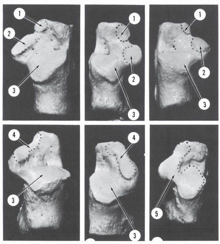
Figure 2.31 Variations of the articular surfaces on the superior aspect of the os calcis. (1, anterior talar articular surface; 2, middle talar articular surface; 3, posterior talar articular surface; 4, fused anterior and middle talar anticular surfaces;5, fused anterior, middle, and posterior talar articular surfaces.)

Padmanabhan, in a study of 277 Indian calcanei, found the following distribution in regard to the anterior and middle talar facets: type A, 35%; type B, 65%.19 In his series there was no single case of type C.

The canalis tarsus separates the middle and the posterior articular facets. It is narrow and oriented obliquely forward, laterally, and inferiorly It is at a higher level than the floor of the sinus taris and has the same inclination as the sustentaculum tali. This angle of inclination relative to the lower border of the os calcis is 46 degrees on average. This canal is not as deep as its counterpart on the talus, and it opens abruptly into the sinus tarsi. The interosseous talocalcaneal ligament or ligament of the tarsal canal makes its insertion on the floor of the canal, joined by the inward extension of the medial root of the inferior extensor retinaculum. Occasionally, a bony crest is seen in the canal, corresponding to this ligamentous insertion. The axis of motion of the talotarsal joint also passes through the canal. From both the anatomic and the physiologic point of view, the canalis tarsi and its contents are of prime importance.

The sinus tarsi, located on the anterior segment of the superior calcaneal surface, is limited posteriorly by the facies articularis talaris posterior and anteriorly by the anterior border separating the superior calcaneal surface from the anterior cuboidal articular surface. Laterally, the sinus tarsi is limited by the crista lateralis; medially, it is limited by the lateral border of the facies
articularis talaris anterior. The posteromedial corner of the sinus tarsi continues with the calcaneal or tarsal canal.

In front of the posterior talar surface is the fossa calcaneus, perforated by multiple foramina leading to the antrum calcanei, an interior space free of cancellous trabeculae. Occasionally, a large, funnel-shaped foramen is seen in the fossa.

The anterolateral segment of the sinus tarsi is occupied by a bony eminence of variable configuration. This surface may be flat, covered only by rugosities, or it may be slightly elevated like a small plateau; occasionally, it is quite prominent in the form of a high tubercle. The sinus tarsi gives attachment to the following structures:


The extensor digitorum brevis, arising from the anterolateral bony eminence and partially from the fossa calcanei.

The intermediate and medial roots of the inferior extensor retinaculum, located medial to the origin of the extensor digitorum brevis.

The cervical ligament, located between the anterior talar articular surface and the origin of the extensor digitorum brevis. A tubercle may indicate the origin.

The dorsal lateral calcaneonavicular and the medial calcaneocuboid ligaments, arising from the anteromedial corner of the surface.

The lateral calcaneocuboid ligament, originating from the anterolateral corner of the surface.

The crista lateralis is a beamlike bony segment limiting the sinus tarsi laterally. It extends from the posterior articular surface to the anterolateral corner of the superior calcaneal surface, where it becomes less distinct.


Inferior Surface

The inferior surface of the calcaneus is triangular, with the base posterior and the apex anterior (see Fig. 2.29). Two tuberosities occupy the base: the medial (which is the larger) and the lateral. The width of the bony heel as measured from the inner border of the medial tuberosity to the outer border of the lateral tuberosity is, on average, 3 cm in the present series of 50 calcanei (maximum, 3.5 cm; minimum, 2.5 cm).

The width of the posterior tuberosities in the present series of 50 calcanei is: medial—average 2 cm, maximum 2.4 cm, minimum 1.6 cm; lateral—average 1 cm, maximum 1.4 cm, and minimum 0.6 cm.

The medial tuberosity is the main weight-bearing bony segment. Rarely is the lateral tuberosity absent. In most of the calcanei, a triangular space separates the two tubercles; this space is directed anteromedially, with the apex located posterolaterally. At times the apical separation takes the form of a groove. Occasionally, there is no intertubercular space and both tubercles are united with a common anterior (nearly) transverse border. Both tubercles have an anteroposterior convex contour.

The midsegment of the inferior calcaneal surface is covered by longitudinal bony striations. The lateral border is oblique and directed anteromedially and many times is less distinct than the rounder medial border, which presents a shallow medial concavity.

An anterior tuberosity is located near the apex of the triangular inferior surface. It is a round eminence measuring 1.5 cm in width on average (maximum 2 cm, minimum 1.2 cm).

On the posterior tuberosities, the aponeurosis plantaris and the flexor digitorum brevis muscle are inserted transversely in a posteroanterior sequence. The medial tuberosity gives origin to the abductor hallucis muscle and the lateral tuberosity to the abductor digiti minimi (which also reaches the medial tuberosity). The triangular surface interposed between the anterior and posterior tubercles gives attachments to the ligamentum plantaris longus. The anterior tuberosity provides insertion to the deep fibers of the longitudinal plantar ligament and to the short plantar calcaneocuboid ligament.

Between the anterior tuberosity and the anterior apex of the sustentaculum tali is a small depression, the coronoid fossa, that gives origin to the inferior calcaneonavicular ligament. A small articular surface in continuity with the anterior cuboidal articular surface is located on the lateral aspect of this fossa and receives the beak or coronoid process of the cuboid.

In the present series of 50 os calcis, a “heel spur” or shelflike anterior bony projection originating from the medial tubercle occurred in 36%.


Lateral Surface

The lateral surface is shown in Figure 2.29. It is high posteriorly and low anteriorly. The posterior third is subcutaneous and is flat, except at the upper segment, where it is slightly convex in the vertical dimension. The middle third presents a tubercle, the eminentia retrotrochlearis, in its lower segment. This is nearly always present.15 It is a large oval eminence of very variable dimensions. Edwards, in a study of 150 dry calcanei, found this eminence to be present in 98% and absent in only 2%.1 Anterior to the retrotrochlear eminence is another tubercle, the processus trochlearis (Fig. 2.32). When present and well delineated, this process is a ridgelike structure located below the angle formed by the lateral border of the sinus tarsi and the lateral border of the facies articularis talaris posterior. This trochlear process is oriented downward and anteriorly, and the long axis makes an angle of 45 degrees with the horizontal.15

The frequency of occurrence of the processes trochlearis in various series is as follows: Gruber,20 39.1%; Stieda,21 33% Pfitzner,22 39.9%; Laidlaw,15 36.5% (prominent, 20.5%; less marked, 16%); Edwards,1 44% present series (50 calcanei), 32%.

Agarwal and colleagues reported on the variations of the peroneal tubercle in 1410 Indian calcanei from the regions of Agra and Lucknow.23 They classified the calcanei into four types according to the pattern of the peroneal tubercles. Type I, with a single peroneal tubercle present anteroinferior to the tubercle for the attachment of the calcaneofibular ligament, occurred in 31.25% (Agra) and 60.11% (Lucknow). Type II, with a single peroneal tubercle incompletely divided into anterior and posterior parts by a smooth, shallow groove running obliquely from above downward occurred in 18.75% (Agra) and 23.66% (Lucknow). Type III, with two peroneal tubercles completely separated by a roughened area in the middle, occurred in 7.50% (Agra) and 13.76% (Lucknow). Type IV represented the absent
peroneal tubercle and occurred in 42.5% (Agra) and 2.42% (Lucknow).


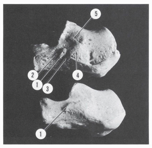
Figure 2.32 Lateral aspect, calcanei. (1, trochlear process; 2, sulcus for peroneus brevis tendon; 3, sulcus for peroneus longus tendon; 4, eminentia retrotrochlearis; 5, tubercle for calcaneofibular ligament.)

The dimensions of the trochlear process are as follows: length—maximum 17 mm, minimum 2 mm; breadth at base— maximum 10 mm, minimum 2 mm; height—maximum 7 mm, minimum 1 mm.1

On the inferior surface of this process glides the peroneus longus tendon. The superior surface is smooth and corresponds to the peroneus brevis tendon. The groove of the peroneus longus tendon leaves a landmark on the lateral aspect of the os calcis in 85%.-1 This groove may be present in the absence of a trochlear process, located then on the anterior aspect of the retrotrochlear eminence or on the lateral aspect of the os calcis.

A cartilage-covered gliding facet may be present on the os calcis along the course of the peroneus longus tendon. Edwards found such facets in 44% of his series, and of these, 10.6% were present in the absence of a trochlear process.1 Those gliding facets are oval, usually not elevated, and located on the posterior slope of the trochlear process or partly on this slope and partly on the lateral surface of the calcaneus.

A definite groove for the peroneus brevis tendon is present in only 2.6%.1 The inferior peroneal retinaculum, bridging both peroneal tendons, attaches to the os calcis above and below the trochlear process and sends a septum to the crest of the process.

Hyer et al.24 investigated the occurrence and characteristics of the peroneal tubercle in 114 calacanei. The peroneal tubercles were “subjectively described as flat, prominenet, concave or tunnel.”24 The occurrence of the peroneal tubercle was then 90.4% with the following distribution: flat in 42.7% (44 specimens), prominent in 29.1% (30 specimens), concave in 27.2% (28 specimens), and tunnel in 1% (1 specimen) (Fig. 2.33). In our interpretation, if “flat” is considered by others as “no tubercle,” then the data of occurrence of the peroneal tubercle is closer to the previously published data. The morphometric data in regard to the peroneal tubercle was reported as follows: length 13.04 mm (range 3.61 to 26.6 mm), height 9.44 mm (range 3.67 to 23.40 mm), and width 3.13 mm (range 1 to 10 mm).


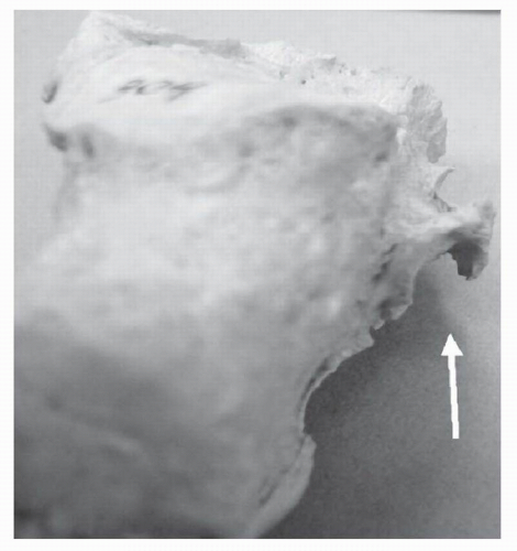
Figure 2.33 Tunnel type peroneal tubercle (arrow). (From Hyer CF, Dawson JM, Philbin TM, et al. The peroneal tubercle: Description, classification, and relevance to peroneus longus tendon pathology. Foot Ankle Inter. 2005;11: Figure 4.)

The tuberculum ligamenti calcaneofibularis is a small tubercle situated behind the midsegment of the facies articularis talaris posterior and is posterosuperior to the eminentia retrotrochlearis. It is present as a well-defined tubercle in 43%.15 The location of this tubercle for the insertion of the calcaneofibular ligament is typical in 64.5% and varies in the remaining 35.5% (anterior location, 25.5%; downward location. 4.5%; posterior location, 5.5%).15

The calcaneal component of the fibulocalcaneoastragalar ligament of Rouvière and Canela Lazaro extends its insertion on the superior aspect of the lateral surface behind the insertion of the calcaneofibular ligament in an oblique linear fashion; the lateral talocalcaneal ligament inserts anterior to it.9 A tubercle (tuberculum ligamenti talicalcanei) may be present for this attachment. Occasionally, a small tubercle for the attachment of the lateral calcaneocuboid ligament is seen in the superior segment of the anterior third of the lateral surface. Morestin describes a second trochlear gliding facet for the tendon of the peroneus longus, located at the anteroinferior corner of the external calcaneal surface.25 He recognizes two varieties: intra- and extra-articular. The intra-articular facet is at the extreme anterior portion of the calcaneus, and the capsule-synovium of the calcaneocuboid joint inserts at its periphery. The extra-articular type is at a distance from the calcaneocuboid articulation and is oval or circular, cartilage-covered, and somewhat elevated above the surrounding parts. When well developed, a sesamoid is found in the substance of the peroneus longus tendon; this sesamoid is different from the sesamoid found in the same tendon further distally as it passes over the tuberosity of the cuboid.

The frequency of occurrence of this second trochlear facet in various series is as follows: Morestin, 5%25 Edwards, δ.6%1 and present series (50 os calcis), 4%.


Medial Surface

The medial surface of the os calcis, similar to the lateral surface, is high posteriorly and low anteriorly (Figs. 2.29 and 2.34). This surface forms a large oblique canal directed downward and anteriorly. It accepts the width of two fingers. The configuration is determined by the medial projection of the sustentaculum tali and the medial extension of the medial calcaneal tubercle. This calcaneal canal is the port of entry from the posteromedial aspect of the ankle to the plantar aspect of the foot.

The sustentaculum tali is a bracketlike projection, triangular with a posterior base and an anterior apex. This surface projects anteromedially and is inclined downward and anteriorly at an average angle of 46 degrees (maximum 60 degrees, minimum 30 degrees) (see Fig. 2.34). The superior surface corresponds to the facies articularis talaris media, described previously. The inferior surface is carved into a groove for the gliding of the flexor hallucis longus tendon and provides attachment to the fibrous tunnel of the tendon. A crest may be present at the attachment site posteriorly. The medial surface of the sustentaculum tali is triangular, with a posterior base and an anterior apex. This surface corresponds to the flexor digitorum longus tendon and its fibrous tendon sheath. The tibiocalcaneal components of the deltoid ligament and the superomedial calcaneonavicular ligament insert on the upper border of the medial surface. The recurrent band of the tibialis posterior tendon inserts on the lower border of the same surface.

The posterior border of the sustentaculum tali corresponds to the medial entrance of the canalis tarsi and gives insertion to the medial talocalcaneal ligament. On rare occasions when the posterior talar articular surface extends medially over the superior surface of the sustentaculum, the opening of the canalis tarsi is on the medial border of the sustentaculum tali (see Fig. 2.31).

The width and length of the sustentaculum tali are variable. The width of the sustentaculum tali, as measured at the base (see Fig. 2.28), was on average 13 mm (maximum 18 mm, minimum 8 mm) in the present series of 50 dry calcanei. The ratio of the sustentacular width to the total width of the os calcis at the same level is on average 0.33 (maximum 0.47, minimum 0.23). These values may be correlated with the supportive function of the sustentaculum tali relative to the talar head. “Incompetent” sustentaculum tali may fall into a group with a minimum value or lower.

The sustentaculum tali may also be classified by length as long or short. A long sustentaculum is continuous through its medial border with the process anterior, which is then in association with a fusion of the facies articularis media and anterior. A short sustentaculum ends suddenly anteriorly, and a notch separates the two articular surfaces (see Figs. 2.31 and 2.34).26 The frequency of occurrence of these varieties, including the intermediary forms, in Laidlaw’s series is: long, about 40%; short, 32% intermediary, 28%.26 In the present series the frequency is: long, 60% short, 34% intermediary, 6%.

The medial surface of the os calcis gives insertion on its inferior two thirds to the medial head of the quadratus plantae. This field of insertion is triangular, with the base posterior. The transverse interfascicular ligament inserts above the quadratus plantae and below the tunnel of the flexor hallucis longus.


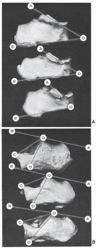
Figure 2.34 (A) Variable inclination of sustentaculum tali—angle AOB. (B) Variations of inclination angle BOD and of tuber-joint angle (Boehler) BOC. (Top, marked inclination; center, moderate inclination; bottom, minimal inclination.)


Posterior Surface

The posterior surface is triangular, with the apex superior and the base inferior (see Fig. 2.29). The medial and lateral borders are well delineated, but the inferior border is ill defined because the surface is continuous with the plantar aspect. The overall contour of the surface is convex. The upper segment is directed upward and anteriorly and is divided into two fields. The lower field is transverse and trapezoidal with an irregular, striated, crenated lower border and a soft and regular superior border. This surface corresponds to the insertion of the Achilles tendon. The upper field is free from tendinous insertion. It is triangular and smooth and corresponds to the pre-Achilles bursa. The lower surface is broad, directed downward and anteriorly; it is striated because of the insertion of the Achilles tendon.


Anterior Surface

The anterior surface is almost entirely articular (see Fig. 2.29). It is saddle-shaped, convex transversely, and concave vertically. The contour of the surface forms a spiral-type groove directed downward and inward. At the posteromedial end of this groove is the calcaneal coronoid fossa, which receives the beak of the cuboid.

The superomedial corner of the articular surface makes a shelflike projection anteromedially This beak or rostrum of the os calcis overhangs the cuboid.


CUBOID

The cuboid is intercalated between the calcaneum and the base of metatarsals 4 and 5. It gives support to the lateral cuneiform and may enter in contact with the scaphoid.

The bone is wedge-shaped or cuneiform rather than cuboid, as the dorsal and plantar surfaces slope toward the narrow lateral surface or border (Fig. 2.35). It presents five surfaces: dorsal, plantar, medial or base, posterior, and anterior, as well as a lateral border or apex.


Dorsal Surface

The dorsal surface is markedly inclined outward and is in continuity with the lateral surface of the calcaneum (Figs. 2.36 and 2.37). Transversely, it continues the curvature of the dorsal surface of the cuneiforms, contributing to the formation of the transverse arch of the midfoot (see Fig. 2.35).

The surface is trapezoidal, covered by rugosities, and crossed by the extensor digitorum brevis and the peroneus tertius tendon. This surface has four borders:


Medial, which represents the base of the trapezoid. The distal segment has a medial projection. It corresponds to the navicular and the third cuneiform.

Lateral, which represents the apex of the trapezoid. This border is short and slightly concave, corresponding to the crossing of the peroneus longus tendon.

Proximal and convex, corresponding to the os calcis. Distal and obtuse, corresponding to the base of the fourth and fifth metatarsals.


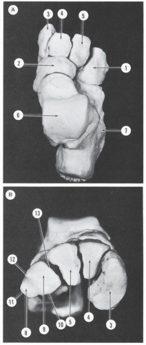
Figure 2.35 (A) Dorsal aspect, tarsus. (B) Transverse arch formed by the cuneiforms and the cuboid, which is also wedgeshaped. (1, cuboid; 2, navicular; 3, medial cuneiform; 4, middle cuneiform; 5, lateral cuneiform; 6, talus; 7, calcaneus. Cuboidal surfaces; 8, 9, lateral and medial aspect of anterior articular surface; 10, inferior border; 11, apex; 12, dorsolateral border; 13, medial border.)


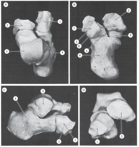
Figure 2.36 Subtalar and midtarsal skeleton. (A) Dorsal aspect. (B) Plantar aspect. (C) Lateral aspect. (D) Anterior aspect. (1, cuboid; 2, navicular; 3, talus; 4, calcaneus [elongated ˜ contour of Chopart joint is seen]; 5, beak of cuboid; 6, sesamoid facet of cuboid; 7, canal of peroneus longus.)

The dorsal surface provides attachment to the following:

Dorsomedial calcaneocuboid ligament, which attaches on a small tubercle located on the posteromedial corner of the surface

Dorsolateral calcaneocuboid ligament, which attaches at least 0.5 cm distal to the proximal border and reaches the middorsal segment

Lateral calcaneocuboid ligament, just above the lateral border

Dorsal cubonavicular ligament, inserting on the medial side of the midsegment

Dorsal cuneo3-cuboid ligament, single or double, inserting in the distal half of the medial dorsal surface

Dorsal cubometatarsal4, cubometatarsal5 ligaments, attached along the distal margin of the bone. The latter has a broader insertion area than the former.


Plantar Surface

The plantar surface faces inferiorly and medially (see Figs. 2.36 and 2.37). It is wider medially and narrower laterally. The medial border is oblique and is directed more posteriorly and slightly medially, while the posterior border is directed more medially and slightly posteriorly. At the junction of these two borders is the beak or coronoid process of the cuboid.


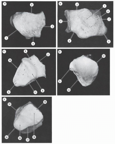
Figure 2.37 Cuboid, right. (A) Dorsal surface. (B) Medial surface. (C) Inferior or plantar surface. (D) Posterior surface. (E) Anterior surface. (1, posterior border of dorsal surface; 2, anterior border of dorsal surface; 3, medial border of dorsal surface; 1′’, posterior border of medial surface; 2′, anterior border of medial surface; 3′, superior border of medial surface; 4, lateral border of dorsal surface; 5, beak of cuboid; 6, groove for peroneus longus; 7, articular surface for third cuneiform; 8, articular surface for scaphoid; 9, tuberosity of cuboid; 10, posterior border of inferior surface; 11, medial border of inferior surface; 12, superior border of posterior surface; 13, inferior border of posterior surface; 14, superolateral border of anterior surface; 15, superomedial border or base of anterior surface; 16, apex of anterior surface; 17, inferior border of anterior surface; 18, articular surfaces corresponding to metatarsals 4 and 5.)


The short lateral border is slightly concave. The anterior border is directed laterally and posteriorly and is divided into a short medial segment and a long lateral segment, which is more inclined posteriorly.

A strong ridge, the tuberositas ossis cuboidei, oriented obliquely anteromedially, divides the plantar surface into a small anterior and a large posterior area. A line extending in the direction of this tuberosity will reach the base of the first metatarsal.

As demonstrated by Stieda and Poirier, there is no cuboidal groove or peroneal groove on the anterior aspect of the plantar surface.21, 27 The tendon of the peroneus longus glides and is reflected over the anterior slope of the cuboidal tuberosity. Furthermore, a gliding facet corresponding to the sesamoid of the peroneus longus tendon is present on the anterolateral aspect of this tuberosity. The sesamoid facet or cuboid facet is present in 93%.1 It is oval (77%), irregularly quadrilateral (18%), or triangular (5%), and is slightly convex.1

Only gold members can continue reading. Log In or Register to continue

Stay updated, free articles. Join our Telegram channel

May 28, 2016 | Posted by in ORTHOPEDIC | Comments Off on Osteology

Full access? Get Clinical Tree

Get Clinical Tree app for offline access