Syndesmology
Shahan K. Sarrafian
Armen S. Kelikian
The distal segment of the fibular shaft and the lateral malleolus are firmly attached to the distal tibia and form a movable articulating system embracing the talar body. The inferior tibiofibular articulation is an integral part of the ankle joint. The subdivision of the ankle joint, as presented in the German literature, into an upper ankle joint (talocrural) and lower ankle joint (subtalar, talocalcaneonavicular) has merit from the anatomic and functional points of view.
LIGAMENTS OF THE INFERIOR TIBIOFIBULAR JOINT
The three ligaments uniting the distal fibular shaft and the lateral malleolus to the distal tibia are the anterior tibiofibular ligament, the posterior tibiofibular ligament, and the interosseous ligament. The lower segment of the interosseous membrane also participates in the stabilization of the distal fibular shaft.
Anterior Tibiofibular Ligament
The anterior tibiofibular ligament (Figs. 4.1 and 4.2) is a flat, fibrous lamina. It originates from the longitudinal tubercle located on the anterior border of the lateral malleolus in front of the upper segment of the articulating surface of the talus and from the lower segment of the anterior border of the fibular shaft. The fibers are directed upward and medially and insert on the anterolateral tubercle of the tibia; some fibers reach the anterior surface of the distal tibia. The fibers increase in length from above downward, the lower fibers being the longest—close to 25 mm.
The anterior tibiofibular ligament is divided into two or three bands or may be multifascicular. Vessels from the anterior peroneal artery penetrate through the interlaminar spaces. During their oblique course, the most inferior fibers cover the tibiofibular corner of the joint and pass over the corresponding segment of the talus. The lowest fibers at their fibular site reach the origin of the anterior talofibular ligament.
Posterior Tibiofibular Ligament
The superficial component originates from the posterior border of the tubercle located above the digital fossa of the lateral malleolus. The origin extends distally to the upper part of the posterior border of the digital fossa and proximally to the ridge separating the lateral and medial fibular surfaces posteriorly. The fibers are directed upward and medially and the major insertion is on the posterolateral tibial tubercle. The remaining fibers continue their course and insert on the distal tibia and they may reach the lateral border of the groove for the tibialis posterior tendon.
The deep component is the transverse ligament. This ligament is thick, strong, and conoid with a twist to its fibers; it originates from the round posterior fibular tubercle located above the digital fossa and from the upper segment of the digital fossa. The fibers are directed upward, medially, and posteriorly. At the posterior border of the tibial articular surface, the fibers change direction and become horizontal or transverse. This ligament inserts on the lower part of the posterior border of the tibial articular surface and reaches the medial border of the medial malleolus. The insertion is the strongest on the outer half. The transverse ligament descends below the posterior tibial margin and constitutes a true posterior labrum deepening the tibial articular surface ( Figs. 4.1 and 4.4). The posterior half of the medial surface of the lateral malleolus is deficient in articular surface but is filled by the transverse ligament, which establishes contact with the talar surface and leaves its imprint as a beveled triangular facet on the posterior half of the lateral border of the superior talar surface.
Interosseous Ligament
The interosseous ligament (Fig. 4.5) is a reddish ligament formed by a dense mass of short fibers intermingled with adipose tissue and vessels. These fibers form a vault over the underlying synovial recess and may be perforated in some specimens. The ligament originates from the anteroinferior triangular segment of the medial aspect of the distal fibular shaft. This area of origin is higher anteriorly and lower posteriorly and the fibers insert on a similar corresponding area on the lateral surface of the distal tibia.
The fibula is in contact with the tibia only through a minute, crescent-shaped, cartilage-coated articular surface in continuity with the articular surface of the lateral malleolus.1 A semilunar cavity is present above this tibiofibular interline and is limited proximally by the concave base of the interosseous ligament. The anterior segment of the cavity corresponds to a synovial recess communicating with the ankle joint through the linear opening. This synovial recess is about 1 cm in height. The posterior part of the semilunar cavity is smaller and occupied by a reddish synovial fringe that originates only from the peroneal surface and descends into the ankle joint between the fibula and the lateral talar surface (Figs. 4.1 and 4.6). In dorsiflexion of the ankle, the synovial fringe retreats toward the upper chamber and, in plantar flexion, it descends into the ankle joint.2, 3
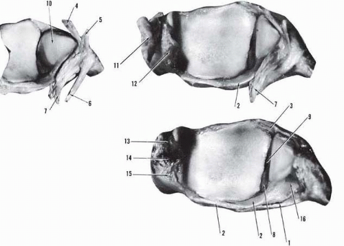 Figure 4.1 Tibiofibular mortise and ligaments. (1, posterior tibiofibular ligament, superficial layer; 2, posterior tibiofibular ligament, deep layer; 3, anterior tibiofibular ligament; 4, anterior talofibular ligament, major component; 5, anterior talofibular ligament, accessory component; 6, calcaneofibular ligament; 7, posterior talofibular ligament; 8, synovial fringe of the tibiofibular recess; 9, tibiofibular recess; 10, lateral malleolar articular surface; 11, superficial deltoid ligament; 12, deep deltoid ligament; 13, anterior colliculus; 14, intercollicular groove; 15, posterior colliculus; 16, digital fossa of the lateral malleolus.) |
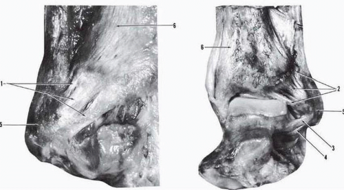 Figure 4.2 Anterior tibiofibular ligament. (1, anterior tibiofibular ligament with three fascicles; 2, anterior tibiofibular ligament with multiple fascicles; 3, anterior talofibular ligament, major component; 4, anterior talofibular ligament, accessory component; 5, lateral malleolus; 6, tibia.) |
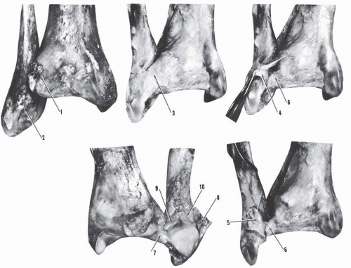 Figure 4.3 Posterior tibiofibular ligament. (1, posterolateral tibial tubercle; 2, lateral malleolus; 3, posterior tibiofibular ligament, superficial layer; 4, partially detached posterior tibiofibular ligament, superficial layer; 5, completely detached posterior tibiofibular ligament, superficial layer; 6, posterior tibiofibular ligament, deep layer, posterior aspect; 7, posterior tibiofibular ligament, deep layer, anterior aspect; 8, anterior tibiofibular ligament; 9, synovial fringe of tibiofibular recess; 10, tibiofibular synovial recess.). |
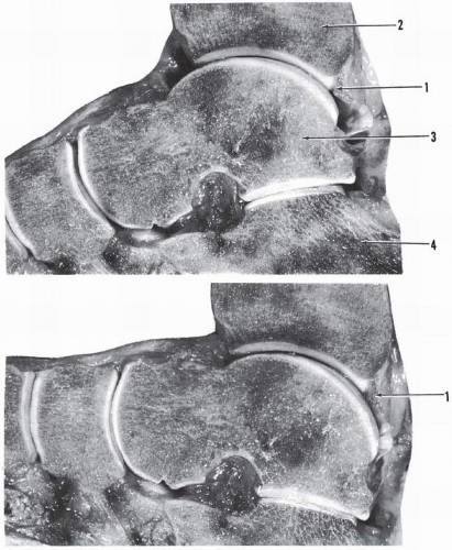 Figure 4.4 Sagittal cross-section of the ankle. (1, tibial attachment of the deep component of the posterior tibiofibular ligament forming a labrum; 2, tibia; 3, talus; 4, os calcis.) |
Interosseous Membrane
The interosseous membrane is in continuity with the apex of the interosseous ligament (Fig. 4.7). The anterior fibers are oblique, directed downward and laterally, whereas the posterior fibers are nearly vertical.
LIGAMENTS UNITING THE DISTAL TIBIOFIBULAR COMPLEX TO THE TALUS, CALCANEUS, AND NAVICULAR
The distal tibiofibular complex is firmly connected to the talus and the os calcis. The talus is anchored to the fibula anteriorly and posteriorly and to the tibia medially. The calcaneus is anchored to the fibula laterally and to the tibia medially. The anterior connection of the tibia to the talus and to the navicular is of lesser magnitude.
Lateral Ligament of the Ankle
Anterior Talofibular Ligament
The anterior talofibular ligament (Figs. 4.8 and 4.9) is a flat, quadrilateral, relatively strong ligament measuring approximately 15 mm × 8 mm × 2 mm, 20 mm × 6 mm × 2 mm,4 or 12 mm × 5 mm × 2 mm.5 This ligament is formed by two distinct bands separated by an interval that allows the penetration of vascular branches. The upper band is larger than the lower. A third band occasionally may be present. The ligament originates from the inferior oblique segment of the anterior border of the lateral malleolus. The upper band reaches the origin of the anterior tibiofibular ligament and the lower band that of the calcaneofibular ligament; in many specimens, these two ligaments are united with arciform fibers at their malleolar origin. The anterior talofibular ligament courses anteromedially and inserts not on the talar neck but on the talar body just anterior to the lateral malleolar articular surface. Two flat tubercles are occasionally seen, corresponding to the insertion of the two bands. The anterior talofibular ligament is in close connection to the capsule of the talofibular joint. In the neutral position of the talus, the ligament is horizontal. In dorsiflexion, the ligament is directed slightly upward. In plantar flexion, the ligament firmly braces the talar body as it stretches over the nearly right angle formed by the union of the anterior and lateral surfaces of the talar body (Fig. 4.10); in this position, the ligament is directed downward, medially, and anteriorly.
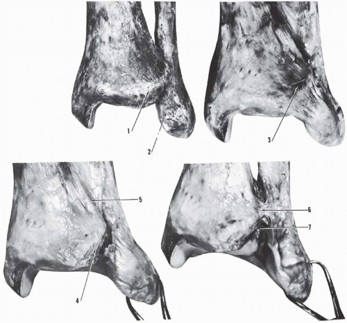 Figure 4.5 Interosseous ligament. (1, anterolateral tibiai tubercle; 2 lateral malleolus; 3, anterior tibiofibular ligament; 4, transected anterior tibiofibular ligament; 5, interosseous membrane; 6, interosseous ligament; 7, tibiofibular synovial recess.) |
Milner and Soames6 investigated the anatomical variations of the anterior talofibular ligament (ATFL) in 26 ankle specimens. They observed single, bifurcate, and trifurcate forms of the ATFL. The single form occurred in 38% (10 occurences with 8 bilateral and 2 unilateral), the bifurcate form in 50% (13 occurences with 12 bilateral and 1 unilateral), and the trifurcate form 12% (3 occurences with all unilateral). “The overall width of the ATFL did not appear to vary greatly irrespective of the number of bands present.”6 In a subsequent study based on the dissection of 40 ankles, the same authors7 provided the following mean measurements to the anterior talofibular ligament: length, 13 mm ± 3.9; width, 11 mm ± 3.3.
Calcaneofibular Ligament
The calcaneofibular ligament (Figs. 4.8 and 4.11) is a strong cordlike or flat oval ligament and measures approximately 30 mm length, 5 mm width, and 3 mm thickness; or length of 20 mm with diameter of 4 mm to 8 mm4; or length of 30 mm to 40 mm with width of 4 mm to 5 mm.3 It originates from the lower segment of the anterior border of the lateral malleolus just below the origin of the inferior band of the anterior talofibular ligament. The origin does not extend to the tip of the lateral malleolus, which is left free. Near the origin, arciform fibers may unite the calcaneofibular ligament and the inferior band of the anterior talofibular ligament. With the foot in neutral position, the ligament courses posteriorly, inferiorly, and medially and is crossed superficially by the peronei tendons and their sheaths, which may leave an imprint on the ligament (Figs. 4.12 4.13, and 4.14). Only approximately 1 cm of the ligament remains uncovered by the crossing peronei.
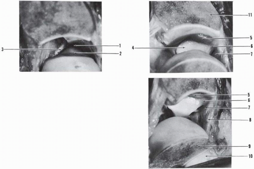 Figure 4.6 Sagittal section of the tibia and talus. (1, talofibular joint; 2, lateral talar articulating surface; 3, anterior talofibular ligament; 4, lateral malleolar articulating surface; 5, tibiofibular synovial fringe; 6, 7, posterior tibiofibular ligament; 8, posterior talofibular ligament; 9, talus; 10, posterior articular surface of calcaneus.) |
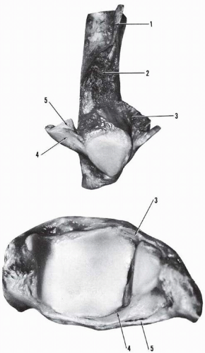 Figure 4.7 Interosseous membrane. (1, insertion of interosseous membrane; 2, insertion zone of tibiofibular interosseous ligament; 3, anterior tibiofibular ligament; 4, posterior tibiofibular ligament, deep component; 5, posterior tibiofibular ligament, superficial component.) |
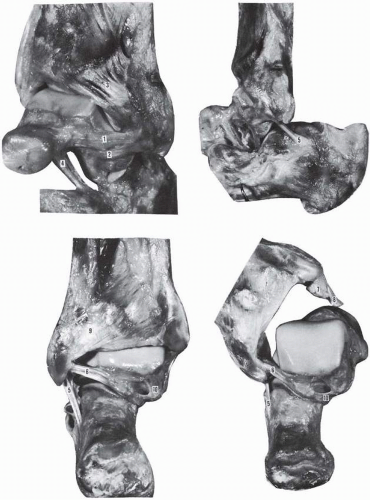 Figure 4.8 Anterior talofibular ligament. (1, anterior talofibular ligament, main component; 2, anterior talofibular ligament, accessory component; 3, anterior tibiofibular ligament, fasciculated; 4, cervical ligament; 5, calcaneofibular ligament; 6, posterior talofibular ligament; 7, deltoid ligament, deep layer; 8, deltoid ligament, superficial layer; 9, posterior tibiofibular ligament; 10, fibrous tunnel of flexor hallucis longus tendon.) |
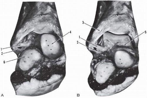 Figure 4.9 Anterior talofibular ligament. (A) Dorsiflexion. (B) Plantar flexion. (1, anterior talofibular ligament, major component. The ligament is oriented upward, medially in dorsification [ A], and downward, medially in plantar flexion [B]; 2, anterior talofibular ligament, accessory component; 3, anterior tibiofibular ligament; 4, navicular articular surface of the talus; 5, talus; 6, cuboidal articular surface of the os calcis.) |
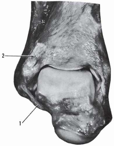 Figure 4.10 Anterior talofibular ligament in plantar flexion. (1, anterior talofibular ligament inserting on the anterior surface of the talar body and under tension in plantar flexion; 2, anterior tibiofibular ligament.) |
The calcaneofibular ligament inserts on a small tubercle (tuberculum ligamenti calcaneo fibularis) located on the posterior aspect of the lateral calcaneal surface, posterosuperior to the peronei processus trochlearis. The insertion of the ligament is variable. Laidlaw, in a study of 750 calcanei, gives the following location of the calcaneal insertion of the ligament: typical location, 64.5%; anterior location, 25.5%; posterior location, 5.5%; downward location, 4.5%.8 The variable insertions result in variable obliquity of the ligament relative to the long axis of the fibula.
Ruth, in a study based on 30 dissected specimens and observations of 55 ankles during surgery, provides the following data with regard to the angle formed by the long axis of the calcaneofibular ligament and the long axis of the fibula (Fig. 4.15): 10 to 45 degrees, 74.66%; 0 degrees, 18.66%; 80 to 90 degrees, 4%; fan shaped, 2.66%.5 The valgus or varus position considerably affects the direction of and the angle formed by the calcaneofibular ligament. The obliquity of the ligament is increased with valgus of the heel and decreased with varus (Fig. 4.16). The obliquity of the ligament is also variable with the position of the ankle joint (Fig. 4.17). The calcaneofibular ligament crosses the talocalcaneal joint and is separated from it by the lateral talocalcaneal ligament. The interval between the two ligaments is filled with adipose tissue.
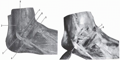 Figure 4.11 Calcaneofibular ligament. (1, calcaneofibular ligament; 2, superior peroneal retinaculum; 3, peronei tendons, brevis anteriorly and longus posteriorly; 4, inferior peroneal retinaculum; 5, stem of inferior extensor retinaculum; 6, tip of lateral malleolus, free of insertion; 7, anterior talofibular ligament, major component; 8, anterior talofibular ligament, accessory component; 9, lateral talocalcaneal ligament.) |
Trouilloud and colleagues9 reported on the variable relationship of the calcaneolibular ligament and the lateral talocalcaneal ligament in 26 ankles. They classified their findings into three categories: A, B, and C. In type A, 35%, the calcaneofibular ligament is reinforced by a lateral talocalcaneal ligament, attached to the former but diverging proximally or distally. In type B, 23%, a lateral talocalcaneal ligament exists anteriorly and independent of the calcaneofibular ligament. In type C, 42%, the lateral talocalcaneal ligament is absent and is replaced by an anterior talocalcaneal ligament, which is parallel now to the interosseous talocalcaneal ligament. In type C, the calcaneofibular ligament acquires more functional significance in providing stability to the subtalar joint.
Milner and Soames7 in a study based on the dissection of 40 ankles provided the following measurements in regard to the calcaneofibular ligament: length, 19.5 mm ± 3.9; width, 5.5 mm ± 1.6.
Posterior Talofibular Ligament
The posterior talofibular ligament (Figs. 4.8 and 4.18) is a very strong ligament situated in a nearly horizontal plane. Trapezoidal in contour, the ligament measures approximately 30 mm in posterior length, 5 mm in width at the fibular origin, and 5 mm to 8 mm in thickness. The ligament originates on the medial surface of the lateral malleolus from the lower segment of the digital fossa (Fig. 4.19). Thick and fasciculated, it courses horizontally toward the lateral and posterior aspects of the talus. The short transverse and intermediary fibers insert along the lateral surface of the talus in a groove along the posteroinferior border of the lateral malleolar articular surface up to its mid segment. The long fibers are directed posteromedially and insert on the posterior surface of the talus. The medial end expands and attaches on the posterolateral tubercle, the trigonal process, or the os trigonum (when present) and contributes to the formation of the floor of the flexor hallucis longus tunnel. When viewed posteriorly, the ligament is triangular, with a lateral apex and a medial base. The upper fibers of the posterior segment are in continuity medially with the superficial talotibial ligament, forming a posterior ligamentous sling.
 Figure 4.12 Lateral talocalcaneal ligament. (From DiGiovanni CW, Langer PR, Nickish F, et al. Proximity of the lateral talar process to the lateral stabilizing ligaments of the ankle and subtalar joint. Foot Ankle Inter. 2007;2:175-180, Figure 3A.) |
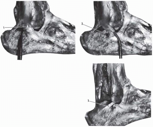 Figure 4.13 Calcaneofibular ligament. (1, adipose tissue covering the posterior segment of the calcaneofibular ligament; 2, exposure of the posterior segment of the calcaneofibular ligament by reflection of the adipose tissue; 3, calcaneofibular ligament exposed after resection of the peronei tendons.) |
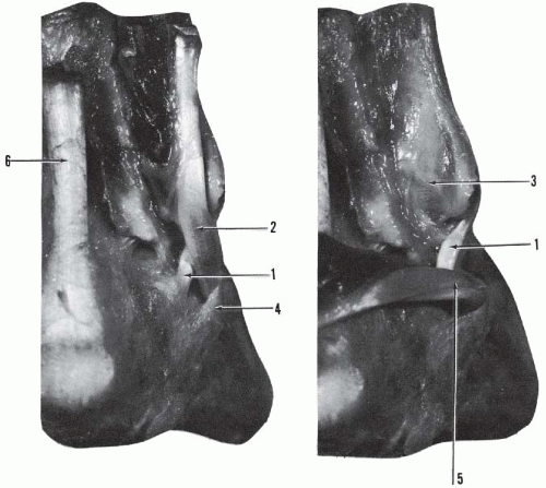 Figure 4.14 Posterolateral aspect of the ankle. (1, calcaneofibular ligament and peronei tendons [2] crossing in X; 3, sulcus of peronei tendons on posterior surface of lateral malleolus; 4, inferior peroneal retinaculum; 5, reflected peronei tendons; 6, Achilles tendon.) |
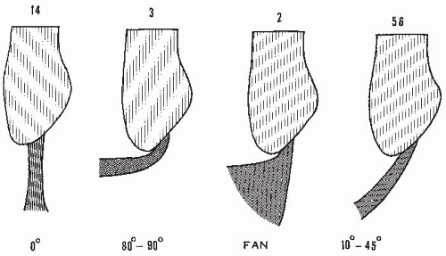 Figure 4.15 Anatomical variations of the calcaneofibular ligament in 75 ankles. Left to right: vertical, horizontal, fan shaped, oblique. (Redrawn after Ruth CJ. The surgical treatment of injuries of the fibular collateral ligaments of the ankle. J Bone Joint Surg [Am]. 1961;43:233.) |
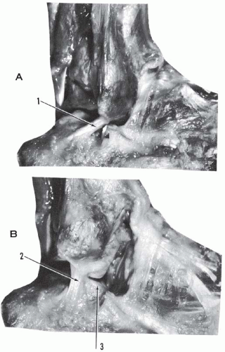 Figure 4.16 The heel in valgus and varus. (A) Heel in valgus; obliquity of calcaneofibular ligament (1) is increased. (B) Heel in varus, obliquity of cancaneofibular ligament (2) is decreased. In this specimen the ligament is vertical. (3, posterior calcaneal surface seen in varus.) |
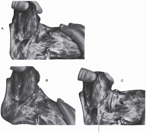 Figure 4.17 Obliquity of the calcaneofibular ligament (arrows). (A) In neutral position of the ankle. (B) In plantar flexion. (C) In dorsiflexion. |
Occasionally a band originates from the superior border of the posterior talofibular ligament near its origin, courses upward and medially, and inserts on the posterior tibial margin, blending with the fibers of the transverse component of the posterior tibiofibular ligament (Fig. 4.20); this insertion may reach the posterior surface of the medial malleolus. Paturet designates this ligament the posterior intermalleolar ligament.10 The posterior talofibular ligament is intracapsular but extrasynovial. The fibular origin is covered by the peronei retinaculum (Fig. 4.21). The superomedial segment is crossed by the tendon of the flexor hallucis longus (Fig. 4.22).
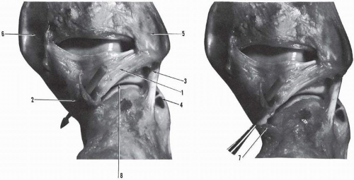 Figure 4.18 Posterior aspect of the ankle. (1, posterior talofibular ligament; 2, fibrous tunnel of the flexor hallucis longus tendon; 3, calcaneofibular ligament; 4, inferior peroneal retinaculum; 5, sulcus of the peronei tendons on posterior surface of the lateral malleolus; 6, sulcus of tibialis posterior tendon on posterior aspect of the medial malleous; 7, posterior talocalcaneal ligament; 8, posterior calcaneal articular interline.) |
Milner and Soames7 in a study based on the dissection of 40 ankles provided the following measurements in regard to the posterior talofibular ligament: length, 23.0 mm ± 7.0; width, 5.5 mm ± 2.5.
Fibulotalocalcaneal Ligament
The fibulotalocalcaneal ligament of Rouvière and Canela Lazaro (Figs. 4.23, 4.24, and 4.25) is an extrinsic ligament that occupies the posterolateral corner of the ankle and posterior subtalar joints. In 1924, Dujarier described briefly and produced a clear illustration of a posterior fibulocalcaneal ligament arising from the posterior aspect of the lateral malleolus and inserting on the posterosuperior aspect of the calcaneus laterally.11 He qualified this ligament as being abnormal and mentioned that, in certain clubfeet, this ligament is considerably developed, forming the posterior fibulocalcaneal ligament of Bessel-Hagen (Fig. 4.26).
In 1932, Rouvière and Canela Lazaro, in a comprehensive study, described the peroneotalocalcaneal ligament.12 This ligament is independent of the capsules and ligaments of the neighboring joints. It originates from the medial border of the peroneal groove located on the posterior border of the lateral malleolus, in common with the origin of the posterior tibiofibular ligament. Inferiorly, the origin may descend to the tip of the lateral malleolus and quite frequently may reach the origin of the calcaneofibular ligament.
At the origin, the peroneotalocalcaneal ligament is in close connection with the fibrous sheath of the peronei but soon separates from it and is directed downward, medially and posteriorly. This flat structure then divides into two fibrous laminae: superomedial and inferolateral. The superomedial lamina or talar component inserts on the posterolateral tubercle of the talus and contributes to the formation of the flexor hallucis longus fibrous tunnel. The inferolateral or peroneocalcaneal lamina is the major component of the ligament. It is directed downward and posteriorly, enlarges, and inserts nearly transversely on the entire width of the superior surface of the calcaneus. At times the calcaneal insertion is oblique, directed forward and medially. In approximately two thirds of the cases, the insertion remains localized to the superior surface of the calcaneus or reaches the lateral surface of the os calcis; the lateral border of the ligament is then separated by a small interval from the insertion of the calcaneofibular ligament. In the remaining one third, the calcaneal insertion clearly extends to the lateral calcaneal surface and blends with the insertion of the calcaneofibular ligament.
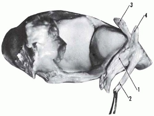 Figure 4.19 Tibiofibular mortise. (1, posterior talofibular ligament; 2 calcaneofibular ligament; 3, anterior talofibular ligament, major component; 4, anterior talofibular ligament, accessory component.) |
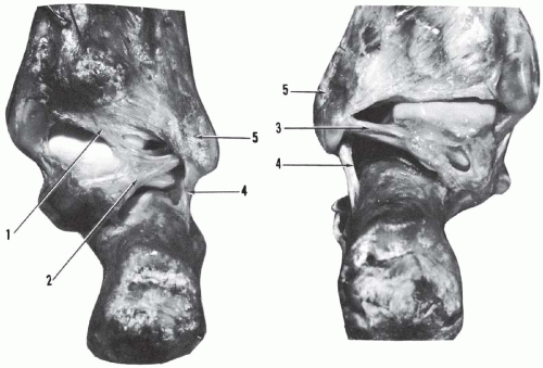 Figure 4.20 Posterior aspect of ankle. (1, posterior intermalleolar ligament; 2, 3, posterior talofibular ligament; 4, calcaneofibular ligament; 5, lateral malleolus.) |
The frequency of occurrence of this ligament is 60% present as a well-defined ligament, 20% present as a thin, weak structure but with ligamentous texture, and 20% absent and replaced by a thin fascia.12 Occasionally both components of the ligament are united, forming a continuous lamina.
On close analysis and as described by Rouvière and Canela Lazaro, the fibulotalocalcaneal ligament is the very thick inferior and suscalcaneal portion of the deep aponeurosis of the leg between the tunnel of the flexor hallucis longus and the tunnel of the peronei.12 This ligament limits the dorsiflexion of the foot.
Medial Ligament or Deltoid Ligament
The medial malleolus provides attachment to the ligaments necessary to stabilize the talus and the naviculocalcaneal complex medially (Fig. 4.27A). The insertion of the talotibial fibers are concentrated on the posteromedial aspect of the talus, whereas the peritalar fibers insert mainly on the sustentaculum tali.
The remaining fibers of the ligament have received variable acceptance as ligaments because of the relative strength to qualify them; this applies particularly to the superficial anterior talotibial ligament.
The interpretation of the disposition of the fibers of the medial ligament as being one or two layers (superficial and deep) has brought forth a plethora of descriptions (Fig. 4.27B-E). On close examination, if one accepts the element of variability with regard to the lesser components of this ligament, the descriptions then fall into a harmonious pattern.
A practical understanding of this ligament requires the consideration of a few points:
The entire medial ligamentous complex is invested, except in the very anterior part, by the deep crural fascia in continuity with the flexor retinaculum (Figs. 4.28 and 4.29).
The anterior border of the ligament is covered by the tibialis anterior tendon with its underlying adipose tissue. This anterior border is in continuity laterally with the thin anterior capsule.
A major segment—mid and posterior—of the ligament is covered by the obliquely crossing tibialis posterior and flexor digitorum longus tendons. The fibrosynovial floors of these tunnels blend with the underlying ligament (Figs. 4.30 and 4.31); minute dissection usually is necessary to separate the two.
The medial ligament, except for its deep talotibial component, is a continuous fibrous lamina and any division into components is usually artificial (Figs. 4.324.33, 4.34). It is only by referring to the insertion of the fibers that descriptive differentiation is possible.
Posteriorly, the ligament is in continuity with the posterior capsule and the posterior talofibular ligament.
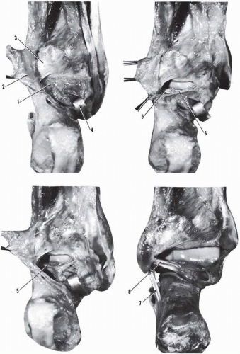 Figure 4.21 Posterior aspect of ankle. (1, adipose tissue and capsule covering the posterior talofibular ligament; 2, ligament of Rouvière and Canela Lazaro; 3, posterior tibiofibular ligament; 4, reflected tendon of flexor hallucis longus; 5, posterior talofibular ligament; 6, reflected posterior capsule; 7, calcaneofibular ligament.) |
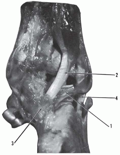 Figure 4.22 Posterior aspect of ankle. (1, posterior talofibular ligament crossed by the flexor hallucis longus tendon [2]; 3, tunnel of flexor hallucis longus; 4, calcaneofibular ligament.) |
The medial ligament is divided into two layers, superficial and deep, each being formed by multiple fascicles. The superficial layer or deltoid ligament is broad and triangular. For the purpose of description, the following components are considered.
Anterior Superficial Tibiotalar Fascicle and Tibionavicular Fascicle
The anterior superficial tibiotalar fascicle and tibionavicular fascicle (Fig. 4.35) have a common origin on the anterior border of the anterior colliculus. The fibers of origin extend to the medial corner of the anterior margin of the tibial quadrilateral inferior articular surface. As specified by Beau, the fibers delineate an anterolateral concave border and further distally divide into two layers.13 The deep fibers insert on the dorsum of the talar neck slightly posterior to the talar head and to the talonavicular capsule; this component is the anterior and superficial tibiotalar ligament. The superficial fibers extend beyond the talonavicular interline and insert on the dorsomedial aspect of the navicular in a curvilinear fashion, extending medially a very short distance from the articular margin; this component is the tibionavicular ligament. The two ligaments overlap except at the most median site, where the talar fibers do not extend onto the navicular.
Tibioligamentous Fascicle
The tibioligamentous fascicle originates from the anterior segment of the anterior colliculus. The fibers present a gentle anterior concavity and insert on the superior border of the superomedial calcaneonavicular ligament. The anterior superficial tibiotalar fascicle, the tibionavicular fascicle, and the tibioligamentous fascicle constitute the broader but weaker component of the medial ligament (Fig. 4.36).
Tibiocalcaneal Ligament
The tibiocalcaneal ligament (Fig. 4.37) is the strongest superficial component. It originates from the medial aspect of the anterior colliculus, descends vertically, and inserts on the medial border of the sustentaculum tali after interlacing its fibers with those of the superomedial calcaneonavicular ligament.14 This ligament is in continuity with the preceding tibioligamentous fascicle. In certain specimens, the origin of the latter overlaps the origin of the tibiocalcaneal ligament, and the interval is filled with adipose tissue. The tibiocalcaneal ligament measures approximately 1 cm in width at the origin and 1.5 cm at the insertion. The average length is 2 to 3 cm and the thickness 2 to 3 mm. It is a substantial structure.
Superficial Posterior Tibiotalar Ligament
The superficial posterior tibiotalar ligament originates from the posterior part of the medial surface of the anterior colliculus and the medial surface of the posterior colliculus (Fig. 4.38). The fibers are directed posteriorly, inferiorly, and laterally and insert on the posteromedial talar tubercle, reaching the flexor hallucis longus tunnel.3, 15, 16, 17, 18, 19 The posterior border of the tibiocalcaneal ligament is distinct from this ligament or the two may be in continuity; if they are in continuity, the superficial layer is represented as a large, fibrous, fan-shaped lamina deserving the “deltoid” denomination. The superficial posterior tibiotalar ligament is separated from the underlying posterior deep tibiotalar ligament with adipose tissue (more so anteriorly) and is in continuity along its superolateral border with the posterior talotibial capsule and the posterior talofibular ligament. The occasional absence of this ligament or its fusion to the deep talotibial ligament accounts for some of the variations in description.
Deep Layer of Deltoid Ligament
The deep layer of the deltoid ligament (see Fig. 4.38) is short and strong. It is formed by a small anterior and a very strong posterior component.9, 16, 18, 19
Deep Anterior Tibiotalar Ligament
The deep anterior tibiotalar ligament originates from the tip of the anterior colliculus and the anterior part of the intercollicular groove and inserts on the medial surface of the talus distal to the anterior segment of the comma-shaped talar articular surface. This ligament is “of variable size in different specimens, sometimes hardly discernible, and in some cases completely absent.”19
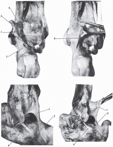 Figure 4.23 Fibulotalocalcaneal ligament. (1, fibulotalocalcaneal ligament of Rouvière and Canela Lazaro; 2, calcaneal insertion of 1; 3, common origin of 1 with the posterior tibiofibular ligaments [4]; 5, connection of 1 with superior peroneal retinaculum; 6, calcaneofibular ligament.) |
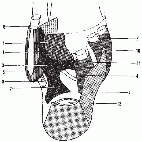 Figure 4.24 Fibulotalocalcaneal ligament. (1, stem of fibulotalocalcaneal ligament of Rouvière and Canela Lazaro; 2, calcaneal component, inferolateral, of 1; 3, talar component, superomedial of 1; 4, deep crural aponeurosis; 5, retinaculum of peronei tendons; 6, posterior tibiofibular ligament; 7, superficial crural aponeurosis; 8, calcaneal insertion of calcaneofibular ligament; 9, tunnel of tibialis posterior tendon; 10, tunnel of flexor digitorum longus; 11, tunnel of flexor hallucis longus; 12, Achilles tendon.) |
Deep Posterior Tibiotalar Ligament
The deep posterior tibiotalar ligament is the strongest component of the entire medial ligament. In most cases, it is conical, with the base superior and the apex posteroinferior. The approximate measurements are 1.5 cm width at origin, 1 cm width at insertion, 1.5 cm length, and 1 cm thickness at origin. The size of the ligament is variable. The thickness varies from 0.5 to 1.5 cm and the width from 1.5 to 2 cm.2, 20
This ligament originates from the intermolecular fosse (which measures about 1 cm in width), the entire anterior surface of the posterior colliculus, and the upper segment of the posterior surface of the anterior colliculus. The fibers are directed downward, posteriorly, and laterally and insert on the medial surface of the talus on an oval elevation located under the tail of the commashaped articular surface. The insertion reaches the posteromedial talar tubercle. This intra-articular but extrasynovial ligament may be fasciculated (Fig. 4.39) or divided into two distinct bands.18 Beau, in his comprehensive study of the ligaments of the ankle, considers this ligament as the homolog of the posterior talofibular ligament and describes it not as a medial ligament but as a posterior ligament of the ankle, bracing the talus posteriorly.13
Anterior Tibiotalar Fascicle, Capsule, and Fat Pad
The anterior capsule of the talotibial joint is thin and reinforced by an oblique fibrous band extending from the medial malleolus to the talar neck. This anterior tibiotalar fascicle (see Fig. 4.35) is narrow in the middle and broader at the origin and the insertion. The tibial attachment of the anterior capsule is 5 to 6 mm proximal to the articular margin (see Fig. 4.4). The talar insertion surrounds the cribriform fossa of the neck and is approximately 8 mm from the anterior trochlear margin. Laterally and medially, the anterior capsular insertion is close to the malleolar facets of the talus.
A prearticular fat pad is present anteriorly, extending transversely between the capsule and the tendons. This fat pad is thicker medially (see Fig. 4.27) and reinforces the anterior capsule.
Milner and Soames,7 based on the dissection of 40 ankles, describe six components to the medial collateral ligament complex: four superficial bands and two deep bands. The four superficial components are the tibiospring ligament (TSL), the tibionavicular ligament (TNL), the superficial posterior tibiotalar ligament (STTL), and the tibiocalcaneal ligament (TCL). The two deep components are the deep posterior tibiotalar ligament (PTTL) and the deep anterior tibiotalar ligament (ATTL). Only three ligaments are constant: PTTL, TSL, and TNL. In 52.5% (21 specimens), there was one additional band present; in 42.5% (17 specimens), there were no additional bands present; and, in 5% (two specimens), two additional bands were present.
LIGAMENTS OF THE CALCANEONAVICULAR JOINT AND ACETABULUM PEDIS
The head of the talus is received into a deep socket or acetabulum pedis formed by the navicular, the anterior and middle calcaneal articulating surfaces, the calcaneonavicular component of the bifurcate ligament, the superomedial calcaneonavicular ligament, and the plantar calcaneonavicular ligament (Figs. 4.40 and 4.41). The flexibility of the acetabulum pedis permits the adaptability in form and size of the containing socket as necessitated by the relative displacements of the talar head calcaneus and navicular.
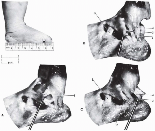 Figure 4.25 Foot of a fetus. (A-C) Ligaments of lateral aspect of the ankle. (1, anterior talofibular ligament lifted by probe in [A]; 2, lateral talocalcaneal ligament lifted by probe in [B]; 3, calcaneofibular ligament; 4, ligament of Rouvière and Canela Lazaro [posterior calcaneofibular] lifted by probe in [C]; 5, cervical ligament.) |
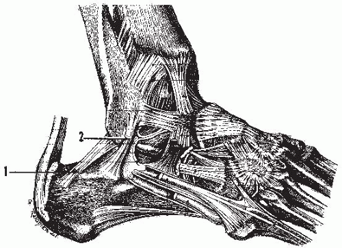 Figure 4.26 Posterior fibulocalcaneal ligament. (1, posterior fibulocalcaneal ligament or ligament of Bessel-Hagen; 2, fibulocalcaneal ligament.) (Dujarier CH. Anatomie des Membres: Dissection—Anatomie Topographique. 2nd ed. Paris: Masson; 1924:399.) |
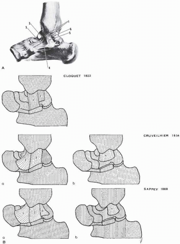 Figure 4.27 (A) Medial or deltoid ligament. (1, deltoid ligament; 2, anterior tibiotalar fascicle; 3, tibionavicular fascicle; 4, tibioligamentous fascicle [insertion on superomedial calcaneonavicular ligament]; 5, tibiocalcaneal fascicle; 6, posterior tibiotalar fascicle.) (Spalteholz W. Hand Atlas of Human Anatomy, Vol 1. Philadelphia: JB Lippincott; 1903: 219.) (B) Interpretations of the deltoid ligament. Cloquet 1822: Origin, apex of medial malleolus and its depression; insertion, medial aspect of talus (2), calcaneus (1); few fibers to fibrous tunnel of flexor digitorum longus. Cruveilhier 1834: (a) Superficial. Origin, apex and borders of medial malleolus; insertion, talus neck (1), scaphoid (2), inferior calcaneoscaphoid ligament (2), calcaneus (3). (b) Deep. Origin, apex and borders of medial malleolus; insertion, entire medial aspect talus under articular cartilage. Sappey 1888: (a) Superficial. Anterior: origin, anterior border of medial malleolus; insertion, scaphoid (1), inferior calcaneoscaphoid ligament (1
Get Clinical Tree app for offline access
Related posts:Stay updated, free articles. Join our Telegram channel
Full access? Get Clinical Tree


|
