Fig. 7.1
Telangiectatic papules and plaques in a patient with subacute cutaneous erythematosus
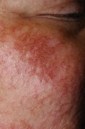
Fig. 7.2
Postinflammatory telangiectasia in a patient with dermatomyositis
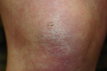
Fig. 7.3
Telangiectatic and avascular patches typical of dermatomyositis
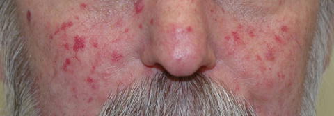
Fig. 7.4
Matted telangiectasias of systemic sclerosis
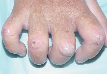
Fig. 7.5
Lacy telangiectasias overlying the proximal interphalangeal joints in a patient with systemic sclerosis. Note active digital ischemic lesion which is associated with this pattern of telangiectasia
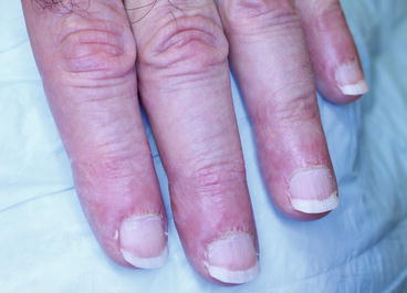
Fig. 7.6
Typical periungual telangiectasias in this patient with lupus erythematosus
Approach to the Patient
In summary, when presented with a patient with mucocutaneous telangiectasias, one should first rule out a primary, congenital syndrome as well as possible secondary causes such as medication or malignancy. The associated cause of the telangiectasias is then decided upon by both configuration and distribution of the lesions.