CHAPTER 68 Synovectomy of the Elbow
INTRODUCTION
Rheumatoid arthritis affects 1% to 2% of the general population.53 The most common cause of elbow arthritis is rheumatoid arthritis in which the involvement of elbow after an average of at least 10 years can be seen in more than half of the patients clinically or radiologically.17,25,27 Medical management is discussed in detail in Chapters 74 and 75. The mainstays of surgical treatment available for the patient with rheumatoid arthritis of the elbow include synovectomy, interposition arthroplasty, and total elbow replacement, in addition to other treatment options with limited application like arthrodesis and resection arthroplasty. Joint replacements and interposition arthroplasty are considered in Chapters 54, 55, and 69. Synovectomy with or without radial head excision is a well-recognized and accepted form of treatment for the rheumatoid elbow. This procedure reduces the amount of joint fluid and removes proliferative synovitis, and thereby decreases intra-articular pressure, relieves pain, and reduces swelling. After a few months, a new synovial membrane is regenerated in the joint5; however, the lack of villous change, vasculitis, and decreased pathologic enzymatic activity in the new synovial membrane and the joint fluid34 indicates a difference in the inflammatory process after synovectomy.38 Still, the role of synovectomy must be defined in the context of the success of total elbow replacement. The primary indication for elbow synovectomy in a patient with rheumatoid arthritis is persistent painful synovitis that is unresponsive to at least 6 months of appropriate medical management as an initial interventive procedure. Despite this, controversy still remains as to the role and method of execution, especially in advanced stages and in patients with instability and stiffness. This chapter highlights the issues of open versus arthroscopic techniques and the resection versus preservation of the radial head during elbow synovectomy.
INDICATIONS AND PATIENT SELECTION
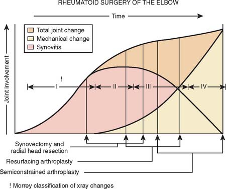
Synovectomy is ideal for the patients who present with uncontrolled synovitis resulting in a painful elbow with limited function. However, a period of nonsurgical treatment including anti-inflammatory agents, steroids, disease-modifying anti-rheumatic drugs in addition to maintaining supple joints with an active range of motion program and splinting for at least 6 months should be attempted before surgery is considered4 (see Chapter 54). Early in the disease, synovitis is a prominent feature (Fig. 68-1). A warm, swollen elbow with a mild flexion contracture and a painful limitation of flexion and extension in addition to pronation and supination characterizes a typical joint. As the disease progresses, the synovitis generally wanes and persistent pain and stiffness results from joint destruction with articular incongruity.
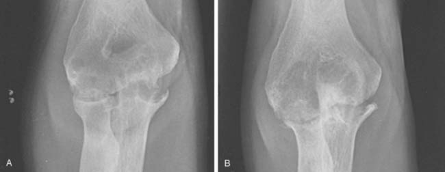
Although classically the disease stage employs the Carson system, for our purposes, it is convenient to describe the radiographic appearance into five categories. Type I: synovitis with a normal appearing joint; type II: loss of joint space but maintenance of the subchondral architecture; type IIIA: alteration of the subchondral architecture; type IIIB: alteration of the architecture with deformity; type IV: gross deformity; type V: radiographic appearance of ankylosis (Fig. 68-2).
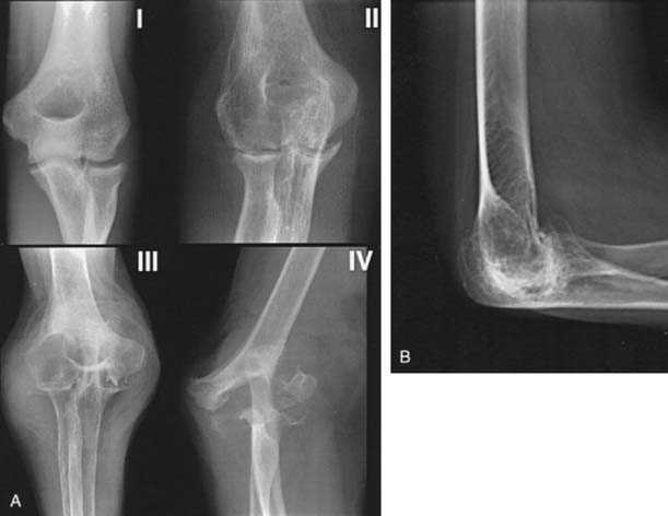
FIGURE 68-2 A, Mayo radiographic classification of rheumatoid involvement of the elbow considers synovitis, articular involvement, and joint distraction (see text). B, A type V radiographic presentation is one of ankylosis, as reported by Connor and associates.6
This classification is helpful in providing a basis for treatment options (see Fig. 68-1). Elbow synovectomy is ideally reserved for the early stages of the disease (type I, type II, and early type IIIA) (Fig. 68-3).
Synovectomy per se is not effective or reliable in restoring motion. As a result, a functional arc (30 to 130 degrees of flexion) should ideally be present.33 In patients with a long-standing and advanced disease, this arc is often not present. Therefore, a total arc of flexion of at least 80 degrees is recommended before the procedure. However, more recently, it has been shown that with anterior and posterior capsular release or capsulectomy, a better outcome may be possible even in those with less than 80 degrees of flexion.28,41
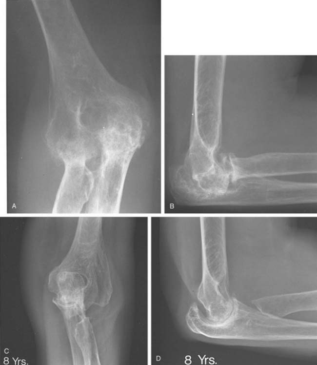
The role of synovectomy in more advanced stages of disease is still debatable in the literature. In advanced stages of the disease, damage to either or both the medial and lateral ligamentous complexes combined with bony and cartilaginous loss in addition to significant erosion and instability of the radial head may result in gross medial-lateral elbow instability, precluding synovectomy. This is an indication for total elbow arthroplasty, which can be reliable even in the presence of instability.6,14,32 Critical assessment of prior reports reveal a small number of articles that report worsening of the results over the long term, especially due to the development of instability.10,50 On the other hand, the majority of reports agree that advanced radiographic stage of the disease might not influence the ultimate outcome* (Fig. 68-4). Furthermore, although instability can be seen in up to 50% of patients in the advanced stages,40 this generally does not seem to cause symptomatic instability requiring an operation.† As a result, synovectomy might be employed with reservations in advanced stages as well if instability is not one of the major complaints of the patient. Nevertheless, those experienced with the techniques of total elbow replacement as well as synovectomy, and those who are familiar with the results of both, favor total elbow replacement to synovectomy in treating later stages of the disease because patients are more satisfied, functional improvement is greater, and the results are more predictable.6,14,32
CONTRAINDICATIONS
One of the major contraindications to synovectomy is severe joint disruption. Synovectomy performed in this situation will not correct instability, and débridement of the joint may actually aggravate instability, particularly when radial head excision is performed. In the series of Rymaszewski and colleagues,40 most of the displeased patients had medial tenderness and pain with attempted valgus stress in the elbow, which was attributed to tension on medial collateral ligaments, mainly due to resection of the radial head. Also, the initial good results may deteriorate with time with increasing instability of the joints due to gradual bone loss.50
Limited elbow motion is the other major contraindication for synovectomy of the elbow, because elbow function cannot predictably be improved, and some patients lose some motion. Severe joint stiffness resulting from inflammatory fibroarthrosis is more commonly seen in juvenile rheumatoid arthritis. Because complete ankylosis can be observed in these patients, grade 5 radiologic classification was added to the original classification of the rheumatoid arthritis6 (see Fig. 68-2). Synovectomy alone cannot be advocated in this subgroup of patients for the sole purpose of improving motion.30 Radiographic involvement of the joint architecture with deformity (type IIIB) is also a relative contraindication.
TECHNIQUES
Synovectomy can be performed nonsurgically (chemical or radiation synovectomy)8,15,37,44 or surgically, which can be performed either open or by an arthroscopic technique.‡
NONSURGICAL SYNOVECTOMY
Chemical and radiation synovectomy has been advocated as a noninvasive procedure. Owing to the size and subcutaneous surface of the joint, rhenium-186,15 yttrium-90,44 and osmic acid37 have been used with caution at the elbow. Although patients with early radiologic stages of elbow involvement have a better success rate, especially with the addition of triamcinolone to radiosynovectomy, the overall success rate is less than 50% with these agents with a very limited follow-up.15,44
The potential for cartilage necrosis and the subcutaneous location of this joint has limited the use of these agents in the United States.16,31 Also, radial head excision, if needed, is not possible. Besides, in marked synovitis in which synovial swelling is above the effective penetration range of the radionuclide, the lower layers of synovium cannot be reached by radiosynoviorthesis and the destruction process and pain will continue.15 However, this technique can be considered as a more conservative treatment option to surgical synovectomy, especially in patients who are unable to undergo surgery.
SURGICAL SYNOVECTOMY
ARTHROTOMY
Although there are an increasing number of reports on arthroscopic synovectomy, open surgery is still commonly performed for elbow synovectomy. This technique is well established and requires less technical expertise than arthroscopic synovectomy, particularly when radial head excision is performed. However, more remarkable postoperative pain, increased risk of wound breakdown, and increased risk of infection, in addition to loss of ligamentous or muscle and tissue supports require a delay in the initiation of rehabilitation, which might potentially result in elbow stiffness.3,21
Technique
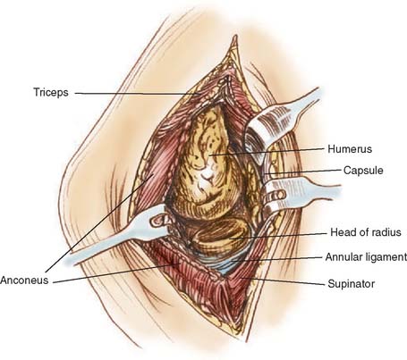
An extensile Kocher approach provides excellent visualization of the lateral joint and preserves the medial collateral ligaments (Fig. 68-5). The radial head is removed if there are significant symptoms with pronation and supination or marked radial humeral joint pain with flexion and extension. The synovitis typically involves the sacriform recess of the radial neck and a thorough synovectomy of this region is required. If the radial head is removed, a very thorough synovectomy can be carried out.2,13,18,22,39,50,54 If radial head is preserved anterior compartment exposure is more difficult but can still be adequately achieved. A second medial exposure is not necessary in our experience and that of most of the investigators.§ The triceps is elevated from the lateral column and the joint is extended. The posterior compartment synovectomy can then be carried out.
Because a complete synovectomy can be achieved through the extensile Kocher approach, separate lateral and medial approaches have been advocated sporadically.7,39,50 The main advantage of adding a medial incision is that radial head need not be resected for adequate synovectomy.7 However, even in bilateral approach proponents, it has been noted that if an extended lateral approach is used, results do not differ.39 In various reports, a medial incision is also added to an extended lateral approach in several cases because of the need for the decompression and translocation of the ulnar nerve.3,26,49,54 The use of Mayo posteromedial triceps reflecting exposure has also been advocated for synovectomy of the elbow.20 It provides for identification of the ulnar nerve and facilitates subsequent skin incision for total elbow replacement. However, surgery is more extensive than the extensile Kocher approach with the added potential for extensor mechanism complications, and we have not found it necessary to use this approach for routine synovectomy. Synovectomy and débri-dement through a transolecranon approach is contraindicated.21
ARTHROSCOPIC SYNOVECTOMY
Arthroscopy is being used with increasing frequency to diagnose and treat elbow disorders. Because elbow arthroscopy has become popular in recent years, concern regarding potential complications, including septic arthritis, superficial skin infection, persistent drainage from portal sites, and most frequently, nerve injuries (transient and permanent), has also risen23 (see Chapters 40 and 41). The risk can be minimized with observation of certain safety precautions. In our series, of all procedures, it was found that two factors were associated with a higher risk of nerve palsy–the performance of capsular release and the diagnosis of rheumatoid arthritis.23 In experienced hands, the advantages of arthroscopic over open synovectomy are obvious. It can be performed on an out-patient basis, causes minimal morbidity (decreased scarring, decreased risk of infection, less postoperative pain), possibly enables a more thorough visualization of the elbow joint than is possible with an arthrotomy during some procedures, results in rapid return of motion, and shortens the recovery period.23 With improved technique and greater experience, a complete synovectomy including excision of the radial head, if necessary, is possible (see Chapter 40).
Technique
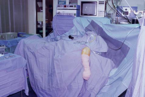
Several strategies can be used and our technique of elbow arthroscopy has evolved.36 Patients are placed in the lateral decubitus position, with the involved elbow over a padded bolster. The forearm is allowed to swing free, and the elbow is flexed to 90 degrees (Fig. 68-6). The arm is then prepared in the usual fashion. The forearm is exsanguinated by elevating the limb. A soft elastic bandage is then wrapped around the hand and forearm to within 10 cm of the olecranon. The tourniquet, which is used routinely, is inflated to 250 mmHg. The elastic bandage is left on until the end of the procedure, to limit the periarticular swelling to the elbow area. At the end of the procedure, the bandage and the tourniquet are removed and any accumulated edema rapidly dissipates into the tissues of the forearm and arm.
Stay updated, free articles. Join our Telegram channel

Full access? Get Clinical Tree


