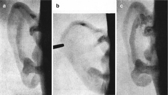
Fig. 17.1
(a) Section of the oval window: unstained histological section, pigmented bone cells on stapes base. (b) Promontorium section: unstained histological section, pigmented cells in periostal layer and on stapes head
In clinical examinations of 17 patients with manifested ochronosis, there have been the following findings in the organ of hearing. Auricular changes are visible, they are typical, and it can be stated that they are one of the most constant symptoms of ochronosis in the course of alkaptonuria. They often lead to the diagnosis of the primary disease. Pigmented cartilage of the auricle appears darker on trans-illumination. Salts are deposited in damaged tissue at affected places, forming rugged blue to violet figures showing through the skin. These changes appear in adult age, usually before the onset of the first joint symptoms. They arise unnoticed so that the patients themselves are frequently not aware of them until other people often warn them of the blue shade. Painless, hard and rough tubers with a wide base looking like infiltrates can be palpated in cartilage. They are firmly bound to the base and show through a fine skin as brown/dark blue/violet figures. The first rugged prominences can be observed on the lower crus of the anthelix, later on the whole anthelix, in the triangular fossa, less frequently on the helix crus and in the cavum conchae, cymba and tragus. In more advanced stages, the whole anthelix and later on the whole auricle are harder on palpation and less elastic and lose their configuration over time. Auricle deformities rarely occur (Fig. 17.2).
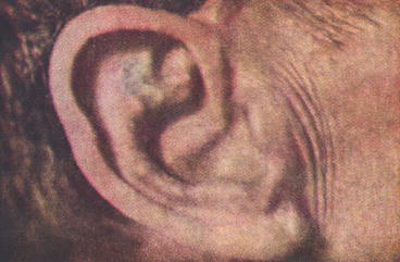
Fig. 17.2
Ochronotic changes on the ear
In our patients, we found blue to violet prominences only on the concave area of the auricle. In this area the perichondrium is bound to cartilage more firmly than on the convex side. Susceptibility to ochronotic changes may be associated with micro-injuries during the cleaning and washing of ears and due to their exposed location. While washing the auricles, fingers frequently touch just the above areas with a predilection for ochronosis. Patients themselves frequently report that some minor injuries like blows, frostbites, etc., preceded pigmentation. Symmetrical localization on both sides is relatively frequent.
Opacities with sharp contours on auricle X-rays resemble calcifications (Fig. 17.3a–c). This finding was present only in the advanced stages of ochronosis when the cartilage was already less elastic and harder on palpation. X-rays were normal in initial stages of the disease as well as in the presence of apparent bluish colouration of the auricles.
 Genetics of Alkaptonuria
Detection of Homogentisic Acid in Plasma and Urine
Coincidence of Alkaptonuric Ochronosis with Other Diseases
Therapy of Alkaptonuria
Clinical Manifestation of Ochronotic Arthropathy in Peripheral Joints
Analysis of X-Ray Symptomatology of Ochronotic Arthropathy in the Area of Peripheral Joints
Genetics of Alkaptonuria
Detection of Homogentisic Acid in Plasma and Urine
Coincidence of Alkaptonuric Ochronosis with Other Diseases
Therapy of Alkaptonuria
Clinical Manifestation of Ochronotic Arthropathy in Peripheral Joints
Analysis of X-Ray Symptomatology of Ochronotic Arthropathy in the Area of Peripheral Joints


Related posts:
Genetics of Alkaptonuria
Detection of Homogentisic Acid in Plasma and Urine
Coincidence of Alkaptonuric Ochronosis with Other Diseases
Therapy of Alkaptonuria
Clinical Manifestation of Ochronotic Arthropathy in Peripheral Joints
Analysis of X-Ray Symptomatology of Ochronotic Arthropathy in the Area of Peripheral Joints
Stay updated, free articles. Join our Telegram channel
Full access? Get Clinical Tree
