Fig. 13.1
Ochronosis. Open knee joint. Deposits of ochronotic pigment give the cartilage the blue-black colour
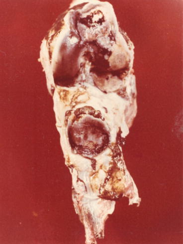
Fig. 13.2
Distal epiphyseal end of femur. Map-like erosions of articular cartilage that has black pigmentation on lateral condyle and facies patellaris. Dorsal area of patella has black pigmentation with smaller erosions of articular cartilage. Pedunculated pigmented corpuscles on apex and base
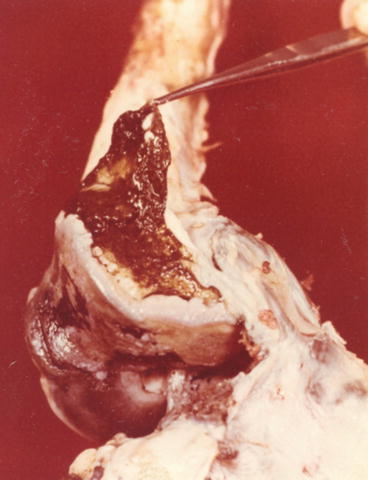
Fig. 13.3
Marked denudation of articular cartilage on lateral femoral condyle. Marginal osteophytes. Brown-black synovial membrane and cartilaginous corpuscles in it
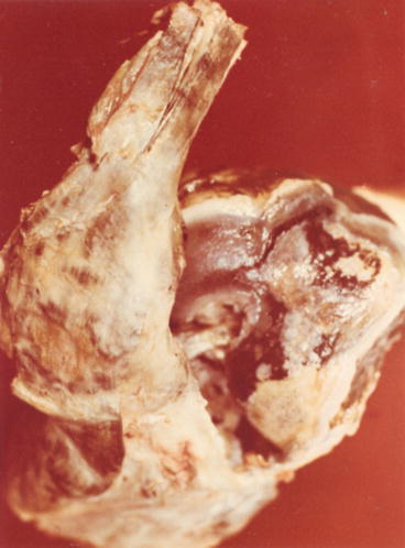
Fig. 13.4
Erosions of black-pigmented cartilage on patellar facet and on lateral femur condyle. Brown to black pigmentation of patellar ligament
Extension is more limited in osteoarthrosis, while mobility limitation in ochronotic arthropathy is caused by flexion reduction. Extension restriction is apparent in the further course of the disease. A complete clinical manifestation of ochronotic arthropathy in the knee area will be marked in 6–12 years from the disease onset. In contrast to the literature data (Zhao et al. 2009; Uebermuth 1928), we have never found an inflammatory character of knee involvement of a rheumatoid arthritis type or clinical signs of pannous synovitis in our patients. No patients complained of pain at rest. Volume expansion of the knee was not caused by inflammatory oedema, but by ‘cold’, slightly painful thickening of soft tissues. We also have not seen any knee deformities characteristic for rheumatoid arthritis. The soft tissues, namely, subcutaneous tissues around the joint, did not exert the symptoms suggesting panniculitis or cellulitis. These conditions are frequent accompanying symptoms of arthritic changes, especially in women. We do not usually detect venous insufficiency of lower extremities.
Shoulders are the second most commonly affected joints. Some patients only have vague muscle pain in the area of pectoral girdle. Nevertheless, in 42.3 % of cases, typical arthropathy of shoulder joints with mobility limitation developed, approximately 10 years after the onset of the first symptoms of spine involvement. The pain initially occurs only during forced movements, and mobility becomes limited only successively and slowly, especially abduction and internal rotation, sometimes by 75 % of original motion range.
Involvement of shoulder joints is usually symmetrical, however not always of the same degree. Extremely affected shoulder joints were observed in a patient in whom long-term and repeated micro-injuries caused concentric symmetrical mobility limitation of shoulder joints by 80 %. X-rays revealed severe cartilaginous and bone changes of shoulder joints with massive hyperplasia of inferior margins of glenoid fossa. This was a clearly mechanically induced intensification of degenerative changes of arthrosis type. When compared to shoulder osteoarthrosis, these joints are affected much more frequently in ochronotic arthropathy. In large statistical sets of osteoarthrosis, shoulder joints are affected in 1–2 % of cases, while in our set of patients with ochronosis it was 42.3 %. The extent of joint affections with functional limitation is also much higher in ochronotic arthropathy. Concentric limitation of mobility can also be observed in some patients with AS, but it is induced by inflammatory process in soft tissues. Hip joints affected in advanced stages of ochronotic arthropathy are directly involved in disability. It is interesting that out of all our patients, nine of them had hip joints affected. Out of this number, five patients were females and four were males, suggesting higher prevalence in females. Occurrence of the first symptoms in hip area is hard to detect because the patients have only slight subjective problems and they cannot recall the time of onset. In contrast to other joint diseases, affection of hip joints in ochronotic arthropathy is rarely associated with mild pain; usually painless, slowly progressive mobility limitation dominates, similarly as in ankylosing hyperostosis. In more detailed analysis of mobility, we find reduction of abduction as the first symptom. Limitation of rotational movements, especially internal rotation, follows. In advanced stages, we can observe concentric limitation of movement excursions. When we compare affection of hip joints in osteoarthrosis, AS and ochronotic arthropathy, we can see a certain analogy in the onset and development of successive limitation of mobility – particularly limitation of abduction, rotation and extension (Siavashi et al. 2009; Forestier et al. 1951; Justensen and Andersen 1984) in all these diseases. The difference consists in nearly simultaneously limited flexion in ochronotic arthropathy, while this movement remains preserved for a long time in coxarthrosis and AS. Early onset of affection of hip joints and more rapid development of the disease with intense concentric mobility limitation that results in ankylosis and premature disability distinguish ochronotic arthropathy from genuine osteoarthrosis of hip joints. In three of our patients, severe flexion contracture developed in this area and caused directly their working disability.
Related posts:
 Genetics of Alkaptonuria
Detection of Homogentisic Acid in Plasma and Urine
Coincidence of Alkaptonuric Ochronosis with Other Diseases
Therapy of Alkaptonuria
Analysis of X-Ray Symptomatology of Ochronotic Arthropathy in the Area of Peripheral Joints
Clinical Manifestation of Ochronotic Arthropathy in Spine
Genetics of Alkaptonuria
Detection of Homogentisic Acid in Plasma and Urine
Coincidence of Alkaptonuric Ochronosis with Other Diseases
Therapy of Alkaptonuria
Analysis of X-Ray Symptomatology of Ochronotic Arthropathy in the Area of Peripheral Joints
Clinical Manifestation of Ochronotic Arthropathy in Spine
Stay updated, free articles. Join our Telegram channel

Full access? Get Clinical Tree


