Fig. 16.1
(a, b) Ochronotic pigmentations on scleras in patients affected by ochronotic arthropathy
According to our findings, the first traces of pigment appear in a horizontal plane 4–6 mm distance from the cornea margin, i.e. approximately at the places where the tendons of the oculomotor muscles are inserted to the scleras. They have a pale grey colour, and they progressively change to dark brown or a violet colour during a slow growth. Pigment colour is dependent on the depth of its deposition in sclera layers. The more superficial the pigmented particles are, the more marked their brown-violet colour is. Pigment spots that are located deeper have grey to blue shade. The colour of pigmented spot is the richest in its centre and paler in marginal parts, suggesting pigment deposition by means of apposition. The shape of the spots is diverse. Round, oval and more or less irregular shapes can mostly be seen. In advanced stages of ochronosis, the whole sclera has a dark grey shade. This phenomenon was sporadically described even without demarcated pigment spots (Sallmann 1926).
In addition to sclera, ochronotic pigment also appears in the conjunctiva and cornea which can often be overlooked during common non-ophthalmologic examination. In conjunctiva, small granules of brown pigment are deposited in the subepithelial area either in an isolated manner or in small groups that are irregularly spread or sometimes forming chain-like figures. In case of more abundant occurrence of pigment granules, they are positioned in circles or semi-circles (Sallmann 1926; Smith 1942). In the cornea, we can observe dark brown granules along temporal and nasal margin in various depths, reaching 1–3 mm to the cornea centre. In contrast to some authors (Smith 1942), we consider ochronotic pigmentation of the cornea as a consistent, not just secondary sign in advanced stages of ochronosis. In this context, we point out that similar pigmentation of the cornea does not occur in any other disease, and therefore, it is pathognomonic for ochronosis (Fig. 16.2).
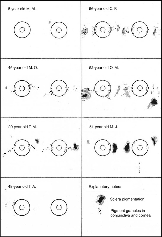
Fig. 16.2
Schematic findings of ochronotic changes in the eye
While examining patients with eye ochronosis, we have gained the impression that the pigment continuously passes from conjunctiva to cornea and is deposited close to the pericorneal network. According to Bürger and Schulze (1953), the mechanisms of development of ochronotic pigmentation of the cornea and gerontoxon in the elderly are similar. In tarsus and eyelid skin, ochronotic pigment is deposited more seldom (Šrámek 1937). In everted eyelids, the pigmented tarsus can be seen through the conjunctiva as a brown shade.
Related posts:
 Genetics of Alkaptonuria
Detection of Homogentisic Acid in Plasma and Urine
Coincidence of Alkaptonuric Ochronosis with Other Diseases
Therapy of Alkaptonuria
Analysis of X-Ray Symptomatology of Ochronotic Arthropathy in the Area of Peripheral Joints
Clinical Manifestation of Ochronotic Arthropathy in Spine
Genetics of Alkaptonuria
Detection of Homogentisic Acid in Plasma and Urine
Coincidence of Alkaptonuric Ochronosis with Other Diseases
Therapy of Alkaptonuria
Analysis of X-Ray Symptomatology of Ochronotic Arthropathy in the Area of Peripheral Joints
Clinical Manifestation of Ochronotic Arthropathy in Spine
Stay updated, free articles. Join our Telegram channel

Full access? Get Clinical Tree


