STT Arthrodesis
H. Kirk Watson
Jonathan R. Sorelle
Ronit Wollstein
E. Aron L. Haass
Indications
Triscaphe/STT Arthrodesis
I (H.K.W.) developed triscaphe/scaphotrapeziotrapezoid (STT) arthrodesis nearly 40 years ago as a management technique for many of the problems involving the scaphoid. The scaphoid is unique in many ways among all of the human skeletal components. It is almost entirely covered by articular cartilage. Blood supply to the bone is a difficult problem, so much so that this is the poorest healing bone in the body, after fracture. The motion demands on the scaphoid are not easily achieved because it crosses both carpal rows. The bone must flex and get out of the way in radial deviation. Along with the lunate, it must carry 100% of the heavy loads transmitted into the radius. The scaphoid is probably protected from avascular necrosis (Preiser disease) because the proximal pole can escape the loads coming through the capitate. Scaphoid stability is primarily dependent on the most commonly damaged ligament in the wrist, the scapholunate interosseous ligament system. Close to 25% of adults normally demonstrate a positive scaphoid shift test associated with some tearing of this ligament (1). The main thrust of all restabilization procedures is to prevent the proximal pole of the scaphoid from escaping from beneath the capitate under load.
Fusing the scaphoid to the lunate provides a long banana-shaped bone with insufficient bone stock to carry the flexing loads. Secondly, there is often a ridge on the articular surface of the radius between the scaphoid fossa and the lunate fossa that the fused unit cannot navigate. Thirdly, the amount of bone between the two is small and technically achieving fusion is very difficult.
The scaphoid can be controlled by fusing its distal pole to the trapezium and trapezoid. Said fusion also allows for an increased blood supply into the scaphoid cancellous bone. Fusing to the trapezium-trapezoid allows motion between the capitate and scaphoid and places responsibility on the capitate-trapezoid ligaments, which are capable of taking such loads. Fusing the scaphoid-trapezium-trapezoid joint allows transfer of loads from the hand through the scaphoid to the radius, circumventing the lunate. This is an ideal treatment for Kienböck disease (2,3,4,5).
We have previously described a radiographic technique that we consider one of the most efficient ways to image the STT joint for both preoperative evaluation of STT pathology, as well as to verify successful postoperative fusion after STT arthrodesis. The wrist is placed in 30 degrees of ulnar deviation so that the thumb is extended fully and in a straight line with the forearm. The thumb pulp is
facing the cassette and the angle between the straight line of the thumb and forearm and the cassette is about 30 degrees. The central ray in this view is directed at the carpometacarpal (CMC) joint. With this technique, we can outline and isolate the trapezoid joint with the least bony overlap (6).
facing the cassette and the angle between the straight line of the thumb and forearm and the cassette is about 30 degrees. The central ray in this view is directed at the carpometacarpal (CMC) joint. With this technique, we can outline and isolate the trapezoid joint with the least bony overlap (6).
We have published a follow-up of 800 STT fusions (7). The following is a breakdown of STT fusions by diagnosis: rotary subluxation of the scaphoid (RSS) 49%, Kienböck disease 13%, degenerative arthritis 12%, static rotary subluxation 11%, midcarpal instability 6%, nonunion of the scaphoid 3.5%, early scapholunate advanced collapse (SLAC) 1.8% persistent symptomatic predynamic RSS with instability 1.8%, and 0.4% for the remaining diagnosis including avascular necrosis of the scaphoid, nonunion scapholunate arthrodesis, symptomatic congenital synchondrosis of the triscaphe joint, and nonunion scaphoid with detached proximal pole (7,8,9,10).
Utilizing the techniques described herein, the nonunion rate is low, and immobilization time is generally 6 to 7 weeks. After fusion, the scaphoid is held firmly beneath the capitate and power use of the wrist is in a normal range. Anecdotally, a professional athlete led the National Basketball Association (NBA) in scoring a year after his STT fusion on his shooting wrist. An orthopaedic surgeon from California won his class in the world wrestling championship in Bulgaria. A woman won the northeastern Induro motorcycle championship a year after her STT limited wrist arthrodesis.
Contraindications
The main contraindication for triscaphe arthrodesis is degenerative changes of the radioscaphoid joint (11,12). If the destruction is restricted to the central portion of the scaphoid in a professional athlete who is highly paid, but for a limited number of years, then an STT arthrodesis may be indicated. This recognizes that as and when SLAC wrist becomes a problem, the STT joint can be osteotomized, the scaphoid removed, and a SLAC reconstruction performed (13). Severe destruction of the radial scaphoid joint or stage II SLAC where the capitate–lunate joint is destroyed are contraindications to an STT fusion and SLAC reconstruction is the preferred approach. A relative contraindication is degenerative arthritis of the STT joint in an elderly person or a person with limited load capacity for other reasons. In this case, the problem can be solved with carpectomy of the trapezium and a properly performed tendon arthroplasty. We mobilize our tendon arthroplasties in 2.5 half weeks and there is considerably less morbidity than 6 to 7 weeks in a cast, some of it long arm and bone graft of an STT fusion.
Technique
Approach the triscaphe joint through a 4-cm transverse dorsal wrist incision just distal to the radial styloid (Fig. 23-1). Use the spreading technique to preserve dorsal veins and branches of the superficial branch of the radial nerve.
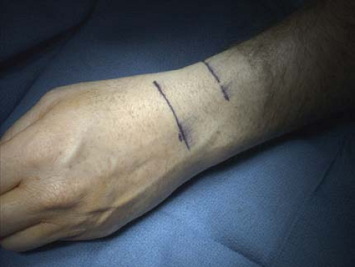
Figure 23-1 This is our typical preoperative marking for scaphotrapeziotrapezoid (STT) fusion and distal radius bone graft.
Figure 23-2 A: Place a spacer between the scaphoid and trapezoid while the pins are run to prevent approximation of bones and loss of scaphoid flexion. B: Volar counter pressure on the scaphoid prevents hyperflexion and maintains the scaphoid within the set parameters as the pins are set. C, D: Typical approach to harvesting a distal radius bone graft.
Open the sheath of the extensor pollicis longus tendon and retract the tendon radially.Related posts:

Stay updated, free articles. Join our Telegram channel

Full access? Get Clinical Tree


