Partial Excision of the Triangular Fibrocartilage Complex
Erich E. Hornbach
A. Lee Osterman
Introduction
Disorders of the triangular fibrocartilage complex (TFCC), which are responsible for ulnar-sided wrist symptoms in a wide variety of patients, can be amenable to effective treatment (1,2,3). They are often caused by a fall on an outstretched hand that results in a combination of extension, pronation, and axial loading to the wrist. The TFCC is a complex anatomic structure with multiple functions. It provides a surface for axial load bearing, and its peripheral ligaments play a key role in stability of the distal radioulnar joint and ulnar carpus. Its central portion is horizontal, avascular, and its thickness is variable (4,5,6). The peripheral portion of the triangular fibrocartilage (TFC) is highly vascular (4) and blends with the ulnocarpal ligaments. It has attachments to the radius, ulna, lunate, triquetrum, and other carpal bones. In 1989, Palmer developed a classification system of injuries to the TFCC (7) (see Table 30-1). Acute traumatic injuries were defined as class I injuries and degenerative injuries were defined as class II injuries. Treatment of class I injuries depends on many factors, but in general peripheral lesions with the capacity to heal are repaired, and central lesions with poor healing capacity are débrided. Class II, degenerative lesions represent an ulnar impingement or impaction process. As noted by the classification, TFC wear deteriorates to an attritional tear, followed by increasing chondral changes on the lunate and distal ulna, eventually resulting in lunotriquetral ligament perforation. Besides TFC débridement, surgical procedures used to treat class II lesions should address the mechanical overload by wafer resection or ulnar-shortening osteotomy.
Indications
Primary indications for partial excision of the TFC are class IA or IIC TFCC perforations. The patient with a class IA tear often presents with a traumatic, twisting or rotation injury to the wrist that results in ulnar-sided wrist pain. Swelling is present, but not dramatic, and the patient notes pain on forceful grasp and rotation. In the subacute phase, audible crepitation may develop with forearm rotation, and intermittent swelling may also be present. Patients with degenerative lesions may have symptoms brought on by an acute traumatic episode, or may report a more insidious onset of pain and symptoms. Often, a history of repetitive motion or previous wrist injury is present. Symptoms and findings are similar to class I tears.
Initially, all patients with suspected tears of their TFC that have normal radiographs and no distal radio-ulnar joint (DRUJ) instability are treated with a regimen of relative rest, with or without immobilization, nonsteroidal anti-inflammatory agents and therapy. Corticosteroid injections can be used for both diagnostic and therapeutic purposes. With resolution of symptoms, patients are
gradually returned to their previous level of activity. If this nonoperative management is ineffective after 3 to 4 months, and the symptoms are sufficiently disabling, surgical treatment is indicated.
gradually returned to their previous level of activity. If this nonoperative management is ineffective after 3 to 4 months, and the symptoms are sufficiently disabling, surgical treatment is indicated.
Table 30-1. Classification of Triangular Fibrocartilage Complex (TFCC) Injuries | ||||
|---|---|---|---|---|
|
Contraindications
Contraindications to arthroscopic treatment include uncontrolled medical problems, open wounds or concomitant infections, lymphedema, and previous open wrist surgery. Treatment of the central TFCC tear alone in an ulnar-positive patient may fail because it does not address the biomechanical cause of the tear.
Preoperative Assessment
The diagnosis of a TFCC lesion begins with a careful history and physical examination. Usually, patients report a twisting injury or traumatic event that leads to persistent ulnar-sided wrist pain. Complaints, such as recurrent pain with activity, swelling, limitation of range of motion, and audible crepitation, suggest TFC complex injury. A differential diagnosis of ulnar-sided wrist pain is listed in Table 30-2. In addition to a comprehensive evaluation of the upper extremity, we routinely include the following findings and provocative tests when assessing ulnar-sided wrist pain: (a) point of maximal tenderness; (b) TFCC grind or ulnocarpal stress test, performed by ulnarly deviating the wrist and applying axial load and rotation (A positive test being pain or painful clicking that reproduces the patient’s symptoms); (c) shuck and lunato-triquetral (LT) ballottement for lunotriquetral ligament
injury; and (d) shear test for pisotriquetral arthrosis. Tests are also done to confirm DRUJ and extensor carpi ulnaris (ECU) stability. Grip strength and wrist and forearm circumference are recorded.
injury; and (d) shear test for pisotriquetral arthrosis. Tests are also done to confirm DRUJ and extensor carpi ulnaris (ECU) stability. Grip strength and wrist and forearm circumference are recorded.
Table 30-2. Differential Diagnosis of Ulnar-Sided Wrist Pain | |||
|---|---|---|---|
|
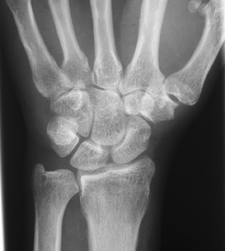 Figure 30-1 Zero-rotation posteroanterior (PA) view. |
Plain radiographs should include a zero rotation posteroanterior (PA) view to determine ulnar variance (Fig. 30-1) (17). Also included are a clenched fist, ulnar deviation view, and a pronated grip view to rule out dynamic ulnar impaction. As with arthrography, magnetic resonance imaging (MRI) is accurate for radial and central TFC tears (Fig. 30-2), particularly if a dedicated wrist coil is used. It provides no information on the size, configuration, or reactive joint synovitis, and poor
information on the lunotriquetral ligament. Unlike arthrography, MRI is noninvasive, and it provides information on ulnar impaction (marrow changes in the lunate, ulnar head, or triquetrum) (Fig. 30-3). Wrist arthrography, although accurate for radial and central TFC perforations, is now rarely used because it provides no information on the size of the tear or the joint reaction to the tear (synovitis). A midcarpal arthrogram may be considered if lunotriquetral ligament integrity is a concern. MRI remains our choice for advanced imaging in the work-up of suspected TFCC disorders.
information on the lunotriquetral ligament. Unlike arthrography, MRI is noninvasive, and it provides information on ulnar impaction (marrow changes in the lunate, ulnar head, or triquetrum) (Fig. 30-3). Wrist arthrography, although accurate for radial and central TFC perforations, is now rarely used because it provides no information on the size of the tear or the joint reaction to the tear (synovitis). A midcarpal arthrogram may be considered if lunotriquetral ligament integrity is a concern. MRI remains our choice for advanced imaging in the work-up of suspected TFCC disorders.
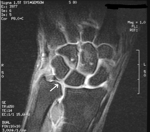 Figure 30-2 T1 fat-suppressed magnetic resonance image (MRI) demonstrating triangular fibrocartilage (TFC) central. |
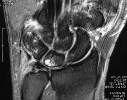 Figure 30-3 Magnetic resonance image (MRI) demonstrating edema within the ulnar portion of the lunate. |
Operative Management
Operative management can be performed either open or arthroscopically. Presently, we perform a diagnostic and therapeutic arthroscopy of the wrist for treatment of the TFCC and associated pathology. Open treatment of these lesions can be performed, and is easily combined with a shortening osteotomy or wafer procedure. Anatomic structures evaluated are presented in Tables 30-3 and 30-4. We generally evaluate all these structures in sequence, prior to treatment of the TFCC lesion. Photographic documentation of the diagnostic arthroscopy is an integral part of the procedure. Currently, we document the findings of the arthroscopy with arthroscopic photographs and occasionally a digital video recorder. The goal of surgical débridement is to remove damaged, loose portions of the TFC while preserving the dorsal and volar radioulnar ligaments. There is no need for an extensive excision of the articular disc.
Table 30-3. Radiocarpal Articulation | |
|---|---|
|
Table 30-4. Ulnocarpal Articulation | ||
|---|---|---|
|
Technique
The procedure is performed under general or axillary block anesthesia. Wrist arthroscopy requires distraction, which can be obtained by the use of a vertical distraction arthroscopy tower (preferred) or by horizontal traction over the hand table.
Apply a tourniquet, but do not inflate it unless the arthroscopic field is obscured by bleeding.
Sterilely prepare and drape the arm and place the patient’s fingers in the finger traps. Two or four finger traps can be used.
When using the arthroscopy tower, place a towel between tower and elbow, as well as the tower and volar forearm. This helps protect the olecranon and ulnar nerve.
With Velcro straps, secure the arm and forearm to the arthroscopy tower.
Flex the wrist 15 to 20 degrees.
Apply 10 to 15 pounds of distraction, draw anatomic landmarks, and identify preliminary portal sites. Because of the change in surface anatomy with the distraction of the joint, it is important to have the traction applied before drawing any landmarks (Figs. 30-4 and 30-5). The landmarks used include the following: Lister’s tubercle, the ECU, extensor pollicis longus (EPL), common extensors, and extensor digiti quinti.
Mark the 3-4 portal 1 cm distal to Lister’s tubercle.
Mark the midcarpal radial portal 1 cm distal to the 3-4 portal in line with the radial border of the long finger metacarpal.
Identify the ECU tendon and mark the 6R portal (primarily used) in the soft spot just distal to the ulnar head at the level of the ulnar styloid. The 6U portal can be marked, but is rarely used.
Reassess landmarks and introduce an 18-gauge needle into the 3-4 portal along the slope of the articular surface of the radius.
With the needle in the radiocarpal joint, inject 10 to 15 mL of saline. Note the pattern of distension, and assess the filling of the midcarpal joint.
Use an 11-blade scalpel to longitudinally incise the skin only.
Using a hemostat, perform blunt dissection down to the level of the capsule.
Next, introduce a blunt trochar or curved small hemostat into the radiocarpal joint along the slope of the articular surface.Related posts:
 Closed Reduction and Percutaneous Pinning
Intrafocal Pinning with “Arum” Pins
Ligamentous Repair for Acute Scapholunate Dissociation
Operative Reconstruction of the Distal Radioulnar Joint
Distal Radio-Ulnar Joint Capsulectomy for Post-Traumatic Limitation of Forearm Rotation
Release of the First Dorsal Compartment
Closed Reduction and Percutaneous Pinning
Intrafocal Pinning with “Arum” Pins
Ligamentous Repair for Acute Scapholunate Dissociation
Operative Reconstruction of the Distal Radioulnar Joint
Distal Radio-Ulnar Joint Capsulectomy for Post-Traumatic Limitation of Forearm Rotation
Release of the First Dorsal Compartment

Stay updated, free articles. Join our Telegram channel

Full access? Get Clinical Tree


