Chapter 57 Stroke


Ischaemic stroke
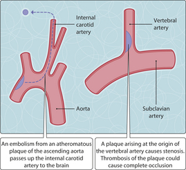
 emboli, usually from atheromatous plaques of the proximal aorta or carotid arteries (Fig. 3.57.1) or from mural thrombi of left atrium or left ventricle; other sources include vegetations of the mitral or aortic valve, and fat embolism following major trauma

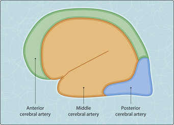
A generalized reduction in cerebral perfusion can occur if systolic blood pressure falls or intercerebral pressure rises. Autoregulation normally keeps the cerebral blood flow relatively constant over a wide range of systemic arterial pressures. However, this flow will fall below a critical threshold and the blood supply will be inadequate if the systolic blood pressure falls below approximately 50mmHg or if the intracerebral pressure rises to a level at which the arterial pressure cannot push blood into the cranial cavity. The neurons are most sensitive to hypoxia and are the first to suffer ischaemic injury. The watershed areas of the brain are particularly prone to hypoxia when there is global ischaemia. The watershed zones represent the boundaries between arterial territories (Fig. 3.57.2). For example, the junction of the areas supplied by the anterior and middle cerebral arteries lies along the superior cerebral convexity. These zones are especially likely to become ischaemic if there is a failure of oxygenation either in hypotension (e.g. shock, arrhythmia) or in respiratory failure. The term ‘watershed infarct’ is used when these border areas become necrotic as a result.
< div class='tao-gold-member'>

Stay updated, free articles. Join our Telegram channel

Full access? Get Clinical Tree


