Chapter 46 Restrictive lung diseases


The defining feature of restrictive lung diseases is reduced compliance (i.e. increased stiffness) of the lungs, thoracic cage or both. The basic defect can be seen on a spirometer tracing (Fig. 3.44.2). Patients with restrictive lung diseases cannot fill their lungs normally, and so the total amount of air they have in their lungs is reduced and the forced vital capacity (FVC), which is the total amount of air that is expelled from the lungs by forced expiration after a full inspiration, is low. However, there is no obstruction to outflow, so they can blow out the air quickly and the forced expiratory volume in one second (FEV1) is in proportion to their lung volume. Therefore, the FEV1:FVC ratio is approximately normal. In contrast, patients with obstructive lung disease (Ch. 44) have normal (or even enlarged) lung volumes, but they cannot blow the air out quickly because of the airway obstruction. Therefore, their FEV1:FVC ratio is reduced.
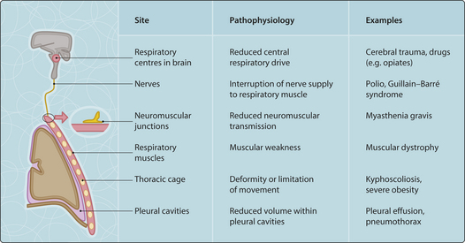
The main causes of restrictive lung disease can be classified according to whether they involve the lungs themselves (pulmonary causes) or structures outside the lungs (extrapulmonary causes) (Fig. 3.46.1).
Pulmonary causes
Acute respiratory distress syndrome

Stay updated, free articles. Join our Telegram channel

Full access? Get Clinical Tree


