heart disease

Chapter 35 Ischaemic heart disease


Myocardial ischaemia is caused by narrowing of the coronary arteries. The overwhelming cause is atherosclerosis, although there are other, rare, causes (e.g. arteritis, dissecting aneurysm of the aorta, syphilis, or emboli from aortic vegetations). Ischaemic heart disease manifests as angina pectoris, myocardial infarction, sudden death or congestive cardiac failure.


Myocardial infarction


Myocardial infarction is usually caused by complete occlusion of a coronary artery by a thrombus overlying a ruptured plaque. The myocardium exhibits coagulative necrosis, but it takes 8 to 12 hours for this to become visible. In experimental animals, electron microscopy demonstrates ultrastructural changes earlier than this, but these changes are of no use in the practical investigation of a sudden death in clinical circumstances. If death occurs within 8 hours of an infarct, the pathologist will probably find no abnormality of the myocardium at autopsy.


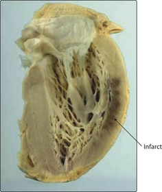
Coronary arteries are end-arteries. The ventricular wall is supplied by perforating branches arising from the epicardial coronary arteries. The subendocardial zone is furthest from the blood supply and it is here that critical ischaemia tends to occur first. As the infarct enlarges, it extends further towards the epicardial surface (Figs 3.35.1 and 3.35.2). The exception is a very narrow band of myocardium immediately adjacent to the endocardium that receives enough oxygen to survive from the blood within the ventricle. The area of infarcted muscle depends on the territory supplied by the obstructed artery. No two hearts are exactly the same, but typical regional infarcts tend to occur in the territories shown in Fig. 3.35.3.


< div class='tao-gold-member'>

Stay updated, free articles. Join our Telegram channel

Jul 3, 2016 | Posted by in MUSCULOSKELETAL MEDICINE | Comments Off on heart disease

Full access? Get Clinical Tree

Get Clinical Tree app for offline access