28
Stiff Joints
Shelly M. Sailer
History and Clinical Presentation
A 44-year-old right hand dominant cabinet maker presented to the emergency room after accidentally cutting the fingers of his right hand with a table saw.
Physical Examination
The patient completely amputated his index finger through the middle phalanx just distal to the proximal interphalangeal (PIP) joint and his small finger through the proximal phalanx. The long and ring fingers were lacerated from the volar aspect through the flexor tendons and distal aspect of the proximal phalanges, but were held on by dorsal skin bridges. They were both avascular and insensate. The amputated portions of the index and small fingers were retrieved, but they were severely mangled and not amenable to replantation. He was taken to the operating room for immediate surgical management.
PEARLS
- Static-progressive and serial static splinting are very useful for resolving contractures with a “hard” end feel.
- Dynamic splinting is most useful in resolving contractures with a “soft” end feel.
PITFALLS
- Dynamic splinting forces are difficult to control, leading to poor splint compliance.
- Dynamic splinting can cause inflammation of tissues when tension is placed too high.
- Static splints do not allow for active motion while splinted, possibly contributing to tendon adhesions.
Diagnostic Studies
Initial radiographs revealed traumatic amputation of the index finger through the middle phalanx, ring finger at the proximal phalanx, and small finger at the proximal phalanx, and a unicortical defect of the long finger at the proximal phalanx (Figs. 28–1 and 28–2).
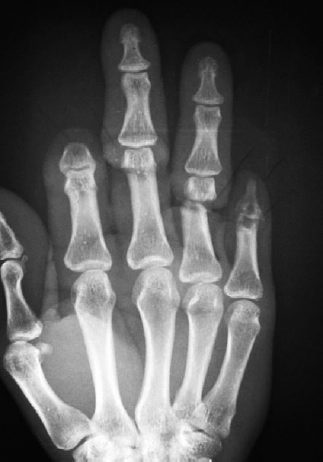
Figure 28–1. Anteroposterior (AP) view of the initial injury demonstrates complete amputations of the index and small fingers and near amputations of the long and ring fingers.

Figure 28–2. Oblique view of the initial injury.
Postoperative C-arm images demonstrate two Kirschner wires (K-wires) anatomically reducing the ring finger proximal phalanx fracture and extending through the PIP joint (Fig. 28–3).
Radiographs taken 8 weeks postoperatively demonstrate union of the ring finger fracture and complete closure of the unicortical defect of the middle finger.
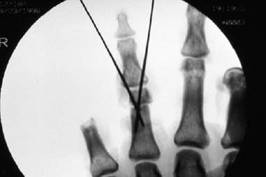
Figure 28–3. Postoperative C-arm image demonstrates fixation of the ring finger fracture.
Surgical Management
The ring finger proximal phalanx level amputation was stabilized with two 0.045-inch crossed K-wires placed across the fracture site. Distally the pins extended through the PIP joint to provide better fixation of the short remaining distal fragment of the proximal phalanx. Despite near amputation, the long finger proximal phalanx fracture was found to be stable and did not require fixation. On both the ring and long fingers, the flexor digitorum profundus (FDP) tendons were repaired and the flexor digitorum superficialis (FDS) tendons were tenodesed to the FDP tendons. The radial and ulnar digital arteries and nerves were repaired. The extensor mechanism was intact in both long and ring fingers. Revision amputations of the index and small fingers were performed.
A second surgery was ultimately required to regain full active range of motion. Five months after the initial injury this patient underwent a flexor tenolysis to the middle and ring fingers after passive range of motion was restored and the scars were soft and mature.
Postoperative Management
Two weeks after initial surgery, the patient was referred to hand therapy to begin active motion of the thumb and wrist and only metacarpophalangeal (MP) motion of the fingers. The MP joints were stiff initially, flexing only 50 degrees, but progressed to 80 to 100 degrees within 4 weeks. Desensitization activities and compressive wraps were initiated for the index and small finger amputations. The volar wounds of the ring and long finger were slow to heal, but they finally healed by 6 weeks postoperatively.
This case was complicated by the development of cellulitis in the ring finger 8 weeks postoperatively, which necessitated removal of the two percutaneous K-wires, and a short course of IV antibiotics followed by oral antibiotics. After the K-wires were removed, it was determined that the proximal phalanx fractures were stable enough to tolerate motion at the interphalangeal joints of the long and ring fingers. It was at this time that the severely stiff interphalangeal joints, the intrinsic tightness, and the lack of active flexor tendon function were brought to the forefront as a therapy challenge.
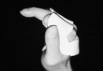
Only 10 degrees of passive motion at the PIP joints was achieved on the first day of mobilization and was slow to improve with range-of-motion exercises alone. There was no active PIP or distal interphalangeal (DIP) flexion as the patient was now consistently contracting the lumbricals, which flexed the MP joints and extended the PIP joints. He could not isolate use of the FDS and FDP muscles as he had not used them functionally for weeks and they were adherent at the digit level. An MP blocking splint, which facilitated flexion at the PIP joints, was fabricated to retrain use of the long flexors (Fig. 28–4). A biofeedback unit was used to assist in training the patient to contract the long flexors and relax the lumbricals. Even though the patient had very little active motion, due to adhesions on the long flexors, it was important to maintain muscle strength and preserve independent function in preparation for a flexor tenolysis when appropriate.