Sports Medicine

Chapter 29 Sports Medicine


Sports medicine involves the care of patients with illnesses and injuries related to sports and exercise. Both orthopedic and medical conditions affect a wide spectrum of patients during sports and recreational activities. The field of primary care sports medicine has emerged over the last 30 years as a leader in the comprehensive care of athletes, teams, and exercising individuals. Although sports medicine is often viewed in the context of professional and elite athletes, the principles and practice of sports medicine apply to athletes of all levels and ages.


The role of a sports medicine physician is to promote the health and safety of exercising individuals, prevent injury and illness, optimize function, and minimize disability that would preclude sports participation. Sports medicine physicians work closely with a multidisciplinary team of health care providers (specialty physicians and surgeons, athletic trainers, physical therapists, nutritionists, psychologists, and conditioning coaches) to satisfy the complete medical needs of athletes.


Sports medicine includes a rapidly expanding core of knowledge related to medical issues in sports and exercise for which the primary care sports medicine physician is uniquely trained. Primary care sports medicine is a subspecialty designation obtained through completion of an accredited sports medicine fellowship and passing the Certificate of Added Qualification examination. Many practicing primary care providers also gain vast experience in sports medicine through office treatment of orthopedic injuries, event coverage, and preparticipation screening evaluations. Musculoskeletal problems account for up to 20% of all primary care office visits (Urwin et al., 1998; Woodwell and Cherry, 2004), and approximately 90% of all sports injuries are treated nonsurgically. Thus, a sound clinical foundation in the evaluation and treatment of musculoskeletal disorders is critical to primary care providers and helps guide appropriate referral to orthopedic and rehabilitative specialists.


This chapter focuses on both the medical aspects of sports medicine and common orthopedic injuries in athletes. Detailed reviews cover the preparticipation physical evaluation, cardiac disorders in athletes, concussion assessment and management, cervical spine injuries, on-the-field assessment of the injured athlete, facial trauma, environmental influences such as exertional heat illness, common infections in athletes, and a systems-based review of dermatologic, pulmonary, hematologic, gastrointestinal, and genitourinary disorders in athletes. Orthopedic areas common in primary care sports medicine are also included, such as athletic low back pain, muscle strains, tendinopathy, shin pain, stress fractures, and issues unique to the pediatric or female athlete. See Chapters 30 and 31 for a comprehensive review of additional musculoskeletal problems relevant to sports medicine and the care of exercising individuals.


Preparticipation Physical Evaluation


Key Points


A preparticipation physical evaluation (PPE), or “sports physical,” is frequently required for medical clearance before participation in organized sports. The Preparticipation Physical Evaluation monograph, first introduced in 1992, provides recommendations for the content and format of the evaluation (AAFP et al., 2005). The third edition of the monograph, updated in 2005, is supported by six national medical societies: American Academy of Family Physicians (AAFP), American Academy of Pediatrics, American College of Sports Medicine, American Medical Society for Sports Medicine, American Orthopaedic Society for Sports Medicine, and American Osteopathic Academy of Sports Medicine. The fourth edition published in 2010 also includes collaboration with the American Heart Association (AHA) regarding the preparticipation cardiovascular evaluation.


The primary objectives of the PPE include the following:


Secondary objectives of the PPE include the following:


The recommended frequency of the PPE varies widely according to the requirements of the specific school, organization, or state. AHA first published consensus recommendations for cardiovascular screening in athletes in 1996 and reaffirmed these recommendations in 2007, which have influenced the format and timing of the PPE (Maron et al., 1996b, 2007). A comprehensive PPE is recommended on entry into middle school, high school, and college before participation in competitive sports. For youth and high school athletes, a comprehensive PPE should be repeated every 2 years. For years not requiring a comprehensive PPE, annual updates consisting of a comprehensive history and determination of height, weight, and blood pressure, along with a problem-focused evaluation of new concerns, illnesses, or injuries, should occur on interval years for youth and high school athletes and on all subsequent years for college athletes.


The PPE history focuses on symptoms related to exercise, such as exertional syncope, lightheadedness, chest pain, palpitations, dyspnea, wheezing, or fatigue with less than expected activity (Box 29-1). A past medical history of preexisting cardiac or pulmonary conditions, murmurs, hypertension, coronary artery disease risk factors, asthma, concussions, illicit drug use, prior orthopedic injuries, or other medical conditions that place an athlete at risk of injury should be noted. Specific questions to identify a family history of premature death, cardiovascular disease, and hereditary cardiac disorders, such as hypertrophic cardiomyopathy, Marfan’s syndrome, and long QT syndrome, are also recommended.


The PPE examination consists of both medical and musculoskeletal components. The cardiovascular examination is a major focus of the medical evaluation and consists of a blood pressure measurement, palpation of the radial and femoral artery pulses, cardiac auscultation with the patient both supine and standing, and recognition of the physical manifestations of Marfan’s syndrome (Maron et al., 1996b; Maron et al., 2007). Any heart murmur detected should be further assessed during the Valsalva maneuver or while moving the patient from a squatting to a standing position. These maneuvers decrease venous return and may accentuate the murmur of hypertrophic cardiomyopathy, the leading cause of sudden cardiac death in young athletes. However, hypertrophic cardiomyopathy is difficult to detect on examination alone because outflow tract obstruction, which causes the harsh systolic murmur, is present in only about 25% of patients with the disorder (Maron, 1997) (Fig. 29-1).
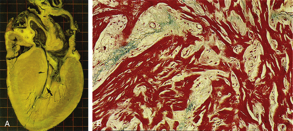


The musculoskeletal assessment serves as a screening evaluation of the spine and upper and lower extremities. Joint range of motion, strength, and stability should be tested. Several functional tests, such as hopping, squatting, and “duck walking,” assess many anatomic areas at once and make the evaluation more efficient. Previous orthopedic injuries can also be evaluated in more detail to detect problems that require further rehabilitation or protective bracing before sports participation.


Limitations of Cardiovascular Screening


Despite efforts to standardize the format and approach to the PPE, several limitations of the screening process still exist. In 2007, only 19% of U.S. states used forms that were considered adequate by AHA, and 35% of states allowed practitioners with limited cardiovascular training to perform the evaluation (Glover et al., 2007). Unfortunately, recommendations to use a comprehensive personal and family questionnaire to guide the PPE are not widely adopted, and recommended screening protocols are often incompletely or inadequately implemented. In addition, the PPE is screening for medical conditions that place an athlete at increased risk of sudden death, and no outcomes-based research has demonstrated that the PPE is effective in preventing sudden death or potentially catastrophic events, or for identifying athletes at risk. A complicating feature of the screening process is that athletes who harbor underlying structural heart disease may be asymptomatic until the sudden cardiac arrest. In a review of 134 cases of sudden cardiac death, only 18% of athletes had symptoms of cardiovascular disease in the 3 years before their death, and only 3% were suspected of having a cardiovascular condition after PPE (Maron et al., 1996a).


Limitations in the traditional screening process have led to widespread debate regarding the addition of noninvasive cardiovascular screening techniques, such as electrocardiography (ECG), to the PPE. In 2007, AHA reaffirmed recommendations against universal ECG screening in athletes, citing a low prevalence of disease, poor sensitivity, high false-positive rate, poor cost-effectiveness, and a lack of clinicians to interpret the results (Maron et al., 2007). In contrast, the European Society of Cardiology, International Olympic Committee, and the governing associations of several U.S. and international professional sports leagues endorse the use of ECG in the screening of athletes (Corrado et al., 2006; Ljungqvist et al., 2009). These recommendations are supported by studies showing that ECG is more sensitive than history and physical examination alone in identifying athletes with underlying cardiovascular disease, including a 77% greater power to detect hypertrophic cardiomyopathy (Corrado et al., 1998).


In 2006, Corrado and associates reported results from screening 42,386 athletes over 25 years from the national PPE program in Italy. Disqualification on the basis of a screening protocol using history, physical examination, and ECG produced a 10-fold reduction in the incidence of sudden cardiac death in young competitive athletes and an 89% reduction of sudden death from cardiomyopathy. Although only 0.2% of athletes were disqualified for potentially lethal cardiovascular conditions, the study reported a 7% false-positive rate and a 2% overall disqualification rate. This raised concerns that adopting such a program in the United States would lead to an unacceptable number of disqualifications in athletes with a low risk of sudden death.


Complex issues regarding feasibility, cost-effectiveness, appropriate physician, and healthy system infrastructure remain before large-scale implementation of ECG screening in the United States can be considered. However, some physicians are using ECG during PPE to improve detection of potentially lethal cardiovascular abnormalities. When used, ECG interpretation must be based on modern criteria to distinguish abnormal findings from physiologic alterations in athletes, to ensure acceptable accuracy and a low false-positive rate (Drezner, 2008). In a study of 2720 competitive athletes and physically active schoolchildren, a U.K. study reported a false-positive rate of 3.7% using history, physical examination, and ECG, with only 1.9% of false-positive results determined by ECG alone (Wilson et al., 2008). Nine athletes (0.3% of those screened) were found to have a cardiovascular condition known to cause sudden cardiac death in young persons, and all athletes were detected by ECG, not by history or physical examination. Physicians interpreting ECGs in athletes should be familiar with common training-related ECG alterations that are normal variants. In contrast, training-unrelated ECG changes suggest the possibility of underlying pathology, require further diagnostic workup, and should be considered abnormal. Current recommendations for ECG interpretation in athletes to distinguish pathologic ECG abnormalities from physiologic ECG alterations have been recently provided (Corrado et al., 2009).


Cardiac Disorders in Athletes


Key Points


Arrhythmias


Cardiac arrhythmias in athletes range from benign to life threatening. Bradyarrhythmias are more common in athletes than in the general population because of an increase in resting vagal tone as a response to regular strenuous exercise. Common bradyarrhythmias include sinus bradycardia, sinus arrhythmia, first-degree atrioventricular (AV) block, and Wenckebach (or Mobitz I) second-degree AV block (Huston et al., 1985; Link et al., 2001). These bradyarrhythmias are usually asymptomatic and should resolve during exercise by the withdrawal of vagal tone and associated catecholamine influx.


Athletes with symptomatic Wenckebach block, such as presyncope or syncope during exertion, require further evaluation by a cardiologist and often placement of a permanent pacemaker and restriction of activities. Higher degrees of heart block, such as Mobitz II second-degree and complete third-degree AV blocks, are always pathologic in any individual, including athletes (Fig 29-2). Mobitz II and complete heart blocks signify marked disease in the His-Purkinje system and are generally accepted as a class I indication for permanent pacemaker placement, even in the absence of symptoms (Link et al, 2001).
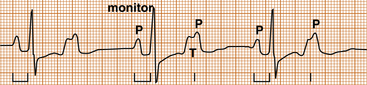


Tachyarrhythmias in the athlete are abnormal and require further evaluation and treatment before participation in strenuous exercise. The treatment of many supraventricular tachyarrhythmias has been greatly advanced by the use of radiofrequency (RF) ablation, which might offer an actual cure and obviate the need for lifelong pharmacologic treatment.


The most common tachyarrhythmia in athletes is atrial fibrillation (AF). Studies have suggested that AF occurs more frequently in athletes than in the general population (Furlanello et al., 1998; Huston et al., 1985). This might be a consequence of increased vagal tone and bradycardia in athletes, which allows dispersion of atrial repolarization and results in a higher susceptibility to AF. Radiofrequency ablation is curative in most cases of paroxysmal AF (Link et al., 2001). If pharmacologic treatment is needed, rate control can be accomplished with beta-adrenergic blockers or calcium channel blockers, and anticoagulation should be considered with aspirin or warfarin, depending on the frequency of AF and other risk factors for thromboembolism. Any athlete receiving anticoagulation therapy with warfarin should be restricted from sports involving collision or bodily contact.


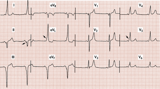
Atrioventricular nodal reentrant tachycardia (AVNRT) is characterized by abrupt onset and termination of symptoms, a narrow QRS complex, and no evidence of atrial activity on the ECG during the tachycardia. AVNRT caused by an accessory bypass tract, known as Wolff-Parkinson-White (WPW) syndrome, may be evident on the ECG by the characteristic delta wave (slurred upstroke of QRS complex), short PR interval, and prolonged QRS complex (Fig. 29-3). Athletes with WPW may be at risk of sudden death and should be strongly considered for RF ablation. Radiofrequency ablation for both AVNRT and WPW offers cure rates higher than 95% (Link et al., 2001; Manolis et al., 1994).


Atrial flutter is an unusual arrhythmia in trained athletes and typically results from an underlying cardiomyopathy.


Ventricular tachyarrhythmias in athletes are life threatening and usually the result of structural heart disease such as hypertrophic cardiomyopathy, anomalous coronary artery, dilated cardiomyopathy, arrhythmogenic right ventricular dysplasia (ARVD), or atherosclerotic coronary artery disease (CAD). Ventricular arrhythmias and sudden death may also occur in individuals with structurally normal hearts. This may occur from ion channel disorders, such as long QT syndrome, catecholaminergic polymorphic ventricular tachycardia (CPVT), and Brugada syndrome, or from commotio cordis. Long QT syndrome is characterized by a prolonged QTc interval, and Brugada syndrome is suggested by an incomplete right bundle branch block and ST-segment elevation in the precordial leads. Exercise may provoke ventricular arrhythmias because of high catecholamine levels and make ventricular fibrillation more difficult to terminate. Athletes who experience a resuscitated sudden death should undergo an extensive workup and treatment with an implantable cardioverter-defibrillator (Link et al., 2001).


Sudden Cardiac Death


Sudden cardiac death in athletes is a catastrophic event and the leading cause of death in exercising young athletes (Maron et al., 2009) The estimated incidence of sudden cardiac death in high school and college athletes is 1 in 100,000 to 200,000 athletes per year (Maron et al., 2009; van Camp et al., 1995). However, these studies are limited by the lack of a mandatory reporting system for juvenile sudden death and their reliance on electronic databases and media reports to identify cases of sudden death. Thus, current reports likely underestimate the true incidence of sudden cardiac death in athletes.


The cause of sudden cardiac death in young athletes (<35 years) is usually a structural cardiac abnormality, with hypertrophic cardiomyopathy and coronary artery anomalies representing 36% and 17% of U.S. cases, respectively (Box 29-2) (Maron et al., 2009). Commotio cordis, involving a blunt, nonpenetrating blow to the chest that leads to a ventricular arrhythmia, accounts for approximately 3% of cases. Commotio cordis is most common in younger athletes (mean age, 13 years) with compliant chest walls (Maron et al., 2002). Commotio cordis occurs most often in sports using a firm projectile, such as baseball, softball, hockey, and lacrosse, but can also occur from contact with stationary field equipment, the ground, or another player. In older athletes (>35), atherosclerotic CAD accounts for more than 75% of cases of sudden cardiac death.


Automated External Defibrillators in Athletic Medicine


Limitations of the cardiovascular screening process, the overwhelming desire to protect young athletes from a tragic event, and the success of early defibrillation programs (Caffrey et al., 2002; Page et al., 2000; Valenzuela et al., 2000) using accessible automated external defibrillators (AEDs) have propelled the placement of AEDs into the athletic setting (Drezner et al., 2005). Recent research suggests an improved survival rate for young athletes with sudden cardiac arrest if early defibrillation is achieved. A retrospective cohort of 1710 U.S. high schools with an on-site AED program found 14 cases of sudden cardiac arrest in high school student-athletes and a 64% survival rate if early cardiopulmonary resuscitation (CPR) and prompt defibrillation with an AED were provided (Drezner et al., 2009).


Comprehensive emergency response planning is needed to ensure an efficient and structured response to sudden cardiac arrest in the athletic setting. This includes establishing a communication system to activate the emergency medical services (EMS) system and alert any on-site response team, training of anticipated responders (e.g., coaches) in CPR and AED use, access to an AED, and practice and review of the response plan. High suspicion of sudden cardiac death should be maintained in any collapsed and unresponsive athlete, with application of an AED as soon as possible for rhythm analysis and defibrillation if indicated (Drezner et al., 2007).


Concussion in Sports


Key Points


In 1966 the Congress of Neurological Surgeons proposed a consensus definition of concussion. Since then, both the definition and our understanding of concussion have been evolving. A concussion is defined as a traumatically induced transient disturbance in neurologic function, usually caused by a direct blow to the head, neck, or face (Aubry et al., 2002).


Symptoms and Incidence


A concussion usually results in the rapid onset of brief neurologic impairment that resolves spontaneously. Symptoms of concussion include loss of consciousness, amnesia, confusion, headache, vision problems, nausea, and balance problems (Box 29-3). A concussion may or may not be associated with a loss of consciousness. Over 90% of concussions are not associated with a loss of consciousness, and unconsciousness is not a marker of the severity of the injury (Lovell et al., 1999). Amnesia can include loss of memory of the events before (retrograde amnesia) or after (posttraumatic amnesia) the concussion, or both, and amnesia appears to be the best predictor of severity in athletic concussion (Collins et al., 2003). Conventional imaging studies such as computed tomography (CT) or magnetic resonance imaging (MRI) are normal in concussion, indicating that concussion is a functional rather than a structural injury (McCrory, 2001).


An estimated 300,000 concussions occur each year from sports-related activity (Centers for Disease Control and Prevention [CDC], 1997). In high school football, there are 40,000 concussions per year, for a 3% to 5% incidence (Powell and Barber-Foss, 1999). High-risk sports include contact and collision sports such as football, ice hockey, rugby, wrestling, and to a lesser extent, soccer and basketball. Women may be more prone to concussion in some sports (Tierney et al., 2005), for unclear reasons, with further research needed. Younger players also may be more prone to concussion because of less developed neck muscles and the higher relative weight of the head compared with the rest of the body. In addition, children may sustain more serious concussions because of their immature nervous system.


Sideline Management


When attending to an athlete on the field with a suspected concussion, attention must first be given to basic first aid. Airway, breathing, and circulation (ABCs) and level of consciousness should be initially assessed, followed by an evaluation for potential cervical spine injuries. If loss of consciousness has occurred, the duration of unconsciousness should be determined. A brief loss of consciousness is generally defined as less than 30 to 60 seconds. If a player is unconscious for more than 30 to 60 seconds or still unconscious by the time a physician reaches the athlete on the field, most would consider this “prolonged.” Patients with concussions involving prolonged loss of consciousness, suspected cervical spine injuries, or gross neurologic impairment should be stabilized and transported to a hospital for further evaluation (see Cervical Spine Injuries: On-the-Field Assessment).


After it is determined that the athlete is stable and safe to be moved, further assessment can take place on the sideline. Physical examination should include evaluation of cranial nerves, pupillary dilation and reactivity, balance and coordination, motor strength, and cognitive function. Orientation to time, place, and person is an incomplete assessment of cognitive function in the sports setting. Memory and recall testing, serial 7s (counting backward by 7 from 100), serial 3s (counting backward by 3 from 100), and listing the months of the year backward and forward are simple sideline tests of cognitive function. Several sideline assessment tools exist to aid medical professionals in the evaluation of an athlete with concussion. The Standardized Assessment of Concussion (SAC) is a validated sideline assessment tool, and the most recent consensus conference on concussion recommended a modified and expanded tool, the Sport Concussion Assessment Tool (SCAT2) (McCrea et al., 1997; McCrory et al., 2009). This includes a symptom scale, standard cognitive assessment, and guided physical examination. Such standardized examinations are useful for serial follow-up and if different medical personnel will be evaluating the athlete. It is important to reassess concussed athletes frequently to monitor for resolution of symptoms or signs of deterioration.


Return to Play


Return-to-play decisions are driven by the concern of preventing potential complications, primarily second-impact syndrome and permanent neurologic deficit. Second-impact syndrome occurs when a player returns to play following a first concussion before the symptoms have completely resolved, and a second, often minor, blow to the head is sustained, which leads to diffuse cerebral swelling, brainstem herniation, and death. The risk for catastrophic injury in athletes returned to play while still symptomatic is of particular concern in children and adolescents.


Long-term cognitive deficits from concussion or repetitive blows to the head are also a concern. Repetitive concussions, especially if severe, might put athletes at risk for permanent neurologic deficits (Guskiewicz et al., 2003). However, no evidence indicates that repetitive nonconcussive blows, such as those sustained from heading a soccer ball, lead to short-term or long-term neurologic impairment.


Most concussion guidelines designed to facilitate return-to-play decisions rely on loss of consciousness and the presence of amnesia to grade the severity of the concussion. This approach makes their usefulness limited because most sports-related concussions do not involve loss of consciousness or amnesia. Current recommendations suggest that management of concussion be based on symptoms and severity. The mildest concussion is one that involves transient symptoms that resolve quickly. More severe concussions involve persistent or prolonged symptoms and cognitive deficits on neuropsychological testing. Management of concussion includes both physical and mental rest, until all symptoms have cleared, and the neurologic examination, followed by a graded and stepwise program of exertion before return to sport (Box 29-4). Repetitive concussions, which take longer to return to baseline or that occur with progressively less trauma, are also considered of greater severity. Complex or recurrent concussions with persistent cognitive impairment, neurologic findings on examination, or prolonged symptoms may warrant advanced imaging and formal neuropsychological testing and should be managed by physicians with specific expertise in sports-related concussion.


Neuropsychological testing in concussion, initially a comprehensive battery of conventional written tests administered by a trained neuropsychologist, has evolved into a variety of computer assessment tools. These neuropsychological assessment tools are gaining acceptance in high-risk sports at the collegiate and professional levels and are most useful when there is a baseline assessment done before injury. Widespread use of these tests in youth and high school sports with limited budgets and medical personnel might not be practical.


In summary, concussion is a common injury in sport, and athletes sustaining a concussion should be medically evaluated by a physician. No symptomatic athlete should be allowed to return to play, and an athlete should not be allowed to return to play in the same game or practice during which an injury has occurred. Once asymptomatic, athletes should follow a graded program before return to play. Referral should be considered in complex or complicated concussions. Further research is needed to provide evidenced-based return-to-play guidelines and preventive strategies.


Cervical Spine Injuries


Key Points


Stingers


Stingers or “burners” are characterized by transient unilateral upper extremity pain and paresthesias resulting from a blow to the neck and shoulders. Stingers are common in American football and have been reported in up to 50% to 65% of college players (Clancy et al., 1977; Sallis et al., 1992). Stingers are peripheral nerve injuries and are considered a transient neurapraxia of the cervical nerve roots, usually involving the upper trunk of the brachial plexus (C5-C6). Stingers typically manifest with dysesthesia (burning pain) that begins in the shoulder and radiates down the arm. They often are associated with transient numbness or weakness, and all symptoms typically resolve in minutes.


Stingers can occur from a tensile or compression overload (Watkins, 1986). In most high school athletes, the mechanism of injury involves a tensile or traction injury when the involved arm and the neck are stretched in opposite directions. This occurs when the neck is forcibly flexed away while the shoulder is depressed. In college and professional athletes, who have a higher likelihood of degenerative changes of the cervical spine, a compression mechanism is more likely. This involves a pinch of the cervical nerve root within the neural foramen as the neck is forcibly extended in a posterolateral direction (Levitz et al., 1997).


Stingers are always unilateral, a distinguishing feature from spinal cord injuries, which involve symptoms in multiple limbs. Athletes are safe to return to play when symptoms have fully resolved and the athlete can demonstrate full cervical ROM and a normal neurologic examination.


Radiography and MRI should be considered in athletes with recurrent stingers to evaluate for cervical degenerative disk disease (Levitz et al., 1997). Rarely, more significant nerve injury involving axonotmesis (axon disruption) occurs, causing persistent weakness. Athletes with significant weakness 24 to 48 hours after injury may benefit from treatment with a short burst of oral corticosteroids. If weakness persists, electromyography performed 2 weeks or more after the injury will assess the distribution and degree of injury. Fortunately, most cases of axonotmesis recover within 1 year (Clancy et al., 1977).


Catastrophic Cervical Spine Injury


Injury to the spinal cord resulting in temporary or permanent neurologic injury is a rare but potentially catastrophic event during sports competition. Cervical spine trauma is most common in contact and collision sports such as American football, rugby, ice hockey, gymnastics, skiing, wrestling, and diving (Cantu and Mueller, 1999; Carvell et al., 1983; Tator and Edmonds, 1984; Wu and Lewis, 1985). Cervical spinal cord injuries are the most common catastrophic injury in American football and the second leading cause of death attributable to football. The National Center for Catastrophic Sports Injury Research reported that the incidence of cervical spinal cord injury in American football between 1977 and 2001 was 0.52, 1.55, and 14 per 100,000 participants in high school, college, and professional football, respectively (Cantu and Mueller, 2003).


Axial loading is the most common mechanism for catastrophic injury to the cervical spine during sports competition (Torg et al., 1979, 1990). Axial loading occurs when a player strikes another player with the top of the head as the point of initial contact (“spear tackling”). In athletes with cervical spinal stenosis, axial loading followed by forced hyperextension or hyperflexion can further narrow the AP diameter of the spinal canal, resulting in compression of the spinal cord and transient or permanent neurologic changes (Eismont et al., 1984; Penning, 1962; Torg et al., 1993).


Recognition of the axial load mechanism as the major cause of catastrophic cervical spine injury in American football resulted in rule changes that banned “spearing,” defined as intentionally striking an opponent with the crown of the helmet, as well as other tackling techniques in which the helmet is used as the initial point of contact. In 1976 the incidence of quadriplegia was 2.24 and 10.66 per 100,000 in high school and college athletes, respectively. In 1977, only 1 year after rule changes that banned spear tackling, the incidence decreased to 1.30 and 2.66 per 100,000 in high school and college athletes (Torg et al., 2002).


On-the-Field Assessment


Medical providers at sporting events must be prepared to assess, stabilize, and transport athletes with suspected cervical spine injuries. Adequate preparation of and anticipation in required personnel and equipment and a well-designed emergency response are critical to the management of catastrophic neck injuries. In general, any athlete with significant neck or spine pain, diminished level of consciousness, or significant neurologic deficits should be immobilized and prepared for transport.


Guidelines for the prehospital care of the spine-injured athlete were established by the Inter-Association Task Force for Appropriate Care of the Spine-Injured Athlete (2001). The initial assessment of an injured athlete begins with a basic assessment of the ABCs and level of consciousness. EMS personnel should be contacted for any concerns regarding basic life support. Unconscious athletes are presumed to have unstable spine injuries until proven otherwise.


The face mask of a protective helmet should be removed as soon as possible, regardless of respiratory status (Inter-Association Task Force, 2001). In football the face mask can be removed with screwdrivers or the loop straps cut with various cutting tools such as pruning shears or a Trainer’s Angel (Knox and Kleiner, 1997). Football helmets and chin straps should be left in place. If the helmet is removed from a downed player wearing shoulder pads, the athlete’s head will hyperextend, which may result in secondary injury to the cervical spine. If the athlete is not breathing, an adequate airway can be established by the jaw thrust maneuver, which allows opening the airway while maintaining the cervical spine in a stable position. Rarely, assisted ventilation may be necessary.


If transport is indicated, the athlete should be immobilized to a spine board. A supine athlete can be transferred to a spine board using a six-plus person lift technique, with one person responsible for stabilization of the head and neck (Inter-Association Task Force, 2001). To transfer an athlete who is facedown, a logrolling technique is recommended. Transport of a spine-injured athlete should be directed to a trauma center or medical facility with diagnostic and surgical capabilities for spinal injury.


Environmental Influences


Exertional Heat Illness


Key Points


Heat stroke is the third leading cause of death in high school athletes (Lee-Chiong and Stitt, 1995). This is tragic because these deaths should be largely avoidable.


The exercising human is an engine operating at about 25% efficiency, resulting in 3 W of heat production for every watt of work, and requires a biologic radiator to avoid overheating. Humans dissipate heat through convection, conduction, radiation, and evaporation, with evaporative sweat loss being the most significant. Higher temperatures limit heat dissipation from convection and conduction, warm sunny days elevate body temperature through radiant heating, and higher humidity decreases evaporative cooling. Thus, the combination of high ambient temperature, radiant heat from the sun, and high humidity works synergistically to create dangerous playing conditions that promote the development of heat illness.


The wet bulb globe temperature (WBGT) index incorporates ambient temperature, relative humidity, and the amount of radiant heat coming from the sun to provide a measure of the risk of overheating. The American College of Sports Medicine Inter-Association Task Force on Exertional Heat Illnesses Consensus Statement (2006) recommends that WBGT readings from 18° to 23° C (64.4°-73.4° F) result in moderate risk, 23° to 28° C (73.4°-82.4° F) in high risk, and more than 28°C (82.4° F) in extreme risk. The cumulative effect of successive days of exercise in the heat must also be considered. In a U.S. Marine Corps, investigators demonstrated that the risk of exertional heat illness was best predicted by considering the current and the previous day’s WBGT index (Wallace et al., 2005).


Because evaporative cooling is the primary mechanism for heat dissipation, adequate hydration is essential to keep the biologic radiator functioning. Losses of 2% to 3% of body weight are common with high-intensity exercise in the heat (Galloway, 1999). Below 5% fluid losses, performance and thermoregulation are impaired, and thirst is an inconsistent stimulus to rehydrate, so regular, planned fluid consumption is essential. Fluid recommendations vary, but experts have suggested about 500 mL of fluid intake 2 hours or less before exercise and then about 250 mL every 20 minutes during exercise (Convertino et al., 1996). Because of differences in sweat rate, acclimatization, intensity of exercise, clothing, protective equipment, and environmental factors, individual fluid requirements vary. Thus, recording an athlete’s nude weight in the morning and evening is an effective method for determining adequate rehydration. If athletes are losing more than 2% to 3% of their body weight with training, they need to consume more fluids during training. If they cannot regain the lost weight before the next morning’s training, they need to consume additional fluids after training and during recovery time. For every kilogram of body weight lost, 1 L of fluid should be consumed. Cooler, flavored fluids are recommended to increase palatability and absorption.


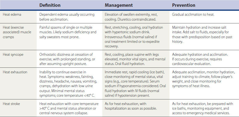
Heat illness is classified as heat edema, heat cramps, heat syncope, heat exhaustion, and heat stroke (Table 29-1) (Binkley et al., 2002; Eichner, 1998). Heat stroke is of the greatest concern, with hallmark features of an elevated core temperature higher than 40.5° C (105° F) and associated mental status changes. Any athlete exhibiting mental status changes and participating in an environment conducive to heat illness requires immediate removal from participation and active cooling. An ice-water tub should be prepared in advance if rapid cooling may be necessary in high-risk events, and an affected athlete should be fully submerged, with only the head above water (Smith, 2005). Other methods of cooling, such as applying ice bags to the neck, axilla, and groin, or using cold-water spray combined with fanning, can be effective, but the rate of core body temperature loss is slower than in ice-water immersion. Close monitoring of mental status and vital signs (e.g., core temperature) is indicated, and athletes should be transported to the hospital if they do not exhibit improving mental status with normalization of vital signs. The National Athletic Trainers’ Association Exertional Heat Illness Position Statement is an excellent reference regarding proper preparedness for heat illness (Binkley et al., 2002).


Exertional Hyponatremia


Key Points


Exertional hyponatremia (serum sodium <130 mmol/L), once considered a rare complication of exercise in the heat, is now recognized as more common and responsible for a number of exercise-related deaths (Almond et al., 2005). Controversy surrounds the exact pathophysiology, but the condition develops in the setting of excessive hypotonic fluid replacement while sodium is progressively lost in sweat (Levine and Thompson, 2005; Noakes, 2002). Typical victims are relatively inexperienced female marathon runners who tend to be light sweaters, finish in over 4 hours, and drink copiously throughout the race. Nonsteroidal anti-inflammatory drugs (NSAIDs) taken before the race may be a contributing factor (Hsieh, 2004). Athletes with a history of exercise-induced hyponatremia do not seem to be predisposed to water overload at rest, although there may be some physiologic mechanism beyond pure water overload that accounts for the condition in some susceptible individuals (Speedy et al., 2001). Symptoms are similar to those of heat exhaustion, including weakness, dizziness, headache, nausea, vomiting, and cramping, but the headache is more prominent and progressively severe, extremity swelling may be noted, and progressive mental status changes occur despite a core temperature lower than 40° C (104° F). Cerebral edema underlies the mental status changes, and pulmonary edema may also occur.


The serum sodium level must be assessed in athletes in whom the diagnosis is suspected, to differentiate hyponatremia from heat stroke. Intravenous (IV) fluids, often indicated in heat stroke, can actually worsen hyponatremia if related to excessive hypotonic fluid intake. Prompt hospitalization is indicated for any athlete with mental status changes or persistently altered vital signs once the diagnosis of hyponatremia has been established. Exertional hyponatremic encephalopathy is treated with 3% hypertonic saline boluses (100 mL) (Hew-Butler et al., 2008). Prevention includes following an athlete’s weight change with exercise to understand the fluid requirements more precisely, not deviating from established fluid intake on race day to avoid overhydration, incorporating sodium/electrolyte-containing fluids, and limiting fluid intake to 1 L/hr unless higher fluid requirements have been established (Gardner, 2002).


Oct 3, 2016 | Posted by in MANUAL THERAPIST | Comments Off on Sports Medicine

Full access? Get Clinical Tree

Get Clinical Tree app for offline access