Care of the Newborn

Chapter 22 Care of the Newborn


Whether present at the delivery or taking care of a new infant in the newborn nursery, the family physician plays an important part in newborn care during the early days of life. At the moment of birth, the newborn infant undergoes many changes and is vulnerable in the new environment. This chapter discusses these physiologic changes as well as the basics of neonatal resuscitation. The initial examination, common benign findings in the newborn period, developmental dysplasia of the hip, and audiology screening are described. Signs and symptoms of concern and common problems in the newborn period, including group B streptococcal infection and hyperbilirubinemia, are discussed, as well as infant safety, parent education, hospital follow-up, and care after discharge from the neonatal intensive care unit.


Preconception and Intrapartum Health


Key Points


A healthy newborn baby begins with a healthy pregnant mother. Family physicians are in the unique position of caring for women as they plan their families, during pregnancy, during childbirth, and after their infant is born. Preconception and intrapartum health care increase the likelihood of delivering a happy and healthy newborn.


Preconception Counseling


Preconception counseling regarding genetic diseases such as cystic fibrosis and Huntington’s disease, as well as the dangers of tobacco, alcohol, and substance abuse, should be addressed with women of childbearing age. All women considering pregnancy should be encouraged to take in 400 μg (0.4 mg) of folic acid per day. Oral folic acid supplementation before conception can significantly decrease the risk of neural tube defects (Czeizel and Dudas, 1992; MRC Vitamin Study Research Group, 1991). Women who have had a previous pregnancy affected by a neural tube defect should take 4000 μg (4.0 mg) of folic acid daily beginning 1 month before pregnancy (Centers for Disease Control and Prevention [CDC], 1992).


During pregnancy, maternal illness can adversely affect the fetus and lead to adverse neonatal outcomes. Maternal hypertension, preeclampsia, alcohol and tobacco use, illicit drug use, and autoimmune diseases can cause intrauterine growth restriction (IUGR) and preterm birth. Children born to women with diabetes mellitus or gestational diabetes are at risk for shoulder dystocia, operative delivery, hypoglycemia, and birth trauma (American College of Obstetricians and Gynecologists [ACOG], 2005). Maternal hyperglycemia at delivery also puts the infant at risk for hypoglycemia. Poorly controlled maternal hypothyroidism and hyperthyroidism are associated with low birth weight (LBW) and preterm delivery (ACOG, 2005). Fetal alcohol syndrome is directly caused by maternal alcohol use and abuse, and other illicit drug use is associated with preterm birth, congenital abnormalities, neurobehavioral abnormalities, and neonatal drug withdrawal syndromes (ACOG, 2005). Box 22-1 lists the more common human teratogens seen in the United States.


Intrapartum care should include regular fundal height measurements to monitor the growth of the fetus. Routine screening obstetric ultrasound is not recommended because of insufficient evidence of benefit or harm (ACOG, 2005). However, ultrasound is a useful and safe diagnostic test for investigating concerns about fetal growth, estimating gestational age, observing fetal anomalies, and studying other intrapartum conditions.


All pregnant women should be offered prenatal screening for trisomy 21 (Down syndrome) and neural tube defects. For most women, maternal serum quadruple analyte screen (quad screen) is the most appropriate screening test. The quad screen measures serum levels of alpha fetoprotein (AFP), human chorionic gonadotropin (hCG), unconjugated estriol, and inhibin-A. Inhibin-A was added to the prior triple screen to improve the detection of trisomy 21. For women younger than 35, the triple screen detects approximately 75% of trisomy 21, 90% of anencephaly, and 80% of spina bifida if measured between 16 and 18 weeks’ gestation (Benn et al., 2003; Graves et al., 2002). Using the quad screen, approximately 85% of trisomy 21 cases can be detected. The quad screen also has a false-positive rate for trisomy 21 of 8.2% after correction of major gestational errors, so a positive quad screen indicates a 1.9% risk for true trisomy 21 (Benn et al., 2003).


Routine prenatal screening for group B streptococci (GBS) significantly reduces early-onset neonatal GBS infection (Schrag et al., 2002b). Women who test positive for GBS or who have had a previous child with invasive GBS disease should receive intrapartum antibiotics for at least 4 hours before delivery (Schrag et al., 2002a). Management of the infant born to a mother colonized with GBS is discussed later.


Transition from Fetus to Newborn


Key Points


During the first hours after delivery, the newborn must adapt to extrauterine life. This adaptation includes a highly coordinated series of physiologic changes during which the infant is particularly vulnerable. Health care providers trained to assess and manage newborns should be available during the delivery and immediate neonatal transition period for all infants.


Circulatory Changes


In utero, a series of central shunts moves oxygenated blood coming from the placenta through the umbilical vein to supply the brain and other organs. Most oxygenated blood bypasses the liver via the ductus venosus and enters the left side of the heart (bypassing the lungs) through the foramen ovale. The poorly oxygenated blood returning from the lungs bypasses the heart through the ductus arteriosus and returns to the placenta to obtain oxygen from the maternal circulation (Moore and Persaud, 1993).


On delivery, the fetal circulation must adapt to self-oxygenation, and the central shunting must cease. When the umbilical cord is clamped and cut, absent blood flow within the umbilical vein leads to the closure of the ductus venosus. The umbilical vessels functionally close by 2 to 3 days of life.


With the initial newborn breath, the aeration of the lungs drives fluid within the air space of the lungs into the pulmonary interstitium. An increase in the partial pressure of oxygen (Po2) within the pulmonary vasculature causes vasodilation and a progressive decrease in pulmonary vascular resistance. Increasing Po2, along with circulating prostaglandins, stimulates the constriction and closure of the ductus arteriosus. Pulmonary blood now flows preferentially to the heart, rather than directly into the aorta through the ductus arteriosus (Moore and Persaud, 1993; Thureen et al., 2005).


In the normal infant the ductus arteriosus might remain partially open during the first several hours of life and cause a soft systolic murmur. It can take the normal infant a few hours to clear the lungs of excess fluid, causing fine crackles to be heard on lung examination and a transient elevation of the respiratory rate. As pulmonary vascular resistance falls, systemic vascular resistance increases. When left atrial and ventricular pressures increase above right atrial pressures, the foramen ovale closes. Poorly oxygenated blood from the superior and inferior vena cava can now flow into the pulmonary arteries (Moore and Persaud, 1993). The foramen ovale typically becomes fully sealed within the first month of life.


Neonatal Resuscitation


The successful transition to extrauterine life depends heavily on the ability of the neonatal pulmonary system to adapt quickly and provide oxygen to the infant. Any illness or injury that interferes with oxygenation puts the infant at great risk and should be identified and treated promptly. It is important that with every delivery, at least one person trained in neonatal care and resuscitation be assigned to care specifically for the infant. Caregivers should have immediate access to the necessary equipment for a complete resuscitation. If there is concern that the newborn will be at high risk for complications (e.g., thick meconium, fetal heart rate decelerations, known fetal anomalies), appropriate equipment should be set up and ready to use immediately (Box 22-2).


The American Academy of Pediatrics and American Heart Association developed specific protocols for neonatal resuscitation (AAP and AHA, 2006). An adapted resuscitation algorithm is given in Figure 22-1. This chapter does not cover the protocol in sufficient detail to produce competency in neonatal resuscitation and should not be used as a substitute for participation in the AAP/AHA Neonatal Resuscitation Program (NRP). The ABCs of neonatal resuscitation are the same as adult resuscitation: clear and position the airway, make sure the infant is breathing (whether spontaneously or with support), and ensure circulation of oxygenated blood.
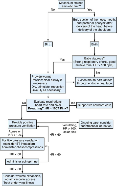


Initial stabilization procedures after every delivery should include warming and drying the infant, removing wet towels quickly, and providing tactile stimulation to encourage vigorous breathing and good muscle tone. After necessary resuscitation is completed, all neonates should receive chemoprophylaxis for ophthalmia neonatorum with 1% tetracycline ophthalmic ointment, 0.5% erythromycin ophthalmic ointment, or 1% silver nitrate aqueous solution. All three medications have similar efficacy; however, silver nitrate is preferred in areas with appreciable incidence of penicillinase-producing Neisseria gonorrhoeae. Silver nitrate is more likely than tetracycline or erythromycin to cause a transient chemical conjunctivitis in the first 1 or 2 days of life (AAP, 2003a).


Infants should also receive 1.0 mg of vitamin K intramuscularly early after delivery to prevent vitamin K–deficiency bleeding (VKDB, previously hemorrhagic disease of the newborn) (AAP, 2003b; Puckett and Offringa, 2000). Concerns about a correlation between intramuscular (IM) vitamin K and childhood leukemia have not been validated, and there is insufficient data to show that oral vitamin K prevents late-onset VKDB; therefore, IM vitamin K should be administered to all newborns (AAP, 2003b).
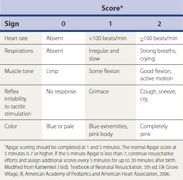


The Apgar Score


The Apgar score is widely used as a part of the early assessment of the newborn (Table 22-1). A score of 0, 1, or 2 is assigned to each of the five physical signs at 1 and 5 minutes after birth. The Apgar score should not be used as a substitute for assessing the ABCs in neonatal resuscitation, and resuscitation efforts should not be delayed or interrupted to assign an Apgar score. However, the Apgar score does allow a quick and consistent way for different providers to describe an infant’s condition. A score of 7 to 10 is considered normal. If the 5-minute Apgar score is abnormal, less than 7, appropriate resuscitation measures should be continued and Apgar scores assigned every 5 minutes until the infant is stabilized.


Although the Apgar score provides a systematic way for different providers to describe an infant’s condition in the first minutes of life, it correlates poorly with future neurologic outcomes (AAP and ACOG, 1996). A poor Apgar score alone cannot be used to diagnose asphyxia in the newborn or predict the development of cerebral palsy. However, the Apgar score is correlated with early infant death. In a large population study of term infants, an abnormal 5-minute Apgar score correlated with a significantly increased risk of death in the first 28 days of life. Even in infants with the lowest scores, however, death within 28 days is uncommon, occurring in 244 of 1000 infants with 5-minute Apgar scores of 0 to 3 (Casey et al., 2001).


Resuscitation of the Infant Born through Meconium-Stained Amniotic Fluid


Meconium staining of the amniotic fluid is a common complication during delivery of term infants, and approximately 5% to 12% of these infants develop meconium aspiration syndrome (MAS) (Wiswell et al., 2000). Risk factors for meconium staining of the amniotic fluid include maternal hypertension, maternal diabetes, maternal chronic respiratory or cardiovascular disease, maternal heavy smoking, preeclampsia or eclampsia, oligohydramnios, IUGR, poor biophysical profile, and abnormal fetal heart rate patterns (Gelfand et al., 2004). MAS is a life-threatening disease for otherwise healthy term newborns, and appropriate resuscitation of the infant at risk for MAS is important (see later discussion). All infants at risk for MAS and showing respiratory distress or bradycardia after delivery should undergo standard neonatal resuscitation procedures. In the depressed infant, tracheal suctioning to remove visible meconium can decrease the incidence and the severity of MAS (AAP and AHA, 2006). However, when the infant is vigorous at birth—defined as heart rate greater than 100, spontaneous respiration, and spontaneous movements or extremity flexion—routine endotracheal intubation and tracheal suction does not prevent MAS (Wiswell et al., 2000).


Initial Newborn Evaluation


Key Points


Once the infant has been stabilized immediately after delivery, maternal and infant bonding should be encouraged. Initiating breastfeeding within the first 60 minutes after birth can increase the chances of successful breastfeeding (Sinusas and Gagliardi, 2001).


Within the first 12 to 18 hours of life, a comprehensive physical examination should be completed and discussed with the parents. During the examination, the infant should be kept in a comfortable environment. Keeping the infant quiet and calm is important for adequate examination in the newborn period. The examination should begin with observation of activity, skin color, muscle tone, and respiratory effort while the infant is quiet. Listening to the heart and lungs should be completed next, disturbing the infant as little as possible. An infant often begins to cry at the touch of a cold stethoscope or cold hands, making the cardiopulmonary examination particularly difficult. The rest of the examination can then be completed in a calm and thorough manner.


As with the examination of adults and children, a complete history should be obtained on every patient. For the newborn, this includes the maternal history, antepartum course, course of delivery and any complications, and any postpartum complications. Specific attention to maternal medication and drug use, maternal medical conditions, traumatic events during delivery, neonatal resuscitation measures required, and maternal postpartum course assist in developing an appropriate assessment and plan for the neonate.


General Appearance


After collecting the history, it is important to observe the calm newborn. The normal term newborn rests with all extremities in some degree of flexion. Breathing should be quiet and effortless, but it can be very irregular. Episodes without breathing lasting 5 to 10 seconds are common and normal in newborns. Skin should be warm to the touch, soft, and smooth. Table 22-2 lists normal vital signs in the first days of life. Box 22-3 lists some common benign findings on the newborn examination. These are generally self-limited and require no further evaluation or treatment in the otherwise asymptomatic newborn.


Table 22-2 Vital Signs in the First Days of Life


Vital sign Normal value
Heart rate 100-180 beats/min
Respiratory rate 24-60 breaths/min
Systolic blood pressure 65-90 mm Hg
Diastolic blood pressure 50-70 mm Hg
Temperature <101.4° F (38.0° C) >96.8° F (36.0° C)

Data from Gunn VL, Nechyba C. The Harriet Lane Handbook, 16th ed, St Louis, Mosby, 2002; Rudolph AM. Kamei RK, Sagan P. Rudolph’s Fundamentals of Pediatrics, 2nd ed. Norwalk, Conn, Appleton & Lange, 1998.


Infants typically lose up to 10% of their birth weight in the first few days but should regain this weight by 2 weeks of age (Thureen et al., 2005). Excessive weight loss can indicate systemic disease or feeding difficulties and should be evaluated before discharge.


Neurologic Examination and Newborn Reflexes


The neurologic examination of the newborn begins with the general appearance of the infant. The newborn should have a strong cry and exhibit symmetric movements; a high-pitched or weak cry can be associated with current illness or neurologic deficits. Asymmetric movements can indicate musculoskeletal or focal neurologic injury. Complete absence or asymmetry of any newborn reflex can indicate neurologic deficit or injury. The following developmental reflexes are present at birth in the normal newborn (Thureen et al., 2005):


Head, Face, and Neck


Examination of the head and neck should include evaluation of cranial sutures, anterior and posterior fontanelles, facial symmetry, hard and soft palate, patency of the ear canal, patency of both nares, position of the nasal septum, placement of the ears, and intercanthal distance of the eyes.


The fontanelles should be flat and soft, and cranial sutures should be slightly mobile. The cranial sutures may override each other, and there may be temporary skull asymmetry after prolonged labor or vaginal delivery. Caput succedaneum and cephalohematoma (subperiosteal hemorrhage) are also common complications of labor and vaginal delivery. A cephalohematoma is a result of bleeding in the subperiosteal space and does not extend across a suture line, whereas caput succedaneum is caused primarily by subcutaneous edema and can involve any amount of the scalp. Caput succedaneum is present at delivery, and cephalohematoma is not evident until a few hours of age.


Many infants have transient skin lesions on the face and neck, and these should be noted and discussed with caregivers. Some birthmarks, however, can be a sign of underlying disease. For example, a port-wine stain occurring in the distribution of the first branch of the trigeminal nerve may be associated with Sturge-Weber syndrome. Hemangiomas are not always present at birth and might not be visible until 1 month of age. Most hemangiomas spontaneously regress during childhood and need no specific management. Periocular hemangiomas, however, should be managed with ophthalmologic consultation and aggressive therapy (Thureen et al., 2005).


The eyes, nose, mouth, and ears should be symmetric. Using a gloved finger, the hard and soft palate should be palpated and found to be fully merged. The oropharynx should be visualized, and the uvula should be single and midline. The ear canals should be inspected with an otoscope and found to be patent, and a small nasogastric tube should pass freely through each nare to the oropharynx. The helix of the ear (top portion) should be above the position of the inner canthi of the eyes.


Genitalia and Anus


The genitalia of the female child should have a visibly patent vaginal orifice, with the labia majora covering the labia minora, clitoris, and vaginal opening. The labia majora should be completely separate from each other (ensuring the absence of labioscrotal fusion). The labia majora should be palpated to evaluate the presence of inguinal hernia or ectopic gonads.


The male infant should have two palpable testicles of similar size within the scrotum. If the infant is cold during the examination, the testicles may be retracted and might even be found at the distal end of the inguinal canal. Each testicle, however, should be able to be easily brought down into the scrotum. The testicles should be similar in size and shape, although presence of significant hydrocele is a common transient finding in newborns. A hydrocele should transilluminate, and any testicular mass that does not transilluminate should be further evaluated.


The glans of the penis is normally covered completely by foreskin, and the urethral meatus should be located at the tip of the glans. A child with the urethral meatus opening on the ventral (hypospadias) or dorsal (epispadias) surface of the penis should not be circumcised until pediatric urologic consultation has been completed.


Most male and female infants urinate within the first 24 hours of life. Delay in urination should prompt an evaluation of the kidneys, bladder and urethra (Tappero and Honeyfield, 2003).


The anus should be inspected for patency during the initial examination, and the infant should pass stool within the first 24 hours of life. The first several bowel movements are meconium stools, dark and black with a tarry consistency. These eventually change to the thin, yellowish stools of the newborn.


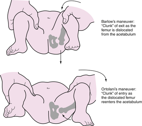
Muscles, Skeleton, and Hips


The initial musculoskeletal examination should be the observation of the infant. All limbs should appear symmetric and move equally, and any deficit should lead to further evaluation. Palpate both clavicles to evaluate for fracture or dislocation that might have occurred during delivery. In the event of a traumatic vaginal delivery, it is important to pay special attention to the potential for a brachial plexus injury. An infant with brachial plexus injury often has the affected arm straight down at the side and might have the wrist slightly bent, the fingers straight, and the forearm pronated (Thureen et al., 2005). Inspect hands and feet for syndactyly (fusion of fingers or toes), polydactyly (extra digits), clubfoot, and other congenital anomalies.


The incidence of developmental dysplasia of the hip (DDH) is estimated to be between 1.5 and 20 cases per 1000 infants (Shipman, 2006). Female gender, breech position, and family history are all reported risk factors for DDH; however, only a minority of cases of DDH are found in infants with identifiable risk factors (Shipman, 2006). In addition, as many as 80% of infants with an abnormal hip examination at birth resolved by 6 weeks, and 90% of mild dysplasia identified by ultrasound resolved between 6 weeks and 6 months (Shipman, 2006). The U.S. Preventive Services Task Force concluded that there was insufficient evidence to recommend routine screening for DDH in newborns (USPSTF, 2006). AAP (2000b) continues to recommend serial clinical examinations of the hip for the first 12 months of life.


There are two maneuvers in the neonatal hip examination: the Ortolani maneuver and the Barlow maneuver (Fig. 22-2). Each hip should be examined separately using gentle but firm pressure. Too much force can injure the hip in a normal infant.


Stay updated, free articles. Join our Telegram channel

Oct 3, 2016 | Posted by in MANUAL THERAPIST | Comments Off on Care of the Newborn

Full access? Get Clinical Tree

Get Clinical Tree app for offline access