Fig. 29.1
Development of the spinal cord at gestational day 19, 20, and 22
Failure of the Ends of the Neural Tube to Close
(a)
Cranially, may cause anencephaly
(b)
Caudally, may cause spina bifida occulta, meningocele, myelomeningocele, and myeloschisis
(c)
Persistence of the neurocentric canal causes diastematomyelia, which is a septum dividing the medullary canal into two parts on sagittal plain.
Gross Anatomy
The adult human spinal cord is made up of 31 segments (12 thoracic segments, 5 lumbar segments, 5 sacral segments, and 1 coccygeal segment). The spinal cord and its surrounding meninges lie within the vertebral canal and reaches down to the space between the first and second lumbar vertebrae. The average length of the spinal cord differs with gender. Average length in humans is 45 cm (18 in.) in males and 42–43 cm (17 in.) in females. It has a width, ranging from 13 mm (0.5 in.) thickness to 6.4 mm (0.25 in.).The average cross-sectional area of the cervical spinal cord in humans is 122 mm2 at C4, while it is 110 mm2 at C2 and 85 mm2 at C7 [7, 35]. The cross-sectional area of the lumbar enlargement is about 50 mm2, the sagittal diameter about 7 mm, and the transverse diameter 9 mm in human cadavers [17]. This cross-sectional area increases from C1 to C6 and then remains constant throughout the middle thoracic segments. It increases again from T12 to L4 and decreases rapidly below S1 [15]. There are four grooves in the spinal cord. The ventral median fissure lies on the ventral surface of the spinal cord as a midline deep longitudinal fissure. In humans, the average depth of the ventral median fissure is 3 mm and is deepest at C5 [7]. On the dorsal surface of the spinal cord lies the dorsal median sulcus. On the lateral side, there is a hollow ventrolateral sulcus and a deeper dorsolateral sulcus.
Meninges
Membranes that cover and protect the brain and spinal cord are named the meninges. There are three meninges according to their anatomical relation with the spinal cord and morphology.
1.
Pia mater: Surrounds the brain and spinal cord, it is the innermost layer of the meninges.
2.
Arachnoid mater: It is located between the more superficial dura mater and the deeper pia mater. It is separated from pia mater by the subarachnoid space. The subarachnoid space contains the cerebrospinal fluid (CSF). CSF exits the subarachnoid space and enters the blood stream via arachnoid villi that are small protrusions through the dura mater into the venous sinuses of the brain.
3.
Dura mater: The outermost and thickest membrane of the three layers of the meninges. It is derived from the mesoderm.
Spinal Cord Blood Vessels
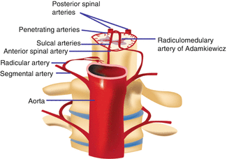
Three arteries, a single ventral spinal artery, and two dorsal spinal arteries supply the spinal cord. The ventral spinal artery originates from the vertebral artery and descends within the ventral median fissure of the spinal cord. The right and left dorsal spinal arteries originate either from the vertebral artery or its inferior posterior cerebellar branch, and descend in the dorsolateral sulcus of the spinal cord. They form anastomoses with the anterior and posterior segmental medullary arteries, which are branches from the vertebral, deep cervical, intercostal, and lumbar arteries (Fig. 29.2). The veins of the spinal cord form a surface plexus, which drain ventrally into the cerebellar veins and cranial sinuses and through the intervertebral veins and external venous plexuses to the azygous system.


Fig. 29.2
Arterial supply to the spinal cord
The actual blood flow caudally through these arteries is inadequate to maintain the spinal cord beyond the cervical segments. The major contribution to the arterial blood supply of the spinal cord below the cervical region comes from the radially arranged posterior and anterior radicular arteries, which run into the spinal cord alongside the dorsal and ventral nerve roots. These intercostal and lumbar radicular arteries arise from the aorta, provide major anastomoses, and supplement the blood flow to the spinal cord. An animal study suggests that critical spinal cord ischemia after complete thoracoabdominal aortic segmental artery sacrifice does not occur immediately, but is delayed 1–5 h or longer after injury [5]. In humans, the largest of the anterior radicular arteries is known as the artery of Adamkiewicz (anterior radicularis magna artery), which usually arises between L1 and L2 [39]. Meticulous surgical technique has to be carried out, since impairment of blood flow through posterior and anterior radicular arteries can result in serious conditions, such as paraplegia due to spinal cord infarction. To further enhance the understanding of spinal cord circulation, a novel monitoring system for spinal cord blood flow has been recently developed in an animal model. The device is told to hold promise to detect imminent ischemia or ensure acceptable blood perfusion in the spinal cord [12].
Structures of the Spinal Cord
The caudal end of the spinal cord narrows and forms the conus medullaris. The conus medullaris runs as a long fibrous strand, called the filum terminale. The filum terminale is an elastic structure composed of longitudinally arranged collagen bundles and elastic fibers [6]. Filum terminale arises from the end of the spinal cord at the level of the L1 vertebra and attaches to the coccyx. In humans, the length of the filum terminale internum is about 15 cm, the mean initial diameter 1.5 mm, and the midpoint diameter is 0.75 mm [26]. At the level of S2 vertebra, it perforates the dura and continues as the filum terminale externum that terminates on the dorsal surface of the first coccygeal vertebra together with the coccygeal ligament. The spinal cord is mainly composed of gray matter and white matter.
Gray Matter
Depending on the level, the gray matter is oriented in the form of the capital letter H in transverse sections. It also looks like a butterfly on the same plain. The gray matter can be divided into dorsal and ventral horns and intermediate region between them. The dorsally projecting arms of the gray matter are called the dorsal horns, and the ventrally projecting arms are called the ventral horns. The central region that connects the dorsal and ventral horns is called the intermediate gray matter. Microscopic analysis of the spinal gray matter reveals a complex structure, characterized by layers of cells from dorsal to ventral. The spinal cord gray matter consists of glial cells, neuronal cells, dendrites, and axons. The spinal gray matter is divided into ten regions in transverse sections. Nine laminae are organized from dorsal to ventral whereas the tenth lamina consists of cells surrounding the central canal. Laminae 1–4 are the main cutaneous receptive regions; lamina 5 receives afferents from the viscera, skin, and muscles. Lamina 6 receives proprioceptive and some cutaneous afferents.
White Matter
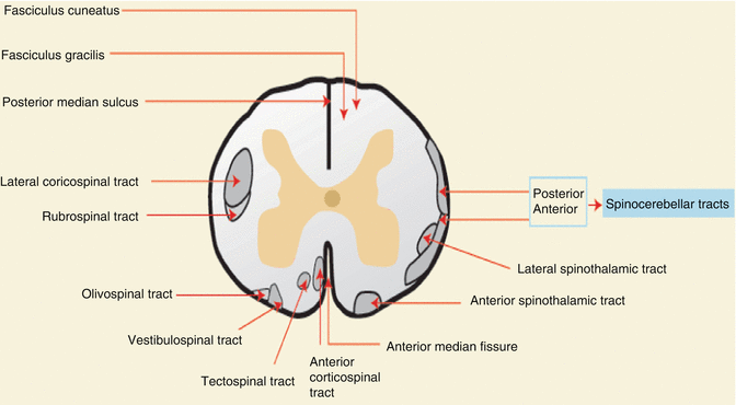
The gray matter is surrounded by white matter. The white matter consists of longitudinally running axons and also glial cells. A large group of axons, which are located in a given area, is called as funiculus. Smaller bundles of axons, which share common features within a funiculus, are called as fasciculus. A group of nerve fibers with the same course, origin, termination, and function is a tract. A group of tracts with a related function is a pathway. The horns of gray matter divide the white matter into three columns: dorsal, lateral, and ventral. The lateral and anterior columns contain ascending and descending fiber groups. The ascending tracts include the spinothalamic, spinocerebellar, and spinotectal tracts and the descending tracts include the corticospinal, vestibulospinal, tectospinal, and reticulospinal tracts (Fig. 29.3).


Fig. 29.3
Ascending and descending pathways of the spinal cord in cross-section
During neural tube closure, the dorsal cells (alar lamina) become primarily sensory (afferent), and the ventral cells (basal lamina) become primarily motor (efferent) in function.
(a)
Dorsal Column: transfer vibration, deep touch sensation, and proprioception
(b)
Anterolateral Column: lateral spinotalamic tract transmits pain and temperature sensation
(c)
Ventral Spinotalamic Tract: light touch sensation
(d)
Lateral Corticospinal Tract: voluntary motor function
Spinal Nerves
Spinal nerves emerge the vertebral column between the adjacent vertebrae through the intervertebral foramina and send motor impulses from the CNS to muscles and target organs and carry sensory information from organs to the CNS. There are 31 pairs of spinal nerves: 8 cervical (C1–C8), 12 thoracic (T1–T12), 5 lumbar (L1–L5), 5 sacral (S1–S5), and 1 coccygeal (Co1). Spinal nerves emerge from the vertebral canal below their respective vertebrae, except from the cervical region. Although the first seven cervical spinal nerves emerge similarly above their respective vertebrae, because there are eight cervical spinal cord segments, but only seven cervical vertebrae, the nerve emerging between C7 and T1 is named the eighth cervical nerve. Each spinal nerve is attached to the spinal cord via anterior and posterior roots. Each root is compromised of seven to nine rootlets. Axons of motor neurons come together and form ventral rootlets whereas axons of sensory neurons form dorsal rootlets. Each dorsal nerve root contains a dorsal root (spinal) ganglion. These cells are the origin of peripheral and central nerve fibers. A transition zone is defined between the CNS and the peripheral nervous system at the attachment point of each rootlet to the spinal cord [9].
Ventral roots: Each ventral root can also be named the anterior root, radix anterior, radix ventralis, or radix motoria. They are attached to the spinal cord by a series of rootlets that emerge from the ventrolateral sulcus of the spinal cord. The ventral roots predominantly consist of efferent somatic motor fibers (thick alpha motor axons and medium-sized gamma motor axons) derived from nerve cells of the ventral column.
Dorsal roots: There are 31 pairs of dorsal roots. Each dorsal root can also be named the posterior root, radix posterior, radix dorsalis, or radix sensoria. They are attached to the dorsolateral sulcus of the spinal cord by a series of rootlets. The dorsal roots contain sensory fibers from the skin, subcutaneous and viscera, skeletal muscles, tendons, and joints. It is estimated that there are 2–2.5 million afferent fibers in human adult dorsal roots on each side [31].
Dorsal root (spinal) ganglia: Each dorsal spinal root comprises dorsal root ganglion (DRG). The DRG consists of pseudounipolar neurons with single axon, cells that are primary sensory. They divide into peripheral and central branches, where the central branch enters the spinal cord via the dorsal roots. The peripheral branch carries sensory information from the body to the DRG cell body, whereas central branch carries information from the cell body of DRG neuron to the spinal cord.
Each spinal nerve consists of somatic and visceral fibers that can be classified according to their functions; as visceral efferent, somatic efferent, somatic afferent, and visceral afferent fibers.
Somatic afferent fibers: Exteroceptive fibers that carry impulses generated by external stimulations and somatic afferent fibers are peripheral continuation of DRG neurons. Their main objective is to carry sensory information from the skin, viscera, subcutaneous tissues, and deep tissues.
Somatic efferent fibers: compromised of axons from the ventral horn neurons. Their main objective is to carry motor impulses to the skeletal muscles.
Visceral afferent fibers: Interoceptive fibers that carry visceral sensation. They are made up of peripheral processes of DRG neurons. They constitute less than 10 % of afferent fibers in dorsal roots.
Visceral efferent fibers: Sympathetic or parasympathetic autonomic nerve fibers that carry motor impulses to glands, cardiac and smooth muscles.
Autonomic Nervous System
The autonomic nervous system is responsible for regulating vegetative functions and maintaining homeostasis in the body. The visceral efferent fibers are autonomic (sympathetic/parasympathetic) nerve fibers that carry motor impulses to glands and cardiac and smooth muscles. Despite visceral efferents that are divided into parasympathetic and sympathetic fibers, afferent fibers of the autonomic nervous system, which transmit sensory information from the internal organs of the body to the CNS, are conducted by general visceral afferent fibers. These are unconscious visceral motor reflex sensations from glands and organs that are transmitted to the CNS. The system is under the control of the hypothalamus and also works concordant with the endocrine system [22].
(a)
Sympathetic: Fibers innervate tissues in almost every organ system. The system is composed of 22 ganglia (3 cervical, 11 thoracic, 4 lumbar, and 4 sacral). In the sympathetic system, two kinds of neurons are involved in the transmission of signals: preganglionic and postganglionic. Preganglionic neurons are shorter and originate from the thoracolumbar region levels, especially T1–L2, and travel to a ganglion. At the paravertebral ganglia, they synapse with a postganglionic neuron. At the synapses within the ganglia, preganglionic neurons secrete acetylcholine, which activates nicotinic acetylcholine receptors on postganglionic neurons. In response, postganglionic neurons secrete norepinephrine, which activates adrenergic receptors on the target tissues. The postganglionic neuron is longer and extends across the body. They provide several regulatory functions. The most prominent ones are pupil dilatation, increase in cardiac rate and force of contraction, dilatation of bronchioles, peristalsis inhibition, sweat secretion activation, and renin secretion activation [23]. Horner syndrome (miosis, ptosis, and anhidrosis) can be seen if damage to the sympathetic nervous system occurs.
(b)
Parasympathetic: The parasympathetic system is responsible for stimulation of activities that occur when the body is at rest and the system uses acetylcholine and cholecystokinin as its neurotransmitter. Nerve fibers of the parasympathetic nervous system arise from the CNS. Parasympathetic preganglionic efferent fibers originate from the neurons of the sacral parasympathetic nucleus. The functions of the parasympathetic system are sexual arousal, digestion, salivation, lacrimation, urination, and defecation. Several cranial nerves have parasympathetic properties: the occulomotor nerve, facial nerve, glossopharyngeal nerve, and vagus nerve. Apart from cranial nerves, three spinal nerves in the sacrum (S2–4) also act as parasympathetic nerves, which are named as the pelvic splanchnic nerves.
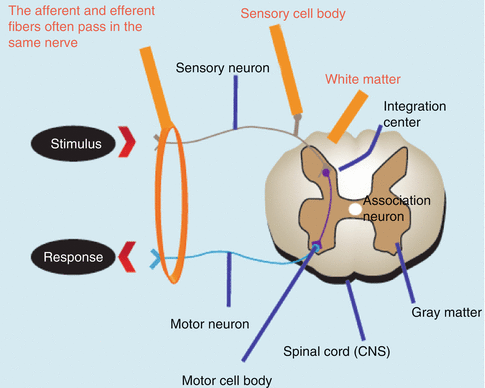
Apart from these functions, sensory and motor fibers of the spinal nerves form a reflex arc. The reflex arc is polysynaptic and functions when afferent fibers impulses end up with a motor reaction before sending information to the brain. This maintains the posture of the body and helps movements to occur in harmony (Fig. 29.4).


Fig. 29.4
Reflex arc of the spinal cord
Dermatomes
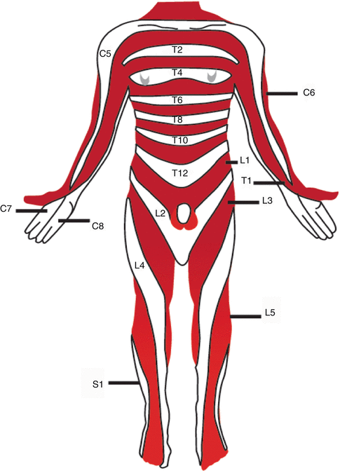
A dermatome is the area of skin supplied by a single spinal nerve. Although there are 31 pairs of spinal nerves in humans, there are only 30 dermatomes. Along the trunk, dermatomes have a segmental distribution represented as narrow bands of skin running almost horizontally, whereas in the limbs, due to the growth and rotation of limb buds during development, they are generally oriented longitudinally along the long axis of the limbs (Fig. 29.5).


Fig. 29.5
Dermatomes of the human body. Areas of the skin supplied by afferent nerve fibers originating from posterior spinal root
Segmental Motor Distribution
Skeletal muscles are innervated by more than one spinal nerve with a segmental motor distribution (Table 29.1). Each spinal nerve supplies the muscles originating from the myotomes of the same segment. Although the segmental distribution is apparent in the distribution of peripheral nerves to the muscles, axons of the spinal nerves are redistributed into peripheral nerves in plexuses in the cervical, brachial, and lumbosacral regions.
Table. 29.1
Motor innervation of the upper lower limbs and associated nerves, nerve roots
Action | Muscles | Nerves | Nerve roots |
|---|---|---|---|
Finger extension | Extensor digitorum, extensor indicis, extensor digit minimi | Radial nerve (posterior interosseous nerve) | C7, C8 |
Wrist flexion and hand abduction | Flexor carpi ulnaris | Ulnar nerve | C7, C8, T1 |
Wrist flexion and hand abduction | Extensor carpi radialis | Radial nerve | C5, C6 |
Elbow flexion | Biceps, brachialis | Musculotaneous nerve | C5, C6 |
Hip flexion | Iliopsoas | Femoral nerve, and L1-L3 nerve roots | L1, L2, L3, L4 |
Knee extension | Quadriceps | Femoral nerve | L2, L3, L4 |
Knee flexion | Hamstrings | Sciatic nerve < div class='tao-gold-member'>
Only gold members can continue reading. Log In or Register to continue
Stay updated, free articles. Join our Telegram channel
Full access? Get Clinical Tree
 Get Clinical Tree app for offline access

|


