Fig. 27.1
Localised ACLE with discrete butterfly rash in a patient with SLE
Malar erythema – the butterfly rash – is the prototypical SLE picture and, not surprisingly, constitutes one of the ACR classification criteria. This type of skin lesions develops in areas with thermoregulatory vessels, i.e. those that turn reddish upon overheating. Thus, the erythema prominently affects the nose and the cheeks, but leaves out the nasolabial folds. It commonly starts with small, symmetrical erythematous macules or papules that then become confluent. The erythematous area is usually palpably indurated. Sometimes, fine scales are found on the lesions [7].
Generalised ACLE often involves the UV-exposed aspects of toes and fingers, characteristically sparing the knuckles and thus resulting in a negative picture as compared to the Gottron papules of dermatomyositis. This subtype can also involve large portions of the integument and occasionally mimic toxic epidermal necrolysis in its most acute forms. Even if less acute, the rash can be pruritic. In addition to the skin changes, ACLE is often accompanied by superficial mucosal ulcers, which constitute yet another ACR criterion. These ulcers typically afflict the hard palate, but may be found anywhere in the oral cavity as well as the nose.
Differential Diagnosis
Several relevant differential diagnoses have to be distinguished from malar rash. Acne rosacea is characterised by small pustules and telangiectasias. The rash of dermatomyositis is more oedematous and has a more violet hue, as well as a slightly different localisation pattern, with predominant involvement of the front and lids. Solar erythema can occasionally be misleading, but so can seborrheic eczema, tinea faciei, perioral dermatitis and erysipelas, in particular, and non-specific erythema, as in febrile patients [7].
Histology
Histologically, ACLE is often less impressive than in the clinical picture. Nevertheless, lymphocyte infiltrates around vessels and adnexes are found in both superficial and deep layers, as well as in the dermoepidermal junction, a picture called interface dermatitis, which is typical for all specific LE skin lesions. Likewise, immune complex and complement split product deposition can be shown.
Subacute Cutaneous Lupus Erythematosus (SCLE)
Definition
Skin lesions of patients with SCLE develop in sun-exposed areas, mostly of the face, the upper extremities and the trunk in a symmetrical fashion, where erythematosus macules or papules evolve into scaly lesions that appear either polycyclic (Fig. 27.2) or psoriasiform and heal with depigmentation, but not scarring [8]. SCLE lesions are not part of the ACR classification criteria, but are arguably covered by photosensitivity within these criteria.
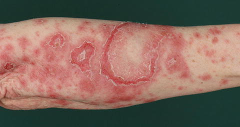
Fig. 27.2
Annular type of SCLE with multiple confluent lesions on the lower arm
SCLE constitutes an isolated cutaneous condition in more than half of the patients, whereas a smaller portion suffers from SLE. SCLE is associated with the HLA haplotype B8 DR3 and with autoantibodies to Ro/SSA and La/SSB. Lesions can be triggered by UV light and several drugs, among them terbinafine, thiazide diuretics and several calcium channel blockers.
Differential Diagnoses
The differential diagnoses of SCLE include tinea corporis, psoriasis, mycosis fungoides, erythema exsudativum multiforme/toxic epidermal necrolysis, erythema annulare centrifugum, erythema gyratum repens, drug eruption, nummular eczema and seborrhoeic eczema [7].
Histology
Histologically, SCLE is characterised by interphase dermatitis leading to keratinocyte necrosis and vacuolar degeneration at the dermoepidermal junction, as well as lymphocytic infiltrates and moderate hyperkeratosis.
Chronic Cutaneous Lupus Erythematosus (CCLE)
Definition
CCLE includes discoid LE (DLE) and lupus panniculitis or lupus erythematosus profundus (LEP). Chilblain lupus is the topic of a specific chapter of this book.
DLE consists of well-defined, coin-shaped erythematous plaques, on which adherent scales develop. Removal of the scale exposes follicle-sized keratotic spikes, the “carpet tack sign” [8]. This form of CCLE can lead to atrophy and scaring (Fig. 27.3). DLE lesions occur in approximately 15 % of patients with SLE, but more than 95 % of patients with DLE lesions suffer from CLE only.
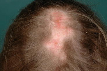
Fig. 27.3
In a patient with SLE, scarring alopecia due to DLE lesions with active erythematous plaques
LEP/lupus panniculitis is characterised by indurated nodules or plaques that will result in deep lipoatrophy. Commonly, LEP is associated with DLE lesions.
Differential Diagnosis
There are several diagnoses that need to be distinguished from DLE, such as tinea faciei, actinic keratosis, lupus vulgaris, and sarcoidosis. LEP/lupus panniculitis needs to be differentiated from other forms of panniculitis, including subcutaneous sarcoidosis, panarteriitis nodosa, malignant lymphoma (in particular subcutaneous panniculitis-like T-cell lymphoma), morphea profunda and subcutaneous granuloma annulare [7].
Related posts:

Stay updated, free articles. Join our Telegram channel

Full access? Get Clinical Tree


