Limited type
Morphea (plaque type)
Guttate morphea
Atrophoderma of Pasini and Pierini
Generalized type
Generalized localized scleroderma (3 or more anatomic sites)
Disabling pansclerotic morphea
Eosinophilic fasciitis
Linear type
Linear localized scleroderma (usually with involvement of the extremities)
Linear localized scleroderma, “en coup de sabre” type
Progressive facial hemiatrophy (synonym: Parry Romberg syndrome)
Deep type
Limited Type of Localized Scleroderma
The most common form of the limited type of LS is the plaque type (morphea). The plaque type is characterized by lesions measuring more than 1 cm in diameter affecting one or two areas of the integument. Characteristic sites of predilection are the trunk – especially the submammary region – and the area between the hip and inguinal regions. The often oval-shaped lesions have an erythematous appearance in the early phases, later hardening increasingly in the center and taking on a whitish or ivory color. Active lesions are characterized by a violet halo surrounding the fibrotic part well known as the “lilac ring.” Fibrosis may be accompanied by the loss of skin appendages and alopecia. Often sclerotic lesions soften over time and even become atrophic, hypo-, or hyperpigmented.
The guttate type of limited scleroderma (morphea guttata) is characterized by multiple yellowish-whitish, small sclerotic lesions that have a glistening surface (<1 cm with a “lilac ring” in clinically active disease) and are located predominantly on the trunk. These lesions may at first simply present as erythematous maculae. Atrophoderma of Pasini and Pierini is possibly an early abortive type of guttate scleroderma. The clinical presentation of this variety, which frequently manifests during childhood, is characterized by symmetrical lesions on the trunk that measure less than 1 cm in diameter. Loss of connective tissue can result in wedge-shaped depressions below the level of the skin. Histology corresponds to late atrophic phases of localized scleroderma.
Generalized Type of LS
The generalized type of LS is diagnosed if three or more anatomic sites are affected. The most commonly affected sites are the trunk, thighs, and lumbosacral region. The plaques are often distributed symmetrically and can coalesce into larger lesions representing different stages of the disease (Fig. 21.1).
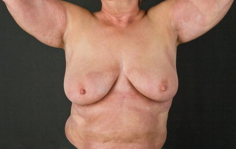
Fig. 21.1
Patient with a generalized form of LS. Please note the involvement of axillary, mammary, and abdominal regions
A rare, severe variant of the generalized type of localized scleroderma is “disabling pansclerotic morphea.” This variant, sometimes in combination with the linear type, leads to extensive skin involvement with only a limited tendency to regression. In these patients contractures and impaired wound healing develop, leading in some patients to ulceration.
Eosinophilic fasciitis (Shulman’s syndrome) is by some experts considered to belong to the disease group of LS and in our opinion belongs to the generalized type.
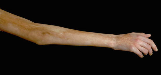
Linear Type of LS
Linear localized scleroderma is characterized by longitudinally arranged linear, band-like, or systematic lesions often following Blaschko’s lines [5]. In mild cases the lesions may heal with hyperpigmentation as the only residuum. However, in many patients the linear type is characterized by the formation of tough, sclerotic bands that sometimes cross joints and may restrict movement. Linear scleroderma can be accompanied by atrophy of the underlying muscle or bone at affected sites (Fig. 21.2). Antinuclear antibodies, usually of low titer and without further specificity, have been reported in up to 50 % of patients with the linear form of LS. The best known type is the “en coup de sabre” form, where the sclerotic tissue passes over the frontoparietal region, usually paramedian from the eyebrows into the hair-bearing scalp resulting in scarring alopecia (Fig. 21.3). Involvement of the underlying central nervous system (CNS), as detectable by MRI, is quite common. Clinically relevant associated neurological changes as, e.g., epilepsy and headache are, however, less frequent. More on the facial involvement of localized SSc is found in Chapter xxx.