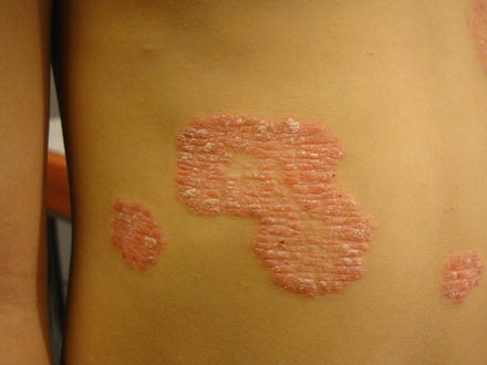
Fig. 13.1
Typical maculopapular rash of systemic-onset JIA
Differential diagnosis of systemic JIA rash is wide. It is usually, however, rather nonspecific, except that it is seen within an overall clinical picture that can help in excluding other diseases (note, of course, that, by definition, JIA is a diagnosis of exclusion). The three conditions most frequently mistaken for systemic JIA are infection, malignancy, and inflammatory bowel disease. The latter condition can have mucocutaneous manifestations as well, but they occur in the form of erythema nodosum, pyoderma gangrenosum, or oral ulcerations. Malignancies can also have multiple types of rashes, but are usually diagnosed on the background of typical laboratory abnormalities. The most difficult situation from a diagnostic point of view is therefore represented by infection or post-infectious illnesses. Rheumatic fever is rare in our climate, and its cutaneous manifestations are erythema marginatum and subcutaneous nodules. This makes the rash of rheumatic fever quite easy to recognize. Particularly difficult to recognize are viral infections such as those caused by adenovirus, parvovirus, and cytomegalovirus; all these infections can mimic systemic JIA with a similar rash accompanied by fever. The course and, occasionally, laboratory features can help distinguish these illnesses.
Psoriatic arthritis occurs in 10–15 % of patients affected with psoriasis. In about half the cases, psoriasis appears years before arthritis although the arthritis can occur, occasionally, before the skin disease. The simultaneous presence of both psoriasis and arthritis is more frequent in children than in adults. The severity of arthritis is not considered to be correlated to any particular type of psoriasis or to the severity/extension of the skin disease. Some clinical features are associated with the psoriatic arthritis, such as nail pitting (Fig. 13.2), dactylitis (Fig. 13.3), a psoriasis-like rash, and a family history of psoriasis. In patients with such a suspected diagnosis, since the rash can be minimal and overlooked, a careful examination of scalp, skin, and nails is necessary. Frequently children affected with psoriatic arthritis have a family history of psoriasis.
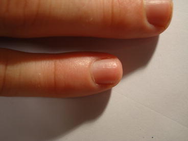
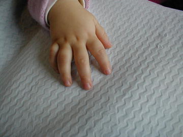
Fig. 13.2
The matrix of nails is often interested. Almost 70–80 % of the patients with psoriatic arthritis show alterations of nails such as nail pitting
Fig. 13.3
Dactylitis in young patients with psoriatic arthritis
The classic lesions of psoriasis consist of round, well-demarcated, red areas of skin inflammation with characteristic grayish or silvery scales (Fig. 13.4). The disorder may present a single plaque or multiple plaques distributed over the whole body. Papules can coalesce and form patches of diameter of >1 cm over very small or wide areas of the body. Psoriatic lesions tend to appear as symmetric eruptions on the elbows, knees, lumbosacral region, the scalp, buttocks, and around genital areas. The distribution on axilla, groin, perineum, central chest, and umbilical region is called inverse psoriasis. The scalp and nails are often involved conjointly. Scalp psoriasis is frequently the earliest presentation, and it is characterized by well-demarcated erythematous plaques with thick, adherent silvery scales. Very small plaques can be confused with seborrheic dermatitis. However, while seborrhea usually remains within the hairline, psoriasis extends to the preauricular, postauricular, and nuchal regions. Onycholysis, transverse ridging, and uniform nail pitting are the three main features of nail involvement. When the diagnosis is based only on nail changes, it is important to distinguish them from trauma or fungal infection (a suspect nail clipping should be sent for microscopic examination).