Fig. 38.1
Palpable purpura in a patient with microscopic polyangiitis. Note the relative sparing of the distal limb
Differential Diagnosis
MPA has only recently been separated from polyarteritis nodosa on the basis of clinical and laboratory features as well as of the size of the vessels involved [1]. Polyarteritis nodosa involves small- and medium-sized vessels. Both conditions may present with similar cutaneous manifestations. Palpable purpura, which reflects vasculitis of the superficial dermis causing damage to the vessels, is more frequent in MPA than in polyarteritis nodosa. Vasculitic purpura must be differentiated from purpura due to trauma, infections, glucocorticoid therapy, systemic amyloidosis, scurvy, and thrombocytopenia [9].
Nodules reflect vasculitis localized in the deep dermis or subcutaneous tissue and tend to occur more frequently in polyarteritis nodosa than in MPA [2] (Fig. 38.2). However, nodules may also be secondary to rheumatoid arthritis, lepromatous leprosy, some skin tumors, secondary deposits, lymphoma, and dermatofibroma [9].
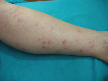
Fig. 38.2
Skin nodules in a patient with polyarteritis nodosa (Image courtesy of Dr. Monica Cattania, Dermatology Department, Arcispedale Santa Maria Nuova, Reggio Emilia, Italy)
Livedo racemosa is a violaceous mottling of the skin. It can be seen both in MPA and in polyarteritis nodosa but also in the antiphospholipid antibody syndrome, the cholesterol emboli syndrome, cryofibrinogenemia, calciphylaxis, and cryoglobulinemic vasculitis [9, 10]. Livedo racemosa should be differentiated from livedo reticularis, which is a functional disorder due to impaired blood flow of the cutaneous vessels leading to a netlike cyanotic pattern in the legs, especially in cold weather [10]. Clues to the diagnosis of livedo racemosa over that of livedo reticularis are a more widespread localization and a shape characterized by irregular, broken circular segments [10].
Skin ulcers can be vasculitic in origin but can also be due to noninflammatory vasculopathy, infections, pyoderma gangrenosum, and malignancies, particularly squamous cell carcinoma [9]. The most common causes of leg ulcers are venous hypertension and atherosclerosis. Venous hypertension typically underlies leg ulcers over the medial malleolus, while atherosclerotic ulcers most commonly occur over the toes and dorsum of the feet [9].
Despite some differences between MPA and polyarteritis nodosa, a distinction between these two conditions cannot reliably be made in the individual patient on the basis of the cutaneous manifestations alone because of a substantial overlap of the different types of skin lesions in these vasculitides [2]. In order to reliably distinguish MPA from polyarteritis nodosa, other features beyond clinical picture and histological alterations need to be taken into account. Both conditions can present with constitutional symptoms, arthralgia, and peripheral nerve disease. However, while MPA mainly causes capillaritis of the lungs and glomerulonephritis, polyarteritis nodosa rarely affects the lungs and kidneys, and when renal involvement occurs, the typical manifestation is of renal infarction instead of glomerulonephritis. In terms of serology, ANCA (usually with a perinuclear pattern) are often positive in MPA, but only occasionally seen in polyarteritis nodosa, whereas polyarteritis nodosa, but not MPA, may be associated with hepatitis B virus. Finally, angiography may show characteristic aneurysms of visceral arteries in polyarteritis nodosa, which are not a feature of MPA.
The vasculitis seen in MPA is often undistinguishable from that observed in Wegener granulomatosis and Churg-Strauss syndrome. All these conditions are associated with circulating ANCA, although ANCA are absent in 60 % of cases of Churg-Strauss syndrome and have mostly a cytoplasmic pattern (c-ANCA) in granulomatosis with polyangiitis (Wegener) [8]. The presence of granulomata and of upper respiratory tract involvement would point to Wegener granulomatosis, while asthma, eosinophilia, and extravascular granulomata would rather suggest Churg-Strauss syndrome.
Related posts:


Stay updated, free articles. Join our Telegram channel

Full access? Get Clinical Tree


