Fig. 41.1
GCA in an 80-year-old woman. Frontal view
Much less frequently severe vascular inflammation in GCA can be associated with ischemic skin lesions such as necrosis of the scalp or tongue (Figs. 41.2, 41.3, and 41.4) [2]. Scalp necrosis usually occurs in active GCA before the initiation of glucocorticoid treatment and is often associated with other ischemic manifestations. In a review of the literature which identified 24 patients with GCA who presented with scalp necrosis, 16 patients suffered visual loss, 4 had tongue gangrene, and 1 had necrosis of the nasal septum [3]. Nineteen patients developed scalp necrosis before receiving glucocorticoids, while in 21 cases out of 24, scalp necrosis occurred before temporal artery biopsy was performed [3]. Temporal artery biopsy was positive in 14/16 cases in which it was performed [3]. Therefore, scalp necrosis appears to be due to active GCA and is not an ischemic complication of temporal artery biopsy.
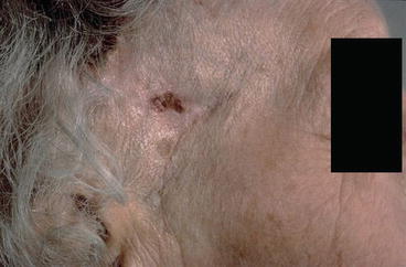
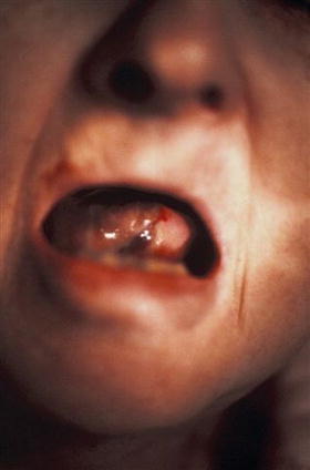
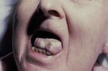
Fig. 41.2
GCA in a 75-year-old woman. GCA of 2 months’ duration. On glucocorticoid treatment for 3 weeks. Two areas of scalp necrosis with loss of surrounding hair can be seen
Fig. 41.3
GCA in an 80-year-old woman. Ischemic necrosis of tongue due to GCA
Fig. 41.4
GCA in an 80-year-old woman. Two months after treatment. There has been sloughing of skin of tip of tongue and healing
Other mucocutaneous changes occasionally seen in GCA are necrosis of the tongue or lips, glossitis, and facial swelling [2, 4, 5]. Necrosis of the tongue and lips is due to ischemia secondary to inflammation, while facial swelling might be related to the underlying inflammation.
Differential Diagnosis
Rarely, the temporal arteries can be involved in ANCA (anti-neutrophil cytoplasmic antibodies)-associated vasculitis [6] and in amyloidosis [7]. Biopsy is diagnostic.
Skin changes due to small vessel involvement such as purpura or nodules are not part of the clinical manifestations of GCA. If such lesions are present, one should suspect a small-vessel vasculitis mimicking GCA [6]. However, skin lesions of several types may occasionally be seen in Takayasu arteritis, a disease similar to GCA [8, 9]. These lesions commonly appear as erythematous nodules over the legs. Biopsy generally reveals vasculitis [8, 9].
Related posts:

Stay updated, free articles. Join our Telegram channel

Full access? Get Clinical Tree


