Fig. 26.1
Objective signs of xerosis (lower limbs)
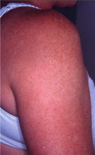
Fig. 26.2
Objective signs of xerosis in SS female patients: roughness, fine scaling
Table 26.1
Primary SS nonvascular skin lesions and their differential diagnosis
pSS skin lesions | Differential diagnosis |
|---|---|
Skin xerosis | Primary xerosis : “idiopathic xerosis,” SS xerosis. Secondary xerosis: (a) dermatological diseases (atopic dermatitis, allergic and contact dermatitis, asteatotic and nummular eczema, autosomal dominant ichthyosis, stasis dermatitis), (b) systemic therapy (hypocholesterolemic drugs, aromatic retinoid, psoralens), (c) topical treatments (retinoid acid, benzyl benzoate (i.e. niacinamide)), (d) systemic diseases (hypothyroidism and autoimmune diseases such as lymphoma, leprosy, AIDS, anemia, sarcoidosis, liver and biliary disease, renal insufficiency, and diabetes mellitus), (e) physiopathological conditions (repeated washings, winter season), and (f) elderly skin xerosis |
Angular cheilitis | Risk factors : inadequate dentures, poor oral hygiene, excessive mouth washing and aggressive dental floss, age, abnormal skin folds at the corner of the mouth, licking, dry skin, and use of tobacco and alcohol. Infectious etiologies: Candida albicans, Staphylococcus aureus, and β-hemolytic streptococci. Noninfectious etiologies: (a) systemic conditions (iron deficiency anemia; riboflavin, foliate, and B12 deficiencies; diabetes mellitus; Crohn’s disease; AIDS; Down’s syndrome), (b) dermatological conditions (atopic and seborrheic dermatitis, contact allergy, perioral dermatitis, and orofacial granulomatosis) |
Eyelid dermatitis | Allergic contact dermatitis, irritant dermatitis, atopic dermatitis, seborrheic dermatitis, urticaria (contact and systemic photo irritation), periocular rosacea, psoriasis, systemic lupus, dermatomyositis, infection (fungal/bacterial), secondary to conjunctivitis, or blepharitis |
Annular erythema | Cutaneous LE (LED, SCLE, and LET), JKLS, REM, Lyme disease (erythema chronicum migrans and linfocitoma benign cutis), rheumatic fever (erythema marginatum), specific paraneoplastic dermatitis (erythema gyratum repens), and hypersensitivity annular erythema (infectious, malignancy; liver diseases; autoimmune thyroiditis) |
Angular Cheilitis. Synonyms: perleche, commissural cheilitis, and angular stomatitis. The term refers to inflammatory conditions that radiate from 1 or both angles of the mouth. Acute angular cheilosis is erythematous or erosive, stinging. Chronic cheilitis is erythemato-squamous and is often asymptomatic [4]. AC has some risk factors and may be present in various systemic conditions and dermatological diseases (Table 26.1) [4]. The most common etiology is infectious. In pSS, AC is related to xerostomia and to oral candidacies (more than 80 % pSS patients) [4].
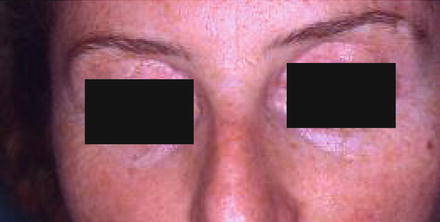
Eyelid Dermatitis (Fig. 26.3). This can be found in various CTDs and particularly in the elderly. ED may be associated with continuous rubbing of the periorbital area due to ocular dryness and a foreign body sensation of xerophthalmia. ED is associated with xerosis in 86 % of pSS patients, typically involved the upper eyelid (types I and II ED), and ranged from slight erythema and infiltration to lichenification and/or brown hyperpigmentation. Moreover, eyelid involvement is frequent in ophthalmological lesions. Atopic dermatitis (AD) is the most common cause of chronic eyelid involvement and patients should be questioned for a personal and family history of asthma, atopy, or eczema; in atopy, it is characteristic of the Dennie-Morgan infraorbital fold of the lower eyelid. In allergic contact dermatitis (ACD) or irritant contact dermatitis (ICD), occupational history, domestic interest, and hobbies are important aspects of diagnosis; moreover, in ACD present in the face and neck, patch-testing positivity improves after allergen removal. Contact urticaria ED is rare and usually includes other symptoms such as rhinitis, edema, gastrointestinal disturbance, and angioedema; a diagnostic feature is the expression of dermatitis after exposure or contact (30–60 min/4–6 h) to an allergen. In ocular rosacea, ED is characterized by eyelid margin telangiectasia rather than by periorbital papules, meibomian inflammation, recurrent chalazia, and the typical facial involvement. In seborrhea ED, the eyelids show greasy-appearing scales, crusting, and erythema. Moreover, the typical nasolabial fold scaling is present. In psoriasis ED, the lid and eyelid present erythematous scaling plaques and in more severe forms edema of the lower lids and ectropion.
Annular Erythema . This is rare in Caucasians. Three types of AE are described [1]: type I, isolated donut ring erythema with an elevated border [2]; type II, SCLE-like polycyclic erythema [3]; and type III or papular-like erythema [6]. The common characteristic is the presence of infiltrated, indurated, non-scaling lesions with central clearing without discromia or atrophy not clearly related to sunlight exposure [1]. AE shares many clinical, histological, and immunological features with cutaneous LE and related LE skin disorders such as Jessner-Kanof lymphocytic infiltrate (JKLI) and reticular erythematous mucinosis (REM). Histopathological findings, rather than clinical findings (see biopsy below), are useful for the differential diagnosis, in particular with respect to subacute cutaneous lupus erythematosus (SCLE) which is considered the corresponding Caucasian form of AE [1, 6]. In SCLE, polycyclic scaling not infiltrating the margins, achromia, and a subacute course are distinctive features [6]. Moreover, a strong association with sun exposure has been detected. Discoid lupus erythematosus (LED) can be distinguished due to the presence of hyperkeratosis and scleral atrophy. Lupus tumidus (LET) is characterized by an intermittent course, high photosensitivity, and edematous lesions [7]. Annular lesions and specific features of the underlying disease should be detected in Lyme disease (erythema chronicum migrans), rheumatic fever (erythema marginatum), and paraneoplastic dermatitis (erythema ageratum reopens). Finally, an annular erythema can be present in various pathological conditions such as a nonspecific hypersensitivity manifestation (Table 26.1) [6].
Related posts:

Stay updated, free articles. Join our Telegram channel

Full access? Get Clinical Tree


