Middle band : is an axillary pouch
Posterior band: form the posterior labrum to the glenoid rim. Not found in all patients
The primary static stabiliser with the arm in flexion and medial rotation, providing posterior stability. It tightens with abduction and moves superiorly with combined internal rotation
Table 17.2 Muscles of the shoulder girdle. Adapted from Horsley (2005) Assessment of shoulders with pain of a non-traumatic origin. Physical Therapy in Sport. 6:6–14 © Elsevier.

Hess (2000) adapted Panjabi’s model proposed for spinal segmental stability (Panjabi 1992) for the glenohumeral joint, which states that joint stability is based on the interaction between the active, passive and neural control subsystems, with the rotator cuff muscles, activating at different positions, compressing the convex humeral head into the concave glenoid, thus resisting the shear force experienced by the humeral head (Lee et al. 2000). Receptors within the joint capsule contribute to a reflex arc, which will cause activation of the muscles which overlie the joint capsule (Guanche et al. 1995).
Overhead athletes suffer repeated microtrauma resulting from repetitive use of the limb at extreme ranges of motions without increasing force. Instability can result from muscle imbalance, contracture, and ligamentous and capsular laxity (Cofield et al. 1993). Range of motion deficits will contribute to injury as this will produce a situation whereby some muscles become tight and some muscles become lax (Baltaci and Johnson 2001). Patients with chronic shoulder pain or instability are sometimes difficult to diagnose and treat. A thorough history and systematic clinical examination followed by a systematic approach to the use of investigating tools such as diagnostic ultrasound or MRI is essential for a successful outcome (Rolf 2008).
Assessment of injury risk
The assessment of posture within the domain of injury rehabilitation has traditionally been performed via visual observation of specific joints/bony landmarks, and the corresponding position they have to one another. Good posture has been described as a state of muscular and skeletal balance that protects the supporting structures of the body against injury or progressive deformity, irrespective of the attitudes in which the structures are resting or working (Kendall et al. 1993). Ideal alignment standards used in clinical practice have previously been highlighted (Kendall et al. 1993; Sahrmann 2002). The widely accepted description of normal standing posture is that proposed by Kendall and McCreary (1983) as a vertical line passing through the lobe of the ear, the seventh cervical vertebra, acromion process, greater trochanter and slightly anterior to the midlines of the knee and lateral malleolus. Deviations outside this theoretical plumb-line have been described as abnormal, and have been linked to numerous problems. Posture deviations frequently found in the cervical and thoracic spine have been suggested to affect the normal function of the glenohumeral joint (Ayub 1991; Kendall et al. 1993; Einhorn et al. 1997; Janda 2002; Sahrmann 2002; Lewis et al. 2005a).
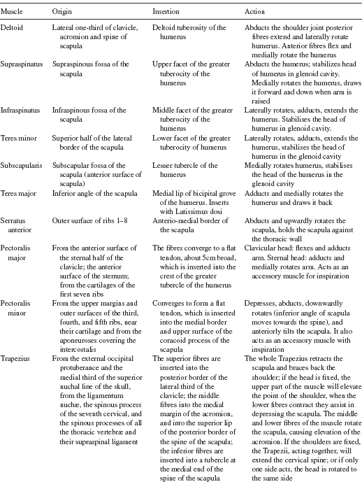
Standing postures associated with a forward head are seen in association with combinations of increased lordosis in the cervical and lumbar regions, an increased kyphosis in the thoracic region, protracted shoulders (with elevation or depression) and abnormal scapula position (Ayub 1991; Greenfield et al. 1995; Grimsby and Gray 1997; McDonnell and Sahrmann 2002; Sahrmann 2002; McDonnell et al. 2005) (Figure 17.1), although not all studies have found this (Raine and Twomey 1997; Hanten et al. 2000). Several authors have suggested that muscle imbalances and shortening can occur in the sternocleidomastoid, upper trapezius and levator scapula with a forward head position. This will lead to elevated and abducted scapula, and increased thoracic kyphosis, increasing the risk of impingement (Ayub 1991; Grimsby and Gray 1997). Subjects with increased thoracic kyphosis have been shown to predispose altered scapular kinematics; when asymptomatic subjects were positioned in a slouched posture when sitting and instructed to elevate their arm, there was a significant reduction in posterior tilt and upward rotation of the scapula, as well as an increase in the amount of scapular elevation and internal rotation (Kebaetse et al. 1999). When subjects who were experiencing sub acromial impingement improved their posture, it was not found to have a significant effect on the intensity of the pain, but increased the range of shoulder elevation before the pain was experienced (Lewis et al. 2005a). Thus thoracic posture needs to be optimised in patients with impingement-like symptoms, during all daily activities, and exercises directed at improving thoracic extension should be considered. Interventions to consider are, amongst others, thoracic spine joint mobilisation (Bang and Deyle 2000), corrective taping of the scapular and thoracic spine (Lewis et al. 2005b), facilitation scapulothoracic musculature (Konrad et al. 2006), and facilitate the activity of the rotator cuff (Magarey and Jones 2003).
Shoulder girdle, scapular and glenohumeral joint position
The role of the scapula is extremely important in providing a stable base from which the glenohumeral joint functions, as well as determining the overall position of the shoulder girdle (Kibler 1991; Paine and Voight 1993; Kibler 1998; Sahrmann 2002; Magarey and Jones 2003). The efficiency of muscular activity is dependent on the position of the scapula and the length-tension relationships of the scapular stabilisers and rotator cuff muscles, which originate on the scapula, cervical and/or thoracic spine (Einhorn et al. 1997; Mottram 1997; Magarey and Jones 2003). The scapula stabilisers, such as trapezius and serratus anterior, can be adversely affected by common abnormal postures, such as increased thoracic kyphosis and forward head positions (Greenfield et al. 1995; Ludewig and Cook 2000; Borstad and Ludewig 2005; Lewis et al. 2005a;). Certain muscle imbalances, particularly shortening, can occur in the sternocleidomastoid, upper trapezius and levator scapula with a forward head position, leading to increased thoracic kyphosis, and elevated or depressed, abducted scapula (Ayub 1991; Grimsby and Gray 1997). This increased thoracic kyphosis causes the scapular to become abducted due to lengthening of the rhomboid and lower trapezius muscles, whilst shortening the serratus anterior, latissimus dorsi, subscapularis, teres major and pectoralis major and minor muscles, and pulling the humerus into an anterior and/or internally rotated position, and further anteriorly tilting the scapula (Ayub 1991; Borstad and Ludewig 2005). This posture alters the scapulohumeral rhythm and perpetuates various forms of impingements, either in the subacromial space or inter-articular, during arm elevation, as the ability of the scapula to tilt posteriorly is inhibited by overactive pectoralis minor (Lewis et al. 2005a).
Functional examination
- Active movements: Active tests do not enable us to differentiate between inert and contractile structures. Active tests inform us about the patient’s willingness to move.
- Passive movements: Test the integrity of the inert structures. Look for pain, range of movement and end-feel.
- Resisted tests (maximal isometric contractions from a neutral, generally mid range, position): Examine the contractile structures, assess pain and muscle strength.
Palpation
Abnormal findings:
- at rest: warmth, fluid, synovial thickening
- on movement: crepitus, end-feel.
End-feel
Normal/physiological:
- hard: e.g. elbow extension, knee extension
- capsular (elastic): e.g. rotations at shoulder, elbow, hip
- extra-articular (tissue approximation): flexion at elbow, hip.
Pathological:
- too hard: e.g. osteoarthrosis
- too soft: e.g. loose body in the elbow joint
- muscle spasm (involuntary muscle contraction): e.g. arthritis
- empty (voluntary muscle contraction, not always the same range): e.g. abscess
- springy block: e.g. meniscus subluxation.
There are many special tests for evaluation of the pathologies arising around the glenohumeral joint, and there have been numerous articles evaluating the sensitivity and specificity, as well the positive and negative likelihood ratios (Dinnes et al. 2003; Hegedus et al. 2008; Munro and Healy 2008). Sensitivity is the ability to identify everyone with a specific condition. Specificity is the proportion of patients without a specific condition who have a negative test. A positive likelihood ratio describes the impact that a positive test has on raising the suspicion that a condition actually exists. High values infer that the condition which is being tested for really exists. Conversely, a low negative likelihood ratio infers that the condition for which is being tested is likely not to exist.
Several authors (Razmjou et al. 2004; Boettcher et al. 2008; Hegedus et al. 2008; Munro and Healy 2009) have analysed the pooled results of studies and have come to the same conclusion; the commonly utilised diagnostic tests for shoulder pathology have a low diagnostic utility.
Below is a description of some of the more common tests for various pathologies arising around the shoulder. Since there are several tests described for the various pathologies, it is indicative that there is no superior test for any single pathology.
Anterior instability
Anterior load and shift test (Hawkins et al. 1996)
The humeral head is grasped with the one hand, while the other hand stabilises the scapula. The humeral head is loaded medially into the joint and then an anterior and posterior shearing force is applied. The direction and translation can be graded using Altchek and Dines classification (1993), a scale of 0 to 3.
Anterior drawer test (Gerber and Ganz 1984)
The patient is placed supine and the arm abducted over the edge of plinth. The examiner stabilises the scapula with one arm whilst the other grasps the humeral head and translates it in an anteromedial direction on the glenoid. Unilateral increases in humeral head translation of the symptomatic shoulder indicate anterior glenohumeral joint instability.
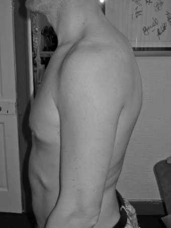
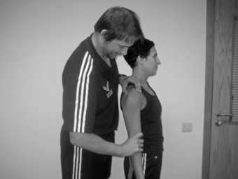
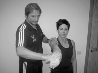
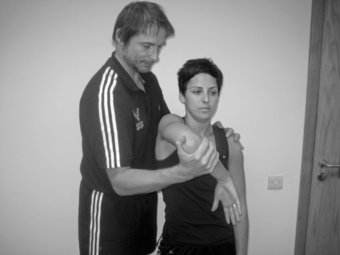
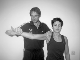
Apprehension test (Jobe et al. 1989)
This is performed with the humerus in 90 degrees of abduction, 90 degrees of elbow flexion and external rotation of the shoulder. The examiner exerts gentle pressure into progressive external rotation (Figure 17.2). A positive test is when the patient feels a sensation of impending dislocation.
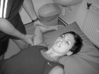
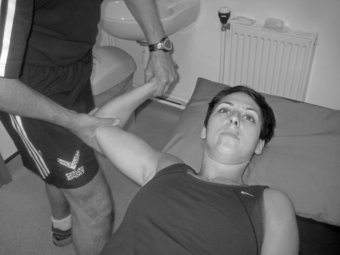
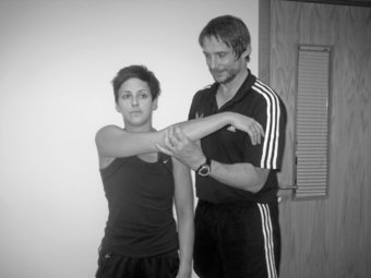
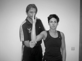
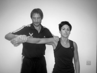
Relocation test
With the patient supine the arm is taken into abduction and external rotation. The test can be augmented by pushing the humeral head anteriorly from behind. The relocation test is performed by pushing posteriorly on the upper part of the humerus (Figure 17.3). The relocation test is positive if the apprehension or pain is relieved.
Posterior instability
Posterior load and shift – posterior drawer test (Gerber and Ganz 1984)
This test is similar to the anterior draw test, and the humeral head is translated in a posterolateral direction. A positive result is a unilateral increase in humeral head posterior translation on the glenoid.
Posterior apprehension test
This is a modification of the posterior draw test described by Gerber and Gantz (1984) where the is arm adducted and flexed to 90 degrees, whilst the examiner imparts an axial posterolaterally directed force to the humerus. A positive result is that of pain, apprehension and often the feeling of a click as the humerus rides over the posterior rim of the glenoid.
Inferior laxity
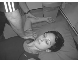
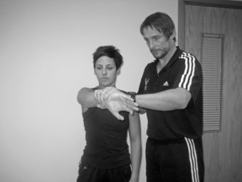
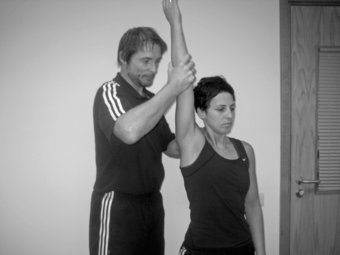
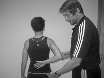
The sulcus sign (Neer and Foster 1980)
This is an examination to determine the extent and/or presence of inferior instability of the glenohumeral joint. This test can be administered with the patient either seated or standing with their arm relaxed at their side. The examiner palpates the shoulder by placing thumb and fingers on the anterior and posterior aspects of the humeral head. The examiner grasps the patient’s elbow with their other hand and applies a downward distraction force. A positive test will result in a sulcus being formed between the acromion and the humeral head as the humeral head moves inferiorly while the force is being applied (Figure 17.4).
SLAP lesions
O’Brien test (O’Brien et al. 1998)
The patient’s shoulder is held in 90 degrees of forward flexion, 30–45 degrees of horizontal adduction and maximal internal rotation. The examiner exerts a downward force distal to the patient’s elbow which the patient tries to resist. The patient is asked to identify, if produced, the location of the pain. The test is repeated in the same position except that this time the humerus is externally rotated and the forearm supinated, so the palm faces up. Once again, a downward force is applied by the examiner, which the patient actively resists, and the patient is asked to identify the location of any pain provoked. The test is considered positive if pain produced during the first part of the test is abolished with the second part of the test (Figure 17.5). For indication of a SLAP tear the pain is located over the anterior aspect of the shoulder, and for AC joint pathology, the pain must be located over the AC joint.
Anterior slide (Kibler 1995b)
The patient stands with hands on hips. One of the examiner’s hands is placed over the shoulder and the other hand behind the elbow. A force is then applied anteriorly and superiorly, and the patient is asked to push back against the force. The test is positive if pain is localised to the front of the shoulder or a click is experienced by the patient.
Posterior slide test
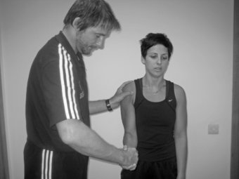
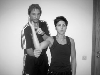
Biceps load test I (Kim et al. 1999)
The test is performed with the patient in the supine position. The examiner sits adjacent to the patient on the same side as the affected shoulder and gently grasps the patient’s wrist and elbow. The arm to be examined is abducted at 90 degrees, with the forearm in the supinated position (Figure 17.6).
The patient is allowed to relax, and an anterior apprehension test is performed. When the patient becomes apprehensive during the external rotation of the shoulder, external rotation is stopped. The patient is then asked to flex the elbow while the examiner resists the flexion with one hand. If the apprehension is lessened, or if the patient feels more comfortable than before the test, the test is negative for a SLAP lesion. If the apprehension has not changed, or if the shoulder becomes more painful, the test is positive.
Biceps load test II (Kim et al. 2001)
The patient is tested in supine. The arm is abducted to 120 degrees, externally rotated maximally, elbow in 90 degrees flexion and forearm supinated. If this test position reproduces pain then perform active elbow flexion against resistance. If the active elbow flexion component of the test increases pain (or produces pain) the test is positive.
Crank test (Liu et al. 1996)
With the patient upright, or supine, and the arm elevated to 160 degrees in the plane of the scapula, joint load is applied along the axis of the humerus with one hand, whilst the other hand performs humeral rotation. A positive test is reproduction of the patient’s overhead symptoms (with or without a click).
Pain provocation test (Mimori et al. 1999)
The patient is seated with the arm is in 90 degrees abduction and 90 degrees external rotation, and the elbow flexed to 90 degrees. The examiner places one hand over the scapula, whilst the other hand holds the patient’s wrist. The patient is then asked to supinate and pronate the forearm. If the pain is worse on pronation, this is indicative of a SLAP tear.
The resisted supination external rotation test (Myers et al. 2005)
The patient is placed in the supine position on the examination bed with the scapula near the edge of the bed. The examiner stands at the patient’s side, supporting the affected arm at the elbow and hand, with the shoulder abducted to 90 degrees, the elbow flexed 65–70 degrees, and the forearm in neutral or slight pronation. The patient then attempts to supinate the hand with maximal effort against the examiner’s resistance. The patient forcefully supinates the hand against resistance as the shoulder is gently externally rotated to the end of range. They are then asked to describe the symptoms at maximum external rotation. The test is positive if the patient experiences anterior or deep shoulder pain, clicking or catching in the shoulder, or reproduction of symptoms that occurred during throwing. The test is negative if the patient described posterior shoulder pain, apprehension, or no pain.
Long head of the biceps
Yergason’s test (Yergason 1931)
The patient is seated or standing with the elbow flexed to 90 degrees and forearm pronated. The examiner resistes active supination and elbow flexion whilst feeling for subluxation of the biceps tendon out of the bicipital groove (Figure 17.7). A positive test is detection of movement of the tendon out of the groove.
Speed’s Test (Bennett 1998)
The patient’s supinated arm is held at 90 degrees elbow flexion and then flexed forwards against resistance (Figure 17.8). Pain felt in the bicipital groove indicates biceps tendon pathology.
AC joint
Anterior/posterior AC shear test (Davies et al. 1981)
With the patient sitting, the examiner cups the heels of both hands, one over the midpoint of the clavicle, anteriorly, and one over the spine of the scapula, posteriorly. With a compressive action both hands are squeezed towards each other. Several repetitions are applied with note being taken of the amount of movement compared with the opposite shoulder. Pain is also considered. A positive test is when the patient complains of superiorly located pain unilaterally.
Cross chest adduction (Scarf/Forced adduction test) (Silliman and Hawkins 1994)
The symptomatic shoulder is flexed to 90 degrees and then forcibly adducted across the chest (Figure 17.9).
Subacromial impingement
Neer impingement test (Neer and Welsh 1977)
In this test, there is forced elevation of the humerus in the scapula plane whilst the shoulder is internally rotated with the other hand on the top of the shoulder girdle to stabilise. A positive test gives rise to pain with passive abduction, which indicates impingement within the subacromial space (Figure 17.10).
Neer impingement injection test (Neer 1983)
The subacromial space is infiltrated with 8–10 mls of local anaesthetic, and the above test is repeated. If there is greater than a 50% reduction in the pain, then this indicates that the probable cause of the pain is the bursa or a rotator cuff tendon.
Hawkin’s-Kennedy test (Hawkins and Kennedy 1980)
The shoulder is placed in 90 degrees of forward flexion and then passive internal rotation of the humerus is applied by the examiner (Figure 17.11). A positive test is provocation of pain around the subacromial space. This test indicates internal impingement of the shoulder as the rotator cuff tendons are compressed by the coracoacromial arch.
Empty can test (Jobe and Moynes 1982)
Standing in front of the patient in order to monitor facial expression during the test, the patient elevates their arm in the scapular plane to 90 degrees with the arm in full internal rotation, so that the thumb is pointing downwards. The examiner then exerts a downward force and asks the patient to resist (Figure 17.12). A positive test produces pain, weakness, or both, and indicates involvement of the supraspinatus tendon.
Full can test
Carried out as the above test except that the thumbs are pointed upwards (Figure 17.13). The test has been shown to isolate the supraspinatus as well as the empty can test (Itoi et al. 1999).
Rotator cuff tear
Supraspinatus: Drop arm test (Hoppenfield and Hutton 1976)
The patient actively abducts the arm in the coronal plane with the thumb pointing forward. From the end of abduction, the patient is instructed to slowly, under control, lower the arm. If there is a lesion within the tendon of Supraspinatus, the patient will be unable to control the descent of the arm into adduction from approximately 90 degrees abduction. If the patient can hold the arm at 90 degrees abduction, then the examiner can lightly apply pressure in a downward direction to the hand, which – if a Supraspinatus lesion is present – will cause the arm to fall into adduction.
Infraspinatus: External rotation lag sign (Hertel et al. 1996)
The examiner stands behind the patient with the elbow flexed to 90 degrees, and elevated to approximately 20 degrees in the plane of the scapula. The examiner passively externally rotates the shoulder, by holding around the wrist, to the onset of capsular tightening, whilst supporting the weight of the arm by placing a hand under the elbow, and asks the patient to actively maintain this position when the examiner lets go of the wrist, but maintaining support at the elbow. A positive test is recorded if the arm falls back into internal rotation, and the magnitude is recorded to the nearest 5 degrees (Figure 17.14).
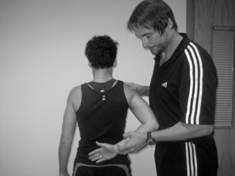
Subscapularis: Internal rotation lag sign test (Hertel et al. 1996)
The patient is asked to position his hand behind his back so that the dorsum of the hand is on the lumbar region. The examiner passively lifts the hand away from the lumbar region, whilst maintaining glenohumeral internal rotation. The patient is then asked to voluntarily maintain this position with only elbow support from the examiner. A positive result is if the hand falls back towards the spine, indicating a lesion of the subscapularis (Figure 17.15). The magnitude of the fall back can be recorded to the nearest 5 degrees.
Gerber’s lift off test (Gerber and Krushell 1991)
The dorsum of the patient’s hand is positioned at the level of the midlumbar spine. The subject is then asked to lift the dorsum of the hand off the back as far as possible, by internally rotating the shoulder (Figure 17.16). The test is considered positive for subscapularis dysfunction if the subject cannot lift the hand off of the back or if the subject performed the lifting manoeuver with elbow or shoulder extension. The test can be repeated whereby the patient is asked to try and push the examiner’s hand away from “hand behind back position”. A positive test is inability with or without pain.
The external rotation lag sign
The patient is seated. The elbow is passively flexed to 90 degrees and the shoulder is held at 20 degrees elevation in the scapular plane in a position of near maximum external rotation (i.e. maximum external rotation minus five degrees to avoid elastic recoil). The examiner supports the elbow and holds the arm in external rotation at the wrist. The patient is asked to hold the position while the examiner supports the elbow but releases the hold at the wrist (Figure 17.17). The degree of movement is estimated and is referred to as the “lag” (i.e. the difference between active and passive ROM).
Related posts:

Stay updated, free articles. Join our Telegram channel

Full access? Get Clinical Tree


