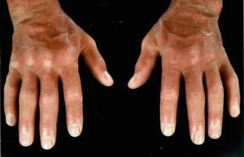
Fig. 32.1
Facial and hand features of scleromyxoedema. (a) Diffuse skin thickening of the face particularly involving the glabella and forehead, with multiple firm papules 1–3 mm on the ears (Reprinted from Lister RK, Jolles S, Whittaker S, et al. Scleromyxoedema: Response to high-dose intravenous immunoglobulin (hdIVIg). J Am Acad Dermatol 2000;43:403–8. With permission from Elsevier). (b) Sclerodermoid appearance of the hands. (c) Involvement of the trunk showing a significant inflammatory component

Fig. 32.2
Contractural changes and skin induration in advanced disease. Skin thickening with marked accentuation of skin folds
Extracutaneous manifestations are common. Over 80 % of cases have a serum paraprotein, usually IgG λ. Its level does not parallel disease activity. Gastrointestinal (oesophageal dysmotility) and neurological manifestations (epilepsy, aphasia, motor impairment, carpal tunnel syndrome, peripheral neuropathy, depression, memory loss, dementia and psychosis) are commonest. Exertional dyspnoea, restrictive or obstructive lung disease, upper airway involvement and more rarely pulmonary hypertension can occur. Proximal muscle weakness, inflammatory myopathy, inflammatory polyarthritis and other haematological disorders are documented [2, 3].
Differential Diagnosis
Scleroderma, scleroedema, nephrogenic systemic fibrosis, eosinophilic fasciitis and pretibial and generalised myxoedema are included in the differential diagnosis (see Table 32.1). The presence of waxy papules giving the skin a cobblestone appearance helps to distinguish scleromyxoedema from scleroedema and scleroderma. Raynaud’s phenomenon, nailfold capillary abnormalities, telangiectases, calcinosis and positive antinuclear autoantibodies (ANA), often with defined specificity and nucleolar staining pattern by immunofluorescence, are typically present in systemic sclerosis but rare in scleromyxoedema (and the other differentials listed). Scleroedema occurs in children and adults and typically affects the upper back, neck and face. The skin induration is nonpitting and hard, and there is no sharp demarcation between the normal and affected skin. Diabetes (typically long-standing and insulin dependent), recent infection (particularly streptococcal) and IgG k paraproteinaemia are associated. Both scleroedema and scleromyxoedema may be associated with paraproteinaemia and with a plasma cell dyscrasia and less commonly with multiple myeloma [4]. NSF and eosinophilic fasciitis commonly involve the extremities and rarely the face. Normal renal function and absence of exposure to gadolinium-based contrast media exclude NSF. In eosinophilic fasciitis there is woody induration of the limbs, usually sparing the hands and feet. At sites of vessels, the overlying skin shows typical guttering. Marked peripheral eosinophilia commonly occurs. Normal thyroid function and absent thyroid autoantibodies exclude both generalised and pretibial myxoedema. A diagnostic skin biopsy, preferably a deep incisional ellipse, is essential. A diagnostic flow chart summarising the key clinical and investigational features that underpin the diagnosis of scleromyxoedema is shown in Fig. 32.3.
Table 32.1
Differential diagnosis of scleromyxoedema: comparative features of related skin diseases
Scleromyxoedema | Scleroderma | Scleroedema | Nephrogenic systemic fibrosis | Eosinophilic fasciitis | |
|---|---|---|---|---|---|
Collagen | Thickened increased collagen clefting | Enlarged eosinophilic collagen bundles orientated parallel to skin surface | Swollen collagen bundles look fenestrated | Thickened increased collagen
Stay updated, free articles. Join our Telegram channel
Full access? Get Clinical Tree
 Get Clinical Tree app for offline access

|


