Fig. 31.1
Appearance of the immunologic response in RA: interleukin-2 (IL2) and interleukin-4 (IL4) cytokines that are released from T-helper lymphocytes proliferate and activate the B lymphocytes. Activated B cells transform into plasma cells that produce immunoglobulins (Ig). Ig combines with the antigens that are presented on the synovial membrane, synovial fluid, and joint cartilage of a RA patient by generating immune complexes. Polymorphonuclear leukocytes (PMNL) and monocytes phagocyte these immune complexes, and as a result, they trigger the production of prostaglandins (PG), leukotrienes (LT), and proteolytic enzymes which cause tissue damage
Synovial tissue is the main site of inflammation. Histologically, synovial cell hyperplasia and proliferation are seen in the joints involved. In the synovia, massive perivascular inflammatory cell infiltration consisting of CD4+ T cells, plasma cells, and macrophages, enhanced vascularity due to angiogenesis, neutrophils and organized fibrin clusters on synovial surface and in the joint space, and pannus tissue containing chronic synovitis characterized by osteoclast activity that causes synovial penetration and bone erosion in the underlying bone are seen [1].
In cellular immune mechanism, immune response begins with the activation of T lymphocytes by triggering antigen via antigen-presenting cell (APC). Here, T-cells are in the form of CD4+ T cells. For the immune response to begin, CD4+ cells need to have recognized the antigen previously and interleukin 1 (IL-1) needs to be released by monocyte and macrophages. Immune response begins with the activation of CD4+ lymphocytes. T cells coming across the antigens alone are not enough to enhance immune response; at least a single co-stimulation is needed. CD80/86-CD28 co-stimulation pathway is well defined. Co-stimulation occurs when the molecules on APC surface (CD80 or CD86) bind to CD28 receptor molecule on T-cell surface. Cytotoxic-T-lymphocyte-associated antigen 4 (CTLA4) is the most important regulator of T-cell co-stimulation and this molecule is expressed by activated T cells and competes with CD28 for binding to CD80/86 on the APC surface, and accordingly, T-cell activation is weakened [7].
T cells play a primary role in the pathogenesis of RA by allowing secondary activation of endothelial cells, macrophages, and osteoclasts. These activated cells stimulate other T lymphocytes, macrophages, and fibroblasts by secreting the cytokines such as interferon gamma (IFN-γ) and interleukin 2 (IL-2). IFN-γ activates monocyte/macrophage cells. Activated macrophages continuously secrete IL-1 and tumor necrosis factor alpha (TNF-α). In RA, IL-1 and TNF-α, which are among inflammatory cytokines, are increased in the synovia, synovial fluid, and systemic circulation and enhance destruction and decrease production resulting in local and systemic bone loss. Among the factors that are effective in osteoclastic differentiation, receptor activator nuclear factor of kappa B ligand (RANKL), IL-1, and TNF-α are found in rheumatic synovia. IL-1 and TNF-α enhance the production of RANKL, which has an important role in osteoclastic differentiation. Fibroblasts and active T cells synthesize RANKL in rheumatic synovia, and accordingly, they enhance osteoclast migration and activation. On the other hand, IL-1 and TNF-α also enhance osteoblast apoptosis and consequently prevent bone formation by increasing bone destruction. IL-1 and TNF-α are the main cytokines that play a role in RA [16].
B lymphocytes, which are activated by helper T lymphocytes, change into plasma cells and secrete Ig and RF. In addition, B cells present antigen to CD4+ T cells. Secreted Igs unite with the antigens in the synovial membrane, synovial fluid, and articular cartilage and form immune complexes. Immune complexes activate complement and cause chemotactic factors to be released. Chemotactic factors allow polymorphonuclear leukocytes and monocytes to accumulate in this region by increasing the vascular permeability. These cells phagocyte immune complexes and allow production and release of prostaglandins, leukotrienes, free radicals, and proteolytic enzymes, which cause tissue injury. They lead to tissue destruction together with activated osteoclasts [1].
Humoral mechanism is responsible for the pathogenesis of secretion of RF and anti-CCP, formation of immune complex, and activation of complement.
Toll-like receptors (TLR) are the molecules that have critical role in natural (innate) immunity. They activate natural immunity against infections. They act as a bridge between natural immunity and adaptive immunity. It has been proven that TLRs play a critical role in the pathogenesis of RA by activating natural immunity [17].
It is believed that synovial macrophages are the inflammation-intensifying cells rather than inflammation-initiating cells. They are abundant in inflamed synovial membrane and cartilage-pannus junction. They act as antigen-presenting cells in the early phase of the disease. Although macrophages secrete substantial amount of inflammatory cytokines and proteases in destruction site, they contribute to tissue destruction by activating fibroblasts rather than being the primary factor [18].
It has been understood that synovial fibroblasts have an active role in the pathogenesis of RA. They cause excessive matrix destruction by secreting matrix destructive enzymes as well as by indirectly influencing osteoclast activation. They have important roles in the activation of synovial macrophages and their cellular differentiation. Synovial fibroblasts of RA prevent programmed cell death (apoptosis) of both T and B lymphocytes. Independent from T cells, synovial fibroblasts play a role in RF production via plasma cell activation. Moreover, it has been suggested that synovial fibroblasts of RA have ability to migrate to different regions and can prevent arthritis to spread to other joints [19].
Clinical Findings
General Findings
Rheumatoid arthritis is the most frequent chronic inflammatory disease of the joints. The onset and progression of the disease vary among individuals. Slow and insidious onset in weeks and months is seen in 55–65 % of RA patients, whereas acute onset is seen in 8–15 % of the patients. The symptoms peak within several days and may be accompanied by fever and extra-articular organ involvement. Symmetrical pattern is less common in acute onset than in insidious onset. Onset is subacute in 15–20 % of the patients. Systemic complications are more common in this group than in the group with insidious onset.
Presenting symptoms may be systemic or articular. Some individuals may develop nonspecific complaints, such as fatigue, weakness, weight loss, low-grade fever, or extensive muscular pain, before articular symptoms.
Articular Symptoms
In anamnesis, patients frequently express involvement of a single joint at first and then the other joints. Typically, joint swelling and tenderness are present and restriction of joint mobility occurs in the long term. Although it is known that classical joint involvement is symmetrical, asymmetrical involvement as well is not rare. Symmetry becomes more prominent in advanced stages of the disease. It is thought that symmetrical joint involvement results from bilateral release of neuropeptides, such as substance P, from terminal nerve ends in the joints. Morning stiffness may appear before pain and is explained by accumulation of edema fluid in inflamed tissue during sleep [2].
Metacarpophalangeal (MCP) joints, proximal interphalangeal (PIP) joints, and wrists are the joints involved most frequently and earliest in RA (Fig. 31.2a, c). Symmetrical fusiform swelling in PIP joints and accompanying swelling in MCP joints are typical involvements in RA. Involvement of distal interphalangeal (DIP) joints alone is almost never seen and it is not the site of initial involvement. Involvement of DIP joint may be due to concomitant osteoarthritis particularly in elder RA patients. Studies, which radiologically monitored the wrist involvement in RA in the long term, have demonstrated that joint injury develops in the first 3 years, particularly in the first year, and that disease progression then slows down. While swelling, pain, and restriction of mobility are initially in the forefront due to synovial hypertrophy, typical deformities for RA are developed over time. Swan neck deformity results from hyperextension of PIP joint and flexion of DIP joint, whereas boutanniere deformity results from flexion of PIP joint and hyperextension of DIP joint (Figs. 31.2a, b, 31.3, and 31.4). Ulnar deviation and volar subluxation are the deformities that can be seen due to the involvement of MCP joints. Dorsal swelling in the wrist due to the inflammation of extensor tendon sheaths is one of the early signs of disease. In further stages, generally radial deviation and volar subluxation are developed (Fig. 31.4). Involvement of distal radioulnar joint causes instability and dorsal subluxation of the ulnar head. This together with erosive changes leads to the rupture of surrounding tendons (Fig. 31.5). Trigger finger is seen due to flexor tenosynovitis, which is encountered frequently [20].
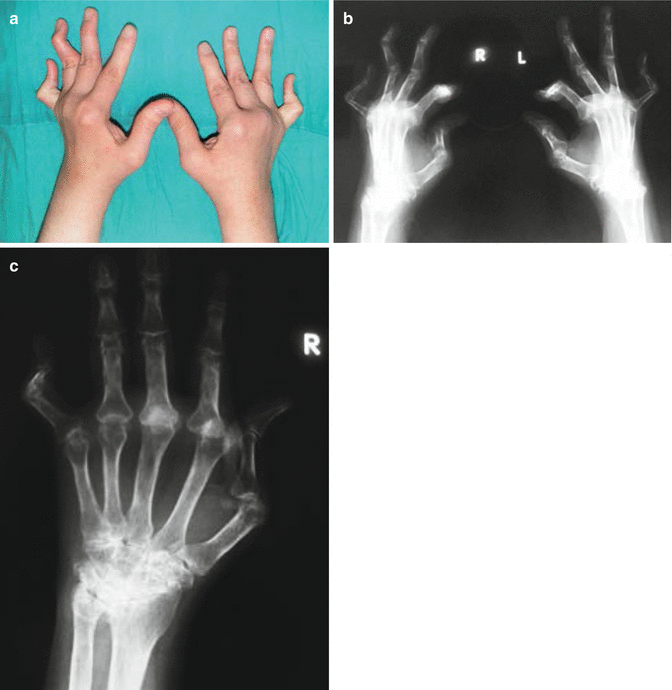
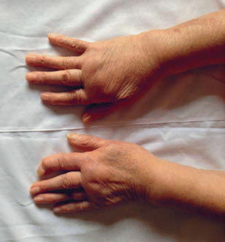
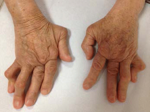
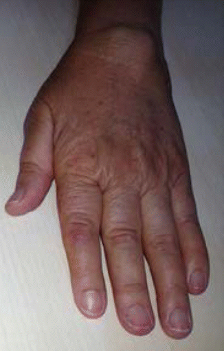

Fig. 31.2
(a–c) Hand involvement in a rheumatoid patient: inflamed synovia causes the tendon rupture and/or dislocation by weakening the ligaments. (a, b) Volar subluxation of the MCP joints, swan neck deformity of the fourth finger of the left hand and boutanniere deformity of the fifth fingers bilaterally. (c) Narrowing, erosion, and cysts of the wrist joint

Fig. 31.3
Fusiform edema of the MCP and PIP joints is the typical involvement in RA accompanying volar subluxation of MCP joints and swanneck deformity of second finger

Fig. 31.4
Ulnar deviation of MCP joints with the swan neck deformity of the fourth and fifth fingers bilaterally in a RA patient

Fig. 31.5
Extensor tenosynovitis on the dorsal surface of the wrist in RA
Among the joints of foot, the most commonly involved one is the metatarsophalangeal (MTP) joint followed by in turn the subtalar and tibiotalar joints. They are the joints involved initially in 20 % of the patients. Involvement of these joints causes more pain and restriction of mobility as compared to the joints of upper extremity because they carry load. Foot deformities include claw toe deformity, hallux valgus, volar subluxation, and cock-up deformities in the PIP joint (Fig. 31.6). Valgus deformity and flattening of the medial longitudinal arch occur with the development of cartilage loss and erosion [2].
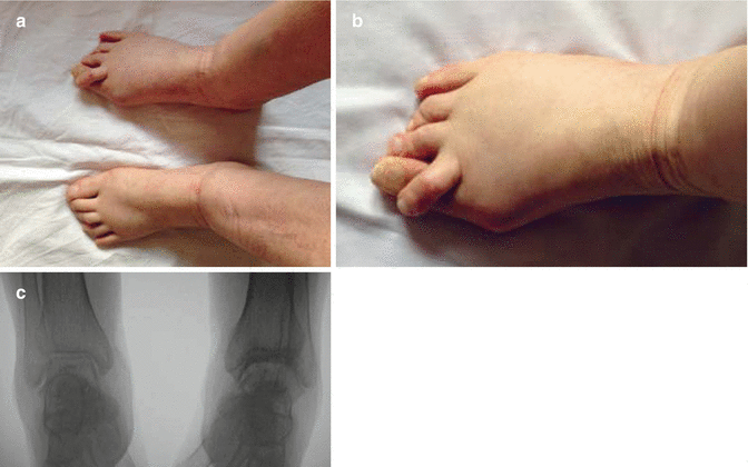

Fig. 31.6
(a–c) RA patient presenting hallux valgus deformity (a), cock-up deformity that is the result of overriding of the second finger by MTP joint extension, adduction, and lateral rotation on the first finger (b). Joint erosion of a patient with the tibiotalar joint involvement on the AP X-ray of the ankle (c)
Complaints related to shoulder joint are seen in two-thirds of RA patients. Synovitis and erosion cause damage both in the glenoid fossa and humeral head. It has been observed that involvement of the shoulder joint is more common particularly in elder patients and in RF-positive cases. RA may involve all components of shoulder joints. Rupture may be seen in the long head of the biceps and the rotator cuff tendons may be involved. Pain is typical in the C4 region in involvement of the glenohumeral joint and in the C5 region in involvement of the rotator cuff.
The most common signs seen in the elbows include failure in complete extension of elbow due to synovitis or effusion, the presence of effusion-related periarticular cysts, and rheumatoid nodules. Limited supination may lead to disability in eating and other daily activities.
Hip joint is rarely involved in RA. Movement restriction may be seen in the abduction and rotation.
Knee involvement is common in RA; it may be the first joint involved in the early period and it is a good indicator of disease activity. Effusion and atrophy and flexion deformity due to dysfunction of quadriceps muscle may be seen in the knees. Baker’s cysts, which can be palpated in the popliteal fossa, are the other common pathology. Baker’s cysts, when ruptured, can cause swelling and pain in the calf. This needs to be discriminated from acute thrombophlebitis. Destruction of the knee joint may lead to involvement of lateral and cruciate ligaments and accordingly instability. Secondary osteoarthritis, which occurs due to progressive synovitis, is the other condition related to the knee joint [2].
Although involvement of the cervical vertebrae is less common, it needs not to be ignored since it may cause life-threatening serious complications. Neck pain with movement and occipital headache are the clinical symptoms of neck involvement. The atlantoaxial joint is a synovial joint and may be exposed to proliferation and instability, similar to the other joints. Subluxation may develop due to erosion and ligament injury. The atlantoaxial subluxation is the enlargement of the space between the odontoid process of the axis and the arch of the atlas, which normally does not exceed 3 mm. In the presence of atlantoaxial subluxation, it is risky for the odontoid process to compress spinal cord during flexion of the neck. Head and neck pain, paresthesia, weakness, transient ischemic attack, and sphincter defect in the bladder and anus may appear. The presence of subluxation should be definitely assessed prior to anesthesia. Lateral cervical radiographs in flexion position are recommended prior to surgery. In case subluxation is determined, preoperative measures that would prevent spinal cord injury should be taken. Sometimes, the odontoid moves to the superior aspect toward to the foramen magnum and may involve the medulla.
Extra-articular Symptoms
Rheumatoid arthritis is a systemic inflammatory disease and has many extra-articular symptoms. These symptoms are more prevalent in patients with high RF-positive titers. Sometimes extra-articular involvement may appear before articular involvement.
Rheumatoid nodules occur in 20–30 % of RA patients. They are usually seen in the extensor surfaces of joints or in other regions that are exposed to mechanical pressure. Extensor surface of the elbow, dorsum of the hand, occipital region, sacrum, and the Achilles tendon are the regions where rheumatoid nodules are seen frequently (Fig. 31.7a, b). They are histologically composed of collagen necrotic part in the center, a middle layer consisting macrophages, and an outer layer consisting granulation tissue (Fig. 31.8). Rheumatoid nodules may be seen in many organs primarily in the lungs, sclera, and heart [2].
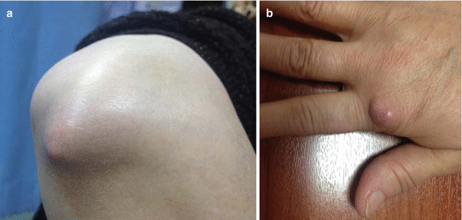
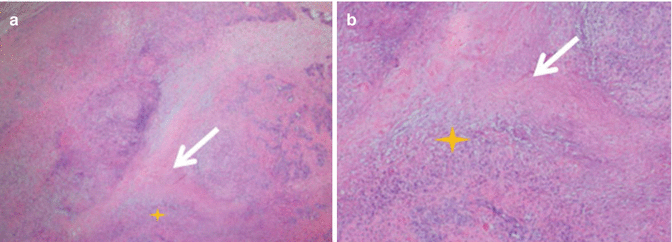

Fig. 31.7
(a, b) Rheumatoid nodules on the elbow extensory side of right elbow (a) and on the left hand (b)

Fig. 31.8
(a, b) Histopathology of rheumatoid nodule: (a) necrotic area (white arrow) settling subcutaneously is surrounded by the histiocytes arranged as palisades (yellow star). HE ×50. (b) The histiocytes and myofibroblasts that show a palisadic order (yellow star) around the necrotic connective tissue (white arrow). HE ×100
Pulmonary involvement of RA most commonly presents with pleurisy and is usually asymptomatic. It may occur either in the early or advanced stages. Pleural fluid is usually exudative and characterized by low glucose concentration. RA-induced interstitial pulmonary disease is more prevalent in individuals with long-term seropositive RA and in smokers. Diffuse interstitial fibrosis is the most common type of parenchymal involvement in RA. Nodular pulmonary disease usually occurs in those with rheumatoid nodules in the other regions of the body and in smokers. Pulmonary hypertension is less common [21]. Pericarditis is the most prevalent complication of cardiovascular involvement. Its prevalence is about 50 % in autopsy series. It is usually observed in seropositive patients with rheumatoid nodules. Myocardial involvement and related conduction defects are less common. Inflammation in RA patients enhances the risk of ischemic heart disease in association with cellular and humoral mechanisms. This risk is considered to be associated with disease severity and the presence of anti-CCP antibodies. Some studies have demonstrated that cardiovascular mortality and morbidity are decreased with methotrexate (MTX) and anti-TNF therapies.
Hematologically, anemia is quite prevalent in RA and is multifactorial. Normochromic and normocytic anemia is the most frequently encountered form. Iron-deficiency anemia may result from gastrointestinal bleeding induced by nonsteroidal anti-inflammatory drugs (NSAIDs) and other drugs. Moreover, macrocytic anemia may develop secondary to folic acid deficiency and drug therapy. Thrombocytosis may be present in those with polyarticular involvement and active disease. Moreover, thrombocytopenia may be seen in patients with RA due to immunosuppressive and cytotoxic drugs or due to treatment with gold, penicillamine, and sulfasalazine [21].
Muscular involvement in RA is usually in the form of atrophy and weakness in the muscles that are adjacent to the joints involved, primarily in the interosseous and quadriceps muscles. Besides, it may develop due to steroid use and neuropathy.
Rheumatoid vasculitis is the late-term finding of RA but not common. It is more prevalent in males, seropositive patients, patients with rheumatoid nodules, and patients with joint destruction. It is classically a vasculitis of the small vessels. Panarteritis is its pathological sign; all layers of the vascular wall are infiltrated by mononuclear cells. Proliferation in the intimal region causes thrombosis. Mostly, thrombosis in the capillary of the perionychium, infarction in the fingertips, and leg ulcers are developed.
There are four main reasons for neurological involvement in RA:
1.
Spinal cord involvement due to cervical vertebral involvement
2.
Entrapment neuropathy due to arthritis and tenosynovitis
3.
Distal sensorial peripheral neuropathy in general
4.
Mononeuritis multiplex resulting from the involvement of vasovasorum due to vasculitis. Mononeuritis multiplex is a sudden and painful involvement of the peripheral nerve [1].
Ocular involvement is seen in the late phase of the disease and in the form of keratoconjunctivitis sicca. Scleritis and episcleritis are rare. Loss of vision due to episcleritis does not occur; however, secondary cataract and related loss of vision may occur.
Renal involvement usually occurs due to amyloidosis, which is a complication of chronic RA, or treatment of the disease with D-penicillamine, cyclosporine, and NSAIDs rather than direct effect of RA.
Gastrointestinal involvement is not directly associated with RA. Duodenal ulcer and liver toxicity may occur due to the use of NSAIDs and some disease-modifying antirheumatic drugs (DMARDs) [21].
Felty syndrome is the presence of splenomegaly and leukopenia together with RA. This may be accompanied by anemia, thrombocytopenia, lymphadenopathies, and liver involvement. It is an important cause of mortality.
Secondary Sjögren’s syndrome is a picture characterized by dry mouth and dry eye, which result from autoimmune destruction of exocrine glands primarily the lacrimal and salivary glands. It is thought to occur in 10 % of RA patients.
Secondary osteoporosis may develop due to inflammation, corticosteroid therapy, and immobility in long-lasting cases, and vertebral and non-vertebral fractures may be seen. The risk is twofold higher in both females and males. Risky patients should be evaluated via bone mineral densitometry.
The risk of cancer is mildly increased particularly in RA patients with lymphoma. It is associated with disease severity and inflammatory activity.
Infections are increased due to the disease itself and to the treatment with glucocorticoids and immunosuppressive and biologic drugs. There is also risk for RA-induced septic arthritis and discrimination from acute arthritis flare-ups is of great importance to prevent joint loss [1].
Laboratory Findings
There is no test specific for the diagnosis of RA.
Rheumatoid factor, which is an autoantibody that reacts with the Fc region of Ig G, is positive in more than two-third (60–80 %) of the patients [22]. RF may be positive in 5 % of healthy subjects; its prevalence increases with age and reaches to 10–30 % over the age of 65 years. Articular and extra-articular symptoms are severer in individuals who have positive high titers. RF positivity is a constant finding in patients with rheumatoid nodules and vasculitis. The important issue in RA is the evaluation of RF together with clinical findings. In addition to RA, some diseases are also associated with RF. These diseases are systemic lupus erythematosus (SLE), Sjögren’s syndrome, chronic liver disease, sarcoidosis, interstitial pulmonary fibrosis, infectious mononucleosis, hepatitis B, tuberculosis, leprosy, syphilis, subacute bacterial endocarditis, visceral leishmaniasis, schistosomiasis, and malaria [22].
Antibodies against citrullinated proteins such as filaggrin and its circular form are called as anti-citrullinated peptide antibody (ACPA). The most widely used one is anti-CCP antibody, the specificity and sensitivity of which are 88–96 % and 48–68 %, respectively, for the diagnosis of RA. When evaluated together with RF, specificity of anti-CCP antibody in diagnosing RA increases up to 98 %. A positivity by 70 % has been detected in early RA. It is well correlated with disease progression. There are studies suggesting that anti-CCP antibody is associated with erosive arthritis development and radiological damage and may be used in diagnosing early RA [23].
Typically, normochromic/normocytic or hypochromic/normocytic anemia may be seen in RA patients. These are chronic anemia diseases. Inadequate production in bone marrow occurs due to suppressing effect of inflammatory cytokines (IL-1, TNF-α, TNF-γ) on bone marrow. Sometimes, iron deficiency may accompany.
Blood leukocyte count is generally high in patients with active RA receiving steroids. Thrombocytes may be increased directly proportional with disease activity.
Erythrocyte sedimentation rate (ESR), C-reactive protein (CRP), and fibrinogen are correlated with the degree of disease activity. CRP is a better indicator of activity than ESR [24].
Hepatic and renal function tests should also be analyzed to evaluate liver and kidney involvement [22].
Radiological Findings
Radiological evaluation is generally performed by direct radiographs. Primarily, anteroposterior hand, wrist, and foot radiographs should be evaluated. Afterward, baseline radiographs are necessary to determine progression. Periarticular soft tissue swelling and enlargement in joint space are seen in the early period. This results from synovial inflammation and fluid production. Periarticular osteoporosis is also one of the early signs. Narrowing of the joint space, subchondral pseudocysts, and erosive changes are seen due to the destruction of cartilage by the pannus. Extreme irregularity on the joint surface, subluxations, general osteoporosis, joint deformities, fractures, degenerative and destructive changes, and bone ankylosis may be encountered in the late period (Figs. 31.9, 31.10, and 31.11a–c) [25]. For standardization of evaluation by direct radiography, indexes were developed first by Larsen and then by Sharp. Atlantoaxial radiograph is valuable in suspected conditions and chest X-ray is valuable for pulmonary involvement [26].
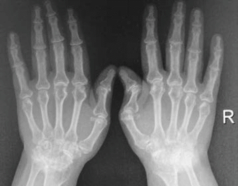
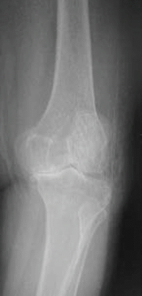
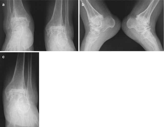

Fig. 31.9
Hand-wrist radiograph of a rheumatoid arthritis patient: narrowing, erosion, and cysts in the carpal joints; narrowing in the proximal interphalangeal joints; and subluxation in the first interphalangeal and metacarpophalangeal joints

Fig. 31.10
Knee radiograph of a rheumatoid arthritis patient: concentric narrowing in the joint space, cysts, and subluxation

Fig. 31.11
(a–c) Ankle joint narrowing and degeneration on AP (a) and lateral radiographies (b)
Ultrasonography (US) is a more sensitive method than conventional radiography to detect small bone erosions and subclinical synovitis in the early period. Inflammation and fluid in the joints and in the bursa and tendon sheaths can be detected by US and enthesopathy can be assessed [27].
Magnetic resonance imaging (MRI) is more sensitive as compared to both physical examination and conventional radiography in detecting synovitis and erosions in the joints of hands and wrists in the early period of the disease. Bone edema is very early determinant of inflammation and can predict further erosions together with synovitis; thus, it has prognostic value. If a bone erosion is initially detected in a region via MRI, the risk of erosion in that region increases sixfold at the end of 1 year. The degree of synovitis is also an important indicator of prognosis in RA [25].
Diagnosis
There is no specific test for the diagnosis of RA. Diagnosis is established with the presence of symptoms and signs of RA and exclusion of other diseases. RA should be considered if RF and anti-CCP antibody are positive in a patient with symmetrical articular pain, swelling in the joints of hands and feet, and morning stiffness.
Diagnostic criteria for RA were first developed in 1958 by ACR (the American College of Rheumatology) and these criteria were then revised in 1987. These either did not establish definite diagnosis of RA or exclude RA. New criteria for RA classification were created in 2010 by EULAR (the European League Against Rheumatism) and ACR [28]. According to the most recent classification, diagnosis of RA is established in those with a score of 6 and higher (Table 31.1) [28].
Table 31.1
The 2010 ACR (the American College of Rheumatology) classification criteria for rheumatoid arthritis
Score | |
A. Joint involvement | |
1 great joint | 0 |
2–10 great joints | 1 |
1–3 small joints (great joint involvement yes/no) | 2 |
4–10 small joints (great joint involvement yes/no) | 3 |
>10 small joints | 5 |
B. Serology (at least one test result is required) | |
Negative RF or anti-CCP | 0 |
Low-positive RF or anti-CCP | 2 |
High-positive RF or anti-CCP | 3 |
C. Acute-phase reactants (at least one test result is required) < div class='tao-gold-member'>
Only gold members can continue reading. Log In or Register to continue
Stay updated, free articles. Join our Telegram channel
Full access? Get Clinical Tree
 Get Clinical Tree app for offline access

| |


