Fig. 11.1.
Rorabeck and Lewis classification: type 1 non-displaced fracture with well-fixed implant; type 2 displaced fracture with well-fixed implant; type 3 non-displaced or displaced fracture with loose implant.
Type 1: Non-displaced and well-fixed implant
Type 2: Displaced and well-fixed implant
Type 3: Non-displaced or displaced with loose implant
11.1.3 Management
Treatment options for distal femur periprosthetic fractures include nonoperative treatment, conventional plate fixation, locking plate fixation, intramedullary nail, external fixation, and revision knee arthroplasty. Nonoperative treatment is typically not advised unless surgery is medically contraindicated. Nonoperative management often leads to high rates of nonunion, malunion, and poor patient satisfaction [5, 6]. External fixation may be a reasonable option in some patients but requires pins to be placed away from the total knee implants given high incidence of pin site infection. This typically requires spanning the knee joint which does not facilitate early motion and contributes to a stiff knee. For these reasons open reduction and internal fixation versus revision arthroplasty are the treatments of choice in distal femur periprosthetic fractures.
The following cases below will discuss the indications for plate fixation, intramedullary nailing, and revision knee arthroplasty.
11.2 Case Presentation #1
A 66-year-old woman presented to the emergency department with right thigh pain and deformity after slipping on a rock while cleaning her pond. She had previous total knee replacement 7 years prior with previously well-functioning and pain-free result. She denies any other injuries. Her comorbidities include morbid obesity, type 2 diabetes, chronic kidney disease, hypertension, and hyperlipidemia.
Physical exam shows isolated pain and deformity to the distal femur with skin intact. Previous surgical wound is well healed with no evidence of skin compromise and leg is neurovascularly intact. Patient is morbidly obese with much of her weight carried in her thigh.
11.2.1 Diagnosis/Assessment
The patient has a closed distal femur periprosthetic fracture with a stable implant, Lewis and Rorabeck type 2 fracture. The displaced component is meta-diaphyseal with a non-displaced fracture extension ending at the level of the implant (see Fig. 11.2).
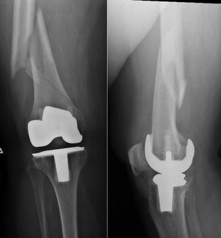
Fig. 11.2.
Displaced distal femur periprosthetic fracture with likely stable implant, type 2, with a non-displaced fracture extension ending at level of implant.
11.2.2 Management
The recommended treatment options for this patient include locking distal femur plate vs. a retrograde intramedullary implant. Both have been shown to provide superior fixation when compared to traditional nonlocking plates [7, 8]. In this case we chose to use an intramedullary nail. The intramedullary nail requires a smaller exposure which is a concern in this obese patient with multiple comorbidities. It also allows distribution of forces over a longer construct in a fracture that extends into the diaphyseal region of the femur [8]. Some studies have shown lower nonunion rates in intramedullary nails when compared to locking plates [9, 10]. Considerations that should be made for an intramedullary nail include adequate bone stock distal to fracture for locking screws, a stable implant, and an implant that will facilitate nail insertion. Relative contraindications include hip hardware (i.e., existing intramedullary nail proximally) or existing hip arthritis that may require hip arthroplasty in the near future. In these cases, retrograde intramedullary implant may be possible if there is an adequate amount of space between the distal tip of any existing fixation in the proximal femur and the proximal tip of retrograde fixation so as not to create a stress riser.
A retrograde intramedullary nail was placed through the intercondylar notch through small portion of previous skin exposure. Careful consideration was taken to protect the polyethylene and femoral component with the soft tissue protector. The nail was then placed in a standard manner. Appropriately sized nail was inserted and locked with 2 screws proximally and 3 screws distally. Patient was made weight bearing as tolerated in the immediately postoperative period (see Fig. 11.3).
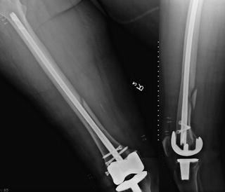
Fig. 11.3.
Type 2 distal femur periprosthetic fracture after reduction and internal fixation with a retrograde intramedullary nail.
11.2.3 Clinical Pearls/Pitfalls
Rorabeck type 1 or 2 fractures
No previous hip arthroplasty or hip arthritis
Adequate bone stock distal to fracture site
Distal femur implant facilitates intramedullary nail
Skin over knee facilitates insertion
Femoral radius of curvature appropriate for insertion point accessibility, heavily influenced by both fixation nail and current arthroplasty implant.
11.3 Case Presentation #2
The patient is a 63-year-old female presented to the emergency department after trip and fall with left thigh pain and deformity. She had previous total knee replacement 6 months ago and was doing well walking without a walking aid prior to the fall. Patient denies any other injuries. Medical problems include hypertension, diabetes, and asthma.
Physical exam shows isolated pain and deformity to the distal femur with skin intact. Previous surgical wound is well healed with no evidence of skin compromise and leg is neurovascularly intact.
11.3.1 Diagnosis/Assessment
The patient has a closed distal femur periprosthetic fracture with a stable implant, Lewis and Rorabeck type 2 fracture. Fracture is at the level of the anterior femoral interface with mild comminution and transverse (see Fig. 11.4).
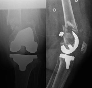
Fig. 11.4.
Displaced distal femur periprosthetic fracture with likely stable implant, type 2, transverse with mild comminution.
11.3.2 Management
The recommended treatment options for this patient include locking distal femur plate, retrograde intramedullary implant, or revision knee arthroplasty. Intraoperative assessment of the distal femur component is necessary to ensure that it remains well fixed. If it does remain well fixed, it appears that adequate bone stock to avoid a revision knee replacement. An intramedullary implant is not a great option due to distal fracture location which will not accommodate adequate distal locking screws. A locking distal femur plate has been shown to be an effective treatment option for this fracture and is superior to nonlocking plates which have significantly higher rates of malunion, nonunion, and reoperation rates [7]. Allograft may be utilized as an adjunct to distal femur plating [11] or even intramedullary fixation [12], when additional stability or biology may be required. The use of allograft strut fixation either alone, or in conjunction with plate and/or intramedullary fixation, is largely surgeon preference but has been shown to provide reliable fracture union, improved alignment, and increased femoral bone stock with a high incorporation rate [13].
Related posts:
 Periprosthetic Infection: Management of Late Acute Hematogenous Infection
Complex Primary Total Knee Arthroplasty: Management of Previous Hardware (Posttraumatic OA)
Revision Total Knee Arthroplasty: Management of Deficient Extensor Mechanism
Complex Primary Total Knee Arthroplasty: Management of Varus Knee
Periprosthetic Infection: Management of Chronically Infected Total Knee Arthroplasty
Complex Primary Total Knee Arthroplasty: Management of Flexion Contracture
Periprosthetic Infection: Management of Late Acute Hematogenous Infection
Complex Primary Total Knee Arthroplasty: Management of Previous Hardware (Posttraumatic OA)
Revision Total Knee Arthroplasty: Management of Deficient Extensor Mechanism
Complex Primary Total Knee Arthroplasty: Management of Varus Knee
Periprosthetic Infection: Management of Chronically Infected Total Knee Arthroplasty
Complex Primary Total Knee Arthroplasty: Management of Flexion Contracture
Stay updated, free articles. Join our Telegram channel

Full access? Get Clinical Tree


