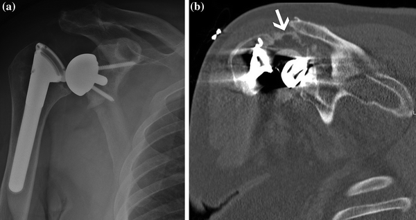
Fig. 24.1
a AP view of dislocated reverse total shoulder arthroplasty with grade III inferior glenoid notching. b Axillary view demonstrating anterior dislocation of reverse TSA. Posterior glenoid bone loss due to notching. Instability in this case was caused by impingement of the humeral component on the glenoid rim
Humeral shortening has been identified in greater than 65 % of cases with instability following RSA [3]. Assessment of the humeral length in relationship to contralateral extremity can help to identify patients with inadequate soft tissue tension [3, 11]. Soft tissue tension has been identified as the most common cause of instability and the most important factor in stability in biomechanical studies [10, 12]. In cases of mild humeral shortening (<15 mm), soft tissue tension can be increased by inserting a thicker polyethylene insert or increasing the humeral neck length which is an option in most RSA systems [3]. In cases of more significant humeral bone loss, the humeral length may need to be restored using an allograft prosthesis composite graft or a tumor prosthesis.
Medialization of the glenoid can also contribute to increased instability [3]. The degree of medialization can be assessed by measuring the distance from the center of the humeral axis to the lateral margin of the acromion [3]. In cases of medialization (humeral axis >15 mm medial to the lateral margin of the acromion), increasing the glenoid size or lateral offset is needed to increase the soft tissue tension and stability of the RSA [3]. Bone grafting of the glenoid may be required in cases of significant bone loss and medialization.
Component position can be evaluated using radiographs or CT scans. Superior placement of the glenoid baseplate and superior angulation of the glenosphere have been identified as risk factors for instability [5]. Correction of glenosphere position can be adjusted using offset components with retention of a well-fixed baseplate in some systems, but frequently will require revision of the glenoid baseplate. Removal of a well-fixed baseplate can be challenging and may result in fractures or glenoid bone loss. Reimplantation of a glenoid baseplate may require bone grafting, and in some, reimplantation may not be possible.
Instability associated with bony or soft tissue impingement can be addressed by surgical excision of the impinging structures or revision of the components to prevent impingement. The use of larger glenosphere sizes and inferior or lateral offset can eliminate bony impingement of the humerus on the glenoid that may be associated with instability and/or notching. Excision of impinging soft tissues should be performed with extreme care due to the close proximity of the axillary nerve. Cases of instability associated with neurologic injury and deltoid dysfunction are not likely to allow successful revision to a reverse prosthesis and are likely to need conversion to a hemiarthroplasty or resection arthroplasty.
Infection
Infection rates in RSA are higher than those in anatomic total shoulder arthroplasty (TSA) with reported incidence of 1–10 % [1, 2]. This finding has been attributed to the increased dead space and hematoma formation that is encountered in RSA. The treatment of an infected RSA in most cases requires component removal, placement of a temporary antibiotic spacer, and intravenous antibiotics, followed by staged reimplantation. Some authors have advocated for the treatment of an early infection with debridement, retention of well-fixed components, and revision of modular components [13]. In recent series, eradication of the infection has been successful in greater than 90 % of cases with removal of the infected prosthesis and reimplantation of a new prosthesis in a single-staged procedure [13–15]. Although the ability to eradicate the infection is reliable, functional outcome following revision RSA for infection is limited [3, 13–15].
Fracture
Fractures following RSA may result in loosening of the humeral or glenoid components. The incidence of postoperative humeral fractures is 1–2 % following RSA [2]. Intraoperative fractures are much more common in cases of revision and have been reported in up to 25 % of cases that involve removal of a humeral stem or cement mantle [4]. Fractures of the humerus distal to a well-fixed component can be treated in the same manner as humeral fracture without a reverse total shoulder with bracing or open reduction and internal fixation. In the case of a loose humeral component, revision of the humeral stem to a longer stemmed component with or without supplemental fixation is required.
Aseptic Loosening
Historically glenoid component loosening was the failure mechanism of the reverse shoulder designs. Lateralized center of rotation and limited fixation resulted in early baseplate failure in 11–40 % of reverse shoulder designs. The adoption of Grammont’s principles of a medialized center of rotation, improved fixation techniques, and bony ingrowth baseplate designs have resulted in a decrease in the aseptic glenoid loosening rates. In most modern series, glenoid component failure has been reported in 0–4 % of primary RSA [1, 2]. Risk factors for failure of a glenosphere include component malposition and inadequate fixation [1, 6]. Scapular notching occurs in greater than 50 % in most Grammont-style reverse shoulder designs [8]. The significance of scapular notching is still unclear with some series reporting poor outcomes and increased failure rates, with others noting no influence of notching on outcome or failure [1, 2, 4, 6, 14] (Fig. 24.2).


Fig. 24.2
a AP radiograph 5 years after reverse total shoulder demonstrating grade II inferior glenoid notching with osteolysis around the inferior screw (red arrow) and breakage of superior screw (white arrow) indicating a loose glenosphere. b Intraoperative photograph after removal of loose baseplate demonstrating inferior glenoid bone loss (white arrow) due to notching. c Intraoperative photograph demonstrating polyethylene wear along the medial aspect of the humeral articulation due to impingement on the glenoid rim
Revision of a loose glenoid component can be challenging due to bone loss and poor-quality bone. Preoperative evaluation of a loose glenoid component should include aspiration and laboratory studies to rule out infection. Imaging studies including a 3D CT scan are important to evaluate the etiology of failure, as well as planning for reimplantation. Glenoid bone defects will often require bone grafting in a single- or two-staged procedure. The use of bone grafting for glenoid defects in primary or revision reverse total shoulder procedures has demonstrated high rates of graft incorporation and good outcomes in early and midterm follow-up [6, 16]. This finding is in contrast to the results of bone grafting with anatomic TSA which has shown high rates of graft resorption and failure [17]. The improved results of bone grafting in RSA are likely due to better graft fixation, ingrowth components, compressive forces, and lack of cement with the glenosphere baseplate.
Aseptic humeral loosening is a rare complication, and the presence of a loose humeral component is an indication of possible subclinical infection [2, 18]. The presence of proximal humeral bone loss has been identified as a risk factor for humeral component loosening [3]. Asymptomatic humeral lucent lines should be monitored, but do not require any immediate treatment. In cases of humeral subsidence or a grossly loose component, revision is indicated. Evaluation for infection with aspiration and laboratory studies is important, but the sensitivity of these studies for infection can be limited. Humeral bone loss may need to be addressed at the time of revision with an allograft prosthesis composite technique or a tumor prosthesis.
Mechanical Failure of Components
Most RSA designs involve modular parts, and mechanical failures have been identified in 1–2 % of RSA [2]. Glenosphere displacement from a well-fixed baseplate, polyethylene disassociation from the humeral component, and failure of a modular humeral stem are the most common mechanical failures [3, 19]. In most cases, these failures can be addressed by replacing the failed components. In some cases, the mechanical failure is associated with malposition of the humeral or glenoid component that will require revision. Proximal humeral bone loss has been identified as a risk factor for failure of modular humeral components [3, 19]. Therefore, in these cases, selection of a monoblock humeral prosthesis is recommended. Assessment of the etiology of the mechanical failure is important to prevent recurrence. Common reasons for failure of the modular components are the presence of bone or soft tissue that blocks the full engagement of the locking mechanism of the glenosphere or humeral polyethylene insert (Fig. 24.3).


Fig. 24.3
a AP radiograph of a dislocated reverse TSA. Attempted closed reduction under anesthesia was unsuccessful. b Arthrogram CT scan of patient after failed attempt at closed reduction demonstrating disengagement of the humeral polyethylene liner from the humeral component preventing closed reduction
Surgical Technique
Preoperative Evaluation and Planning
The evaluation of a painful or failed reverse TSA should include imaging and laboratory studies to evaluate for the etiology of the failure. Common reasons for failure of a RSA have already been outlined in the previous section of this chapter. In preparation for a planned revision, a CT scan with 3D reconstruction and metal artifact elimination sequences should be obtained to assess for component position, loosening, and glenoid or humeral bone defects that may need to be addressed as part of the procedure. An evaluation for infection is a part of the author’s routine work-up for any revision procedure and includes laboratory studies (CBC, ESR, C-reactive protein), aspiration sent for fluid analysis (nucleated cell count), and culture. The cultures are held for 14–21 days due to the slow growth of Propionibacterium acnes. Recent interest has developed in the use of additional markers for infection including IL-6 and ɑ-defensin; however, these markers are still unproven and not used as part of our routine infection screening (Fig. 24.4).


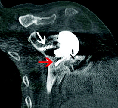
Fig. 24.4
Coronal view of CT scan demonstrating radiolucency around inferior screw and breakage of the superior screw indicating a loose glenosphere baseplate
Preoperative planning should include assessment of the fixation of the glenoid and humeral components. In revision procedures with well-fixed components, the humeral stem or glenoid baseplate can often be left in place with revision of the modular components which significantly decreases the complexity of the procedure. In situations of chronic infection or grossly malpositioned components, removal is often required. Availability of the appropriate tools for component extraction and cement removal is essential in revision procedures. The instruments we commonly use include a router tipped burr, thin osteotomes, square-tipped impactor, threaded Steinmann pins, cement removal punches and gauges, and an ultrasound cement removal device. Prior to performing revision of a RSA, the surgeon should have a full understanding of the implanted prosthesis and contact the manufacturer for specialized instruments that may be required for component removal.
Planning for subsequent reconstruction of the RSA is also important. The most challenging part of the reconstruction process is often dealing with glenoid or humeral bone deficiency. Oftentimes, bone defects can be seen on preoperative imaging studies, and a plan for bone grafting can be made prior to the procedure. However, in many cases, intraoperative fracture or bone loss during the removal of components can result in the need to deal with an unanticipated bone defect. Having available bone graft sources at the time of every revision procedure is important. Bone graft sources include autograft (proximal humerus, iliac crest, or distal clavicle) and allograft (femoral head or iliac crest) bones, depending on type and the amount of bone graft needed.
Surgical Approach
The deltopectoral approach is our preferred approach for all revision procedures. This approach allows for good exposure of the entire glenoid and proximal humerus and allows for extension to an anterolateral approach to the humerus which may be needed to perform windows for cement removal or in cases of intraoperative fracture. Assessment of prior incisions including location and healing as well as evaluation of the deltoid for dehiscence or atrophy from axillary nerve injury is important prior to proceeding with the procedure. The deltopectoral interval can often be difficult to find, and the cephalic vein may not be present due to injury during prior operations. The deltoid can often be scarred to the humerus, and carful dissection staying on the humerus laterally will avoid injury to the axillary nerve on the deep aspect of the deltoid muscle. If the subscapularis is present, it is released from the humerus along with an inferior capsular release along the medial humeral calcar. Identification of the axillary nerve and careful retraction with subperiosteal dissection with electrocautery will help prevent nerve injury. After proximal humeral exposure and soft tissue releases, the reverse shoulder prosthesis can be dislocated.
Humeral Component Removal
Removal of a well-fixed humeral component and cement mantle can be challenging, and high rates of intraoperative fracture have been reported [4]. In our experience, the best method for removal of a well-fixed humeral component involves first removing any modular components to allow access to the interface between the bone and the humeral stem. The next step is freeing the component proximally using a router tipped burr. This technique works for both cemented and uncemented components. After the stem has been circumferentially released proximally, a square-tipped impactor is placed on the medial neck of the component and the stem is dislodged from the humerus with a mallet. In components without a flange along the medial aspect, a notch can be created using a burr to allow for seating of the impactor tip. Most systems also have an extraction device with a slap hammer that can be used to remove the stem. In the majority of cases, this technique is successful; however, in some cases, the stem is unable to be extracted, and further release of the stem fixation is required utilizing a longitudinal osteotomy or window techniques (Fig. 24.5).


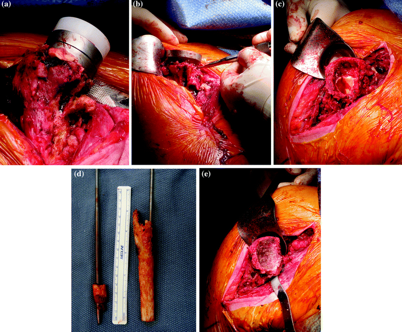
Fig. 24.5


Technique for removal of a cemented humeral stem. a Exposure of the proximal humerus and soft tissue release to allow for anterior dislocation. b Removal of modular components and use of a router tipped burr for release of proximal cement mantle. c Extraction of stem from distal cement mantle using impactor placed on the medial aspect of the component or use of an extraction device with a slap hammer from above. Note the presence of the distal cement mantle remaining in the humeral shaft. d Technique for removal of the distal cement mantle utilizing a threaded Steinmann pin through the cement mantle distally and a vice grip and slap hammer to extract the cement. The use thin osteotomes, gauges, and ultrasound cement removal instruments can also be useful. e Humerus after extraction of the cement mantle with loss of the cancellous bone of the metaphyseal region
Stay updated, free articles. Join our Telegram channel

Full access? Get Clinical Tree


