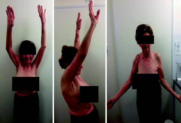
Fig. 19.1
Proximal migration of the humeral head leads to a lack of deltoid tension
In the most severe form, part or all of the deltoid muscle may be completely absent. Such permanent impairment is rare but may be observed following deltoid muscular flap transfer (for irreparable rotator cuff tears, Fig. 19.2) [26–28] or following tumor resection (Fig. 19.3).
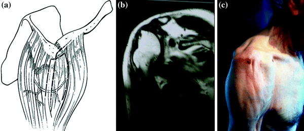
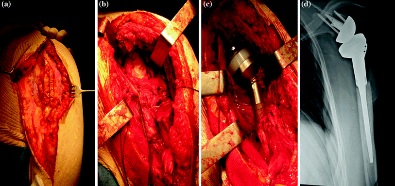

Fig. 19.2
Status after a left deltoid muscular flap transfer for irreparable rotator cuff tears. a Schematic drawing of the surgical technique (with permission of Gazielly D). b Frontal MRI demonstrates absence of the deltoid muscle laterally. c Clinical photograph demonstrating atrophy of the anterior and middle deltoid

Fig. 19.3
a Intraoperative view of a left anterior deltoid resection in the context of proximal humerus neoplasm demonstrating isolation of the anterior deltoid through which an open biopsy had previously been performed. b Resection of the entire anterior deltoid and proximal humerus. c Intraoperative view following implantation of a RSA. d Postoperative anterior–posterior radiograph
One of the most common forms of deltoid impairment seen clinically is disruption of the muscle origin (without removal of the entire muscle belly). This most commonly occurs in the postsurgical setting after an open rotator cuff repair in which a deltoid split approach is used and part of the deltoid origin is take-down to gain exposure (Fig. 19.4) [29]. Failure of the deltoid to heal back to the acromion can easily be appreciated clinically by a defect to palpation. Additionally, deltoid insertion disruption can occur through chronic attritional rupture as in chronic rotator cuff arthropathy with anterosuperior escape [30, 31] or following trauma (Fig. 19.5) [32, 33].
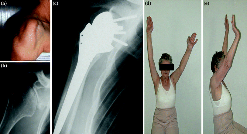
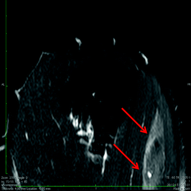

Fig. 19.4
Sequelae of a right open rotator cuff repair involving violation of the deltoid insertion. a Clinical appearance with an anterior deltoid with severe atrophy. b Anterior–posterior radiograph demonstrating rotator cuff arthropathy. c Postoperative anterior–posterior view of the RSA. d, e Coronal and sagittal views of postoperative anterior forward elevation

Fig. 19.5
Evaluation in an acute phase of left posterior deltoid insertion disruption on T2-weighted fat-saturated MRI arthrogram sagittal sequences revealed an edema (red arrows) propagating into the muscle
The deltoid muscle may be globally impaired in the setting of persistent denervation [34], grade 3 or 4 fatty infiltration [35], previous surgical approach (Fig. 19.6), trauma (Fig. 19.7), postradiation syndrome, or myopathy (myositis, Parkinson, Duchenne muscular dystrophy, etc.) [36].
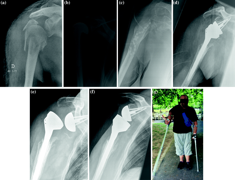
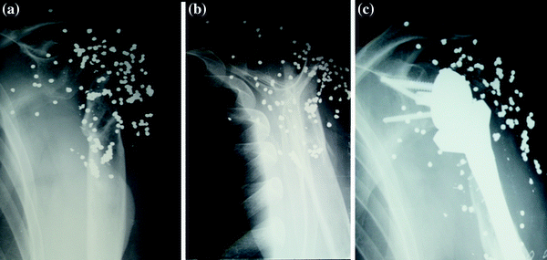

Fig. 19.6
Case illustration. A 54-year-old man with a previous bilateral below knee amputation due to diabetes mellitus sustained a motor vehicle accident. a A hemiarthroplasty had been initially implanted for a four-part fracture of the right proximal humerus. b The patient developed a deep infection that finally required removal of the prosthesis 3 years later. The patient suffered from consequent pain and was unable to walk with crutches. c A RSA was implanted a year later. d A lack of anterior structures (subscapularis, pectoralis major, conjoint tendon) led to an episode of instability. e X-ray after revision surgery with placement of a thicker polyethylene spacer. F) At one-year follow-up, the patient was able to walk with two crutches. Active anterior elevation was limited to 40°, but the patient was satisfied with the result

Fig. 19.7
Preoperative (a anterior–posterior and b lateral scapular views) and post-RSA (c anterior–posterior view) of a right shoulder after a gunshot in a patient that presented post-traumatically with global neurological impairment including the axillary nerve
Finally, in the largest series on deltoid impairment with RSA published to date, the cause of impairment remained obscure/undetermined in 3 of 49 patients (Table 19.1) [37].
Table 19.1
Etiologies of deltoid impairment in the series of Lädermann et al.
Demographics | Shoulders (N = 49) |
|---|---|
Diagnosis* | |
Trauma sequelae | 13 (27 %) |
CTA (Hamada and Fukuda 3 to 5) | 9 (18 %) |
RCT (Hamada and Fukuda 1 or 2) | 8 (16 %) |
Revision shoulder arthroplasty | 13 (27 %) |
Dislocation arthropathy | 6 (12 %) |
Reason(s) for deltoid impairment (multiple reasons possible)† | |
Post-traumatic without surgery | 4 |
Postsurgery | 25 |
Axillary nerve lesion | 9 |
Resection or muscular flap transfer | 5 |
Postradiation | 2 |
Deltoid avulsion | 3 |
Unknown | 3 |
Once the etiology is determined, the deltoid impairment should be then classified according to its location and extent. Lädermann et al. [37] proposed a classification for deltoid impairment based on location: Type 1 corresponds to an impairment localized anteriorly, type 2 is an anterior and middle one, type 3 involves only the middle deltoid, and type 4 is a global impairment. As discussed subsequently, this classification related to prognosis with type 4 in particular having a poorer function following RSA.
Results
Glanzmann et al. [27] first published a case report of the results of a RSA after deltoid muscle flap transfer. At two-year follow-up, the patient was satisfied and had a Constant score [38] of 62 points. Tay and Collin [28] described results of a RSA implanted in the setting of an irreparable spontaneous deltoid rupture. No intraoperative or postoperative complication was noticed. At two-year follow-up, the patient was pain free and had active anterior elevation of 150°, and the Constant score was 65 points. A similar report of a RSA combined with a latissimus dorsal transfer with satisfactory results has also been described [39].
In a multicentered study, Lädermann et al. reviewed 49 patients (49 shoulders) at a mean of 38 ± 30 months postoperative following RSA in the setting of deltoid impairment. The indications and etiologies are summarized in Table 19.1. Postoperative complications occurred in nine (18 %) patients, including two postoperative dislocations and two acute postoperative neurological lesions. Five (10 %) patients required additional surgery. The functional results are summarized in Table 19.2.
Table 19.2
Preoperative versus final follow-up measures
Pre-op | Post-op | Gain | P † | |
|---|---|---|---|---|
Range of motion in degree‡ | ||||
AFE | 50 ± 38 | 121 ± 40 | 64 | <0.0001 |
AER at side | 5 ± 21 | 12 ± 16 | 7 | 0.8438 |
Constant score‡ | 24.8 ± 12.1 (2–51) | 58.0 ± 16.7 (16–83) | 33.2 | <0.0001 |
Satisfaction (%) | NA | 98 % | ||
SANE N = 29‡ | NA | 70.9 ± 17.0 (10–95) | ||
Active forward elevation and Constant score improved significantly. However, these values were significantly lower for patients suffering from global deltoid impairment (type 4) compared to type 1 through 3. The mean postoperative forward elevation was lower in the setting of global deltoid impairment (70°) compared to partial impairment (127°, 136°, and 125°, groups 1–3, respectively) (P = 0.002). The postoperative Constant score was lower in the setting of global impairment (41) compared to partial impairment (57, 63, and 68, groups 1–3, respectively) (P = 0.006). Overall, the rate of patient satisfaction was 98 % at final follow-up.
More recently, Schneeberger et al. retrospectively reviewed the outcome of 19 patients treated with RSA after failed deltoid flap reconstruction [40]. They noticed a high rate of complication (37 %), including one instability. Nonetheless, at a mean follow-up of 4.5 years, only two patients had moderate to severe pain, all patients regained anterior active elevation above 90°, and 15 of 19 patients were very satisfied.
In summary, the literature demonstrates that RSA is feasible in the setting of deltoid impairment. The complication rate and technical complexity are significant as most patients have had previous surgery. However, functional improvement is attainable, particularly if the deltoid impairment is only partial.
Preoperative Planning
A meticulous examination should be performed prior to considering RSA in the setting of deltoid impairment. Inspection is performed to assess for previous scars, deltoid dehiscence, and atrophy to estimate the extent to which the deltoid is impaired. Strength testing examination includes the deltoid and rotator cuff. The subscapularis (belly press [41] and bear hug [42]), infraspinatus, and teres minor (hornblower’s maneuver) [43] are assessed. Notably, in our series of patients, the remaining deltoid had to have manual muscle strength (measured in abduction) of at least 3 out of 5 [44] in the sitting position in order to be considered a candidate for RSA. Patients with grade 2 or less are not considered candidates for RSA [45].
The imaging workup should quantify radiographic evidence of grade 3 or 4 fatty infiltration on a computed tomography (CT) scan [35] or magnetic resonance imaging (MRI) study (Fig. 19.8) [46], at the level of the infraglenoid rim according to Greiner et al. [47].
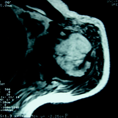

Fig. 19.8
Axial MRI of a right shoulder demonstrates grade 4 infiltration of the anterior deltoid and grade 3 of the posterior and middle deltoid at the level of the infraglenoid rim
Finally, a preoperative electrodiagnostic study is often useful in the setting of deltoid impairment. This can help determine the etiology and severity of deltoid impairment when a neurological lesion is suspected.
Surgical Approach and Implant Design
The surgeon faces three specific challenges with RSA in the setting of preoperative deltoid impairment: respect of the remaining deltoid, protection of the axillary nerve, and avoidance of postoperative instability.
Either a deltopectoral or transdeltoid approach (superior approach) [22, 48] may be used, and each has its advantages and disadvantages [49]. The situation is fairly different in case of a compromised deltoid, which will be the future motor of the shoulder. The transdeltoid approach has the advantage of obtaining better postoperative stability, in particular, because the subscapularis tendon and the anterior ligament complex are preserved. This approach may be appealing because postoperative instability is a concern in the setting of preoperative deltoid impairment. However, one must consider the potential for additional trauma to the deltoid, and consequently, we do not recommend this approach for cases with preoperative deltoid impairment. A compromise recommended by the author is to respect the deltoid by performing a deltopectoral approach, but then perform the RSA without taking the subscapularis down (subscapularis preserving technique) [50] for RSA; this effectively combines the deltopectoral and superior approaches and respects the subscapularis, one of the keys for postoperative stability [51] and postoperative function (Fig. 19.9) [4]. Additionally, since the subscapularis is respected, the patients are allowed to move freely and actively the day after the surgery (Fig. 19.10).
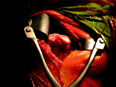

Fig. 19.9
Lateral view from a right shoulder. a Deltopectoral approach has been performed. Using a Brown retractor, the superior head then is exposed, allowing for continuation through a superior approach and respecting thus the deltoid and the intact subscapularis
