Fig. 6.1
Pigmented purpuric dermatosis (progressive pigmentary dermatosis [Schamberg disease]) occurring on the leg as reddish-brown patches with superimposed pinpoint petechiae
2.
Intermediate macular purpura (5–9 mm in diameter, flat, noninflammatory, type of “simple hemorrhage”): Hypergammaglobulinemic purpura of Waldenström and other causes of macular petechiae and macular ecchymoses.
3.
Macular ecchymoses (≥1 cm in diameter, flat, noninflammatory, type of “simple hemorrhage”): Hemophilia, anticoagulants, vitamin K deficiency, hepatic insufficiency, actinic purpura, corticosteroid therapy (topical or systemic; Fig. 6.2), vitamin C deficiency (scurvy), systemic amyloidosis, and Ehlers-Danlos syndrome.
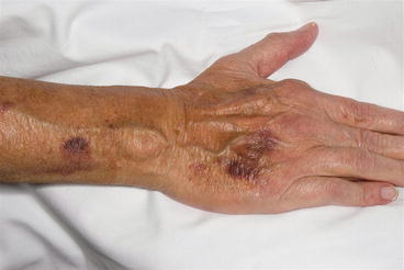
Fig. 6.2
Macular ecchymoses of the dorsal hand and forearm. These were due to long-term systemic corticosteroid therapy for rheumatoid arthritis
4.
Palpable purpura (inflammatory purpura with prominent early erythema; round, port-wine color; partially blanches with diascopy indicating the presence of both inflammation and hemorrhage): The most important consideration in this category is cutaneous small-vessel vasculitis (e.g., leukocytoclastic vasculitis; Figs. 6.3 and 6.4). ANCA-associated vasculitides and vasculitides that affect both small- and medium-sized vessels can also present with palpable purpura. Targetoid lesions with a central purpuric component can be seen with IgA vasculitis and erythema multiforme. Pityriasis lichenoides et varioliformis acuta (PLEVA) can also manifest palpable purpura.
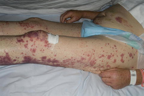
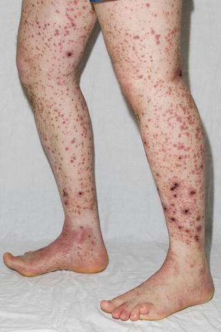
Fig. 6.3
Palpable purpura in a patient with cutaneous small-vessel vasculitis manifesting as purpuric papules and plaques on the lower extremities. Note the presence of both hemorrhage (purpura) and erythema (inflammation). Skin biopsy demonstrated leukocytoclastic vasculitis
Fig. 6.4
Palpable purpura manifesting as purpuric papules and plaques (some with overlying necrosis) on the lower extremities. Skin biopsy demonstrated leukocytoclastic vasculitis, and direct immunofluorescence microscopy of lesional skin revealed the presence of IgA within superficial dermal blood vessels. Evaluation for associated causes of this patient’s IgA vasculitis revealed the presence of renal cell carcinoma
5.
Get Clinical Tree app for offline access

Noninflammatory retiform purpura (minimal early erythema; some lesions are palpable; typically due to microvascular occlusion): Calciphylaxis (Figs. 6.5 and 6.6), heparin necrosis, warfarin necrosis, antiphospholipid antibody syndrome, cryoglobulinemia (monoclonal type only; mixed cryoglobulinemia usually presents with palpable purpura and demonstrates leukocytoclastic vasculitis on histology), hypercoagulable disorders (e.g., protein C or S deficiency), disseminated intravascular coagulation, cocaine use (levamisole-tainted), oxalosis, cholesterol emboli, livedoid vasculopathy, vessel-invasive infection (e.g., Aspergillus, ecthyma gangrenosum), malignant atrophic papulosis (Degos’ disease), and marantic endocarditis.
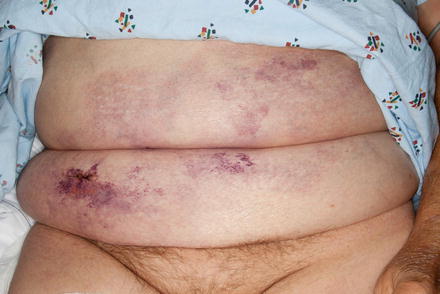
Fig. 6.5


Noninflammatory, retiform purpura manifesting as purpuric patches and indurated, exquisitely tender plaques on the abdomen. Skin biopsy demonstrated findings consistent with calciphylaxis
Related posts:

Stay updated, free articles. Join our Telegram channel
Full access? Get Clinical Tree