Fig. 14.1
Image of a patient with typical thick and white scaling psoriatic plaques
Other forms of psoriasis include guttate, inverse, seborrheic, erythrodermic, pustular and nail psoriasis.
Guttate psoriasis is an eruptive form of psoriasis commonly seen in children and young adults, classically preceded by a streptococcal throat infection. It is characterized by an eruption of dozens to hundreds of ‘drop-like’, 0.5–1 cm, well-defined psoriatic papules. It is often a self-limiting condition. However, individuals are more prone to the development of chronic psoriasis vulgaris in the future.
Inverse psoriasis is distinguished by its predilection for the intertriginous areas of the body – axilla, groin and neck. Inverse psoriasis can be found in the gluteal cleft and genital mucosa. It is characterized by well-demarcated, erythematous plaques, but they are usually non-scaly due to maceration from heat and moisture.
Sebopsoriasis is characterized by erythematous plaques and greasy, yellow scale in the scalp, nasolabial folds and presternal areas.
Erythrodermic psoriasis is an acute, generalized form of psoriasis characterized by erythema and superficial scale that covers more than 90 % of the total body surface area. Individuals are predisposed to the loss of heat and electrolytes, and this condition can be complicated by secondary infection, high-output cardiac failure and even impairment of renal and hepatic function [3].
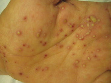
Pustular psoriasis also has several different variants that can be seen on the skin. The Von Zumbusch pattern occurs as an acute-onset, generalized, erythematous and pustular eruption. It is often accompanied by systemic symptoms, including fever, chills, headaches and malaise. The annular pattern occurs when small pustules surround an erythematous, scaly plaque of psoriasis. This type of reaction pattern is occasionally accompanied by systemic symptoms. The exanthematous pattern is an acute eruption of small pustules following a recent infection or medication exposure. Localized pustulosis of the palms and hands is also a very common psoriatic eruption characterized by dozens of small, sterile, yellow-brown pustules on the palms and soles. This condition can be both chronic and debilitating, and these individuals can also have coexistent plaques of psoriasis vulgaris as well (Fig. 14.2).


Fig. 14.2
Palm of patient with dozens of pustules and red scaly patches commonly seen in pustular and palmoplantar psoriasis
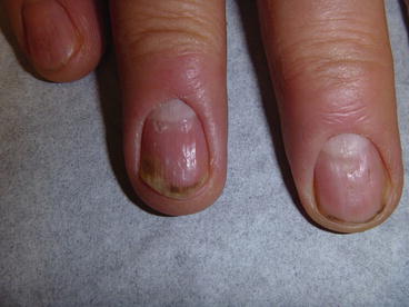
Nail changes in psoriasis are also very common and seen in up to 40 % of patients. Because it is so common, nail involvement is not an accurate predictor of the future development of psoriatic arthritis. However, the frequency of nail involvement is higher in those individuals that are older, have a longer history of psoriasis and have psoriatic arthritis. The most common nail findings include nail pitting (0.5–2 mm, asymmetric depressions in the nail plate), oil spots and salmon patches (translucent, yellow-brown discoloration beneath the nail plate), onycholysis (lifting of the nail plate) and subungual hyperkeratosis (crumbling debris underneath the distal edge of the nail plate). Other potential findings include leukonychia (a white nail plate), crumbling nails, splinter haemorrhages and anonychia (total loss of nail plate) [4] (Fig. 14.3).


