39
Proximal Phalangeal Shaft Fractures
Kevin D. Plancher
History and Clinical Presentation
A 37-year-old right hand dominant high school teacher and coach of the football team comes to the office because he got his finger caught in a football helmet during a drill. He said that he felt a crack and looked down and noticed his long finger on his right hand was pointing toward his thumb. He reports taking hold of it and pulling back straight and had the team trainer tape it to the index finger. He has no history of diabetes or vascular disease, and is presently not on any medications.
Physical Examination
The tips of all fingers have good capillary refill and the patient’s two-point sensory discrimination, as determined by the Weber two-point discrimination test, using a dull pointed eye caliper applied in the longitudinal axis of the digit without blanching the skin, is within normal limits of all fingers. The hand and fingers show no sign of vascular compromise. Fingers are noted to be swollen with no obvious deformity and no open wounds. Patient was able to actively flex and extend all other fingers but felt pain in the little finger between the metacarpophalangeal (MP) and proximal interphalangeal (PIP) joints and was limited on his motion. He was sent for radiographs.
Diagnostic Studies
Diagnostic studies include anteroposterior, splay lateral, and oblique radiographs of the hand. Oblique splay films are especially helpful in assessing intraarticular fractures and in removing the overlap from adjacent digits.
PEARLS
Interosseous Wiring Stable Fracture Patterns (Transverse)
- Treat STABLE FRACTURE PATTERN conservatively.
- When a stable fracture pattern becomes unstable it needs to be surgically stabilized.
- A K wire is utilized to stabilize the fracture after the interosseous wire is placed.
- Preserve periosteum for closure.
- Leave the twisted wire on the noncontact side of the finger. (Start wire from that side.)
- Leave at least 5 mm bone bridge from fracture line to wire.
PITFALLS
- Do not treat an unstable fracture pattern conservatively (spiral oblique).
- If you leave prominent the twisted interosseous wire there will be soft tissue irritation.
- Do not stabilize the fracture with K wire prior to tightening the stainless-steel wire when using the interosseous wire technique.
PEARLS
Screw Fixation
- Fracture edges are inspected and the optimal position for screw fixation (perpendicular) is selected.
- Anatomic reduction is ideal for maximal compression.
- Ensure the fracture fragment is at least three times the thread diameter of the screw for fixation.
- Long oblique and spiral fracture patterns may be fixed with 1.5-mm screws if the fracture is two to three times diameter of the phalanx.
- Loupe magnification is helpful when using 1.1– or 1.5-mm screws.
PITFALLS
Screw Fixation
- Failure to clean and free fracture edges of soft tissue or hematoma will not allow anatomic reduction.
- The screw head many irritate the soft tissues when prominent.
- Early range-of-motion exercises without stable, rigid fixation will cause the loss of anatomic reduction and possible malalignment or malunion.
Differential Diagnosis
Fracture of the phalanx
Intraarticular
Extraarticular
Comminuted
Tumor of proximal phalanx
Diagnosis
Extraarticular Oblique Proximal Phalanx Fracture
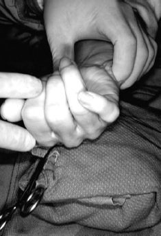
Many types of phalangeal fractures are seen, and the examining physician must note the resting posture of the injured hand in each case. When the fingers of the hand are flexed, a line should be drawn from the dorsal aspect of the PIP joint to the fingernail. This line should converge, not to a single fixed point, but rather directly to the area of the scaphoid from the radial artery to the palmaris longus. One of the most common causes of malunion is a malrotation of a phalangeal fracture, and it is diagnosed by physical examination, not by x-ray (Fig. 39–1).
Indications for operative intervention for phalangeal fractures are based on concerns about shortening or malrotation. Spiral and short oblique fractures are usually more susceptible to instability, and a few millimeters of shortening, although acceptable in the metacarpal, is not acceptable in the phalanx. Malrotation is never acceptable, and operative intervention is required for intraarticular fractures, open fractures, fractures with bone loss, and multiple phalangeal fractures that are associated with a neurovascular or tendon injury. Extensive soft tissue injury may require stable fixation to allow early mobilization and skin loss with hand fractures associated with polytrauma needs operative repair.
Many factors determine the selection of treatment, including fracture location, intraarticular or extraarticular deformity, angulation, rotation, shortening, open or closed injury, associated neurovascular and soft tissue injuries, and the nature of the fracture (i.e., transverse, spiral, comminuted, or oblique). Additional considerations for operative reduction or internal fixation include the surgeon’s skill with the fixation device, the patient’s ability to work postoperatively with an appropriate plan, and the patient’s occupation.
The operative goal is always to avoid prolonged immobilization because of the risk of permanent deformity and stiffness. However, soft tissue damage, infection, the need for a second procedure, and technique failure can occur when moving the fractures aggressively if fixation is not rigid. Operative fixation must ultimately have a better outcome than nonoperative management, and the risks and benefits must always be explained to the patient.