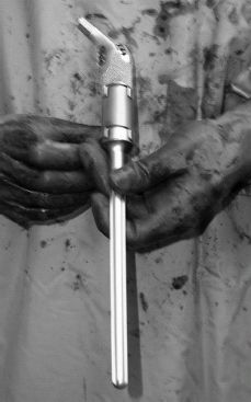
FIGURE 29-1. Acetabulum and reamed femur.

FIGURE 29-2. As the use of these megaprostheses has expanded into the realm of nonneoplastic conditions, refinements in design such as the introduction of modular prostheses that improve intraoperative flexibility and the introduction of provisions to allow for improved abductor soft tissue reattachment have ensued.
INDICATIONS
In general, all metal megaprostheses are best reserved for elderly or sedentary patients with massive quantitative or qualitative proximal bone loss. This may be due to failed total hip arthroplasty, previous periprosthetic sepsis, and recalcitrant and multiply failed nonunion of the proximal femur, or prior resection arthroplasty. In physiologically younger patients who have improved capacity to heal and in whom the restoration of bone stock for future revisions is of paramount importance, the allograft prosthetic composite is typically preferred. In either case, unless a total femoral replacement is planned, one absolute requirement is a distal length of bone that is adequate and suitable for fixation of the proximal femoral replacement.
Elderly patients with multiple medical comorbidities and the inability to follow postoperative weight bearing restrictions constitute a relative indication to consider megaprosthesis reconstruction megaprosthesis reconstruction. Usually, the surgery is less technically demanding and requires shorter anesthesia time compared with more complex reconstructive procedures for massive bone loss.3,9,10
SURGICAL TECHNIQUE
In most cases, the preferred surgical technique for the placement of a tumor prosthesis is the direct lateral approach (Hardinge). If the greater trochanteric bone is viable and substantial, a greater trochanteric slide can be used to mobilize the abductors and vastus lateralis anteriorly and expose the anterolateral aspect of the femur. If, however, massive lysis of the greater trochanter exists and it provides no biomechanical benefit, then it can be split longitudinally in line with the exposure. Meticulous soft tissue handling is important in maintaining the viability of the proximal musculature in order to optimize healing. Even in patients not suspected of being infected, intraoperative culture and frozen section are obtained in all surgical cases. As in all cases of revision total hip arthroplasty, thorough irrigation and debridement should be performed to minimize future potential third body wear by polyethylene and metal debris that may be present in and around the hip joint.
Careful preoperative planning and intraoperative assessment is essential in determining the length of the femoral component to be used and the location of the transverse femoral osteotomy. Preoperative radiographs of the contralateral limb with markers are often helpful, but may be difficult to interpret in patients with prior reconstructive procedures on the contralateral side. My preferred method is to place a Steinmann pin in the iliac crest and measure to a fixed point on the femur prior to dislocation. This distance can then be recreated, plus or minus any increase or decrease in leg length that was desired based upon preoperative limb-length discrepancy. However, because instability is a substantial risk, particularly in light of abnormal abductor muscle attachments, soft tissue tension ultimately determines what length and offset is appropriate in most cases. In some cases, a constrained liner may be necessary when soft tissue tension to gain hip stability cannot be gained while maintaining an acceptable leg length. It is important to recognize that with the abductors detached from the construct, there is little check-rein to stretching of the sciatic nerve, and therefore, care must be taken to avoid over-lengthening.
The transverse femoral osteotomy is then made in the host bone at the most proximal area of adequate circumferential quality bone as templated preoperatively and confirmed intraoperatively. Subperiosteal dissection and placement of a curved or malleable retractor is useful to protect the soft tissues and neurovascular structures prior to osteotomy with a saw. The surgeon should strive to maintain the maximum length of native femur possible because some studies have shown that outcome is related to the length of femur remaining postoperatively.9 Component removal should be carried out as described in previous chapters. The femur is prepared in standard fashion for the implant system being utilized. This typically involves reaming of the femoral canal to at least 2 mm greater than the selected stem size. If the acetabulum is not to be revised, trial components are then inserted and the stability of the hip can be examined. If acetabular bone loss and/or loosening is present and requires reconstruction, this should be performed as outlined in Chapters 9 to 18 of this text. If instability is a concern after trialing, a constrained liner may be used. During trial, the hip should generally be able to be flexed to 90 degrees and extended to 15 degrees. If this is difficult, or if the knee does not easily bend to 90 degrees, then the leg may be overlengthened and this should be carefully examined. After adequate trial and confirmation of stability and femoral anteversion, the femoral component is cemented into place. Third generation cement technique and hypotensive anesthesia, if not contraindicated, should be performed. We often use antibiotic impregnated imprepriated cement in these complex reconstructions. A cement restrictor can be used if feasible, but often the stem tip lies in the capacious distal metaphyseal region of the femur, making the restrictor of little value. If an implant with a porous section or extramedullary bridges is used, ensuring that the porous-coated portion of the stem is in contact with the diaphyseal bone with no interpositioning of cement will improve results. This junction can be optimized by the use of a reamer that matches the flair at the proximal aspect of the stem prior to cementation. A Steri-Strip or a piece of wet gel foam wrapped around the porous surface of the body on the stem can help protect this interface. Once the cement begins to cure, this can be peeled away to minimize contamination of the porous surface adjacent to the host bone with extruded cement. Protecting the porous coating over the implant at the host–implant junction will allow for extracortical bone bridging at this site. This theoretically will decrease the load transfer to the bone-cement interface of the femoral component and may improve loosening rates. Finite element analysis has confirmed that this bony bridging can decrease the stresses at the bone-cement interface of the femoral component.11 This bridging bone may also protect the bone-cement interface from exposure to polyethylene debris by isolating it from the effective joint space. Many routinely bone graft this junction.
Stay updated, free articles. Join our Telegram channel

Full access? Get Clinical Tree


