Revision total hip arthroplasty is complex and demanding. The stakes are high. The risks of intraoperative and postoperative complications are increased compared to primary total joint arthroplasty. To maximize the chances of success on the day of surgery, one must be prepared and must try to anticipate all possible surgical scenarios. A smooth operation depends on identifying the problem to be addressed and carefully crafting a surgical plan (Plan A) and contingencies (Plans B, C, D, etc.) that will be executed to solve the identified problem. This chapter discusses an approach to evaluating a patient who presents with a failed total hip arthroplasty for a revision and how one may go about planning and making decisions in preparation for the day of surgery.
PATIENT EVALUATION
Patients often have complex histories of problems relating to their total hip arthroplasty, multiple prior surgeries, and multiple prior providers. Success in revision surgery begins with the initial encounter. A careful and thorough history is critical.
Some baseline information is essential in the initial evaluation. In addition to the general aspects of the patient history, one can ask questions relating to the Harris hip score (1), as these give a uniform baseline to compare the severity of symptoms across patients. The orthopedic surgeon should also elicit details of all prior hip surgeries and information on the specific implants and bearing surfaces used. Past medical history specifically relating to diseases that may relate to bone quality or immune status is important. A history of metal allergy or venous thromboembolism should also be elicited. One should ask specifically about the original postoperative course and complications, particularly relating to wound drainage and postoperative antibiotic administration. Finally, one should note any recent trauma, hospitalizations, or infections that were temporally related to the onset of symptoms.
What Is the Chief Complaint?
To treat the patient, one must understand what has brought the patient in for evaluation. In failed total hip arthroplasty, the most common presenting complaints are pain, instability, infection, periprosthetic fracture, and metal-related adverse local tissue reaction, with or without pain.
Pain
For the painful total hip arthroplasty, it is important to obtain a history of the pain as this may suggest a diagnosis. In particular, it is important to understand the temporal onset, severity, site, character, and location of the pain. There have been a number of excellent articles describing the evaluation of a painful total hip arthroplasty (2,3). It is helpful to review these articles when faced with a diagnostic challenge. Pain can be either intrinsic to the hip or extrinsic. A list of intrinsic and extrinsic causes of hip pain is listed in Table 21-1. It is obviously important to avoid revising a total hip arthroplasty for pain that is extrinsic to the hip.
It is important to understand the temporal onset of pain. Persistent pain since surgery, new onset of pain following a pain-free interval, severe activity-related pain relieved by rest, or constant pain, including pain at rest and pain at night, all suggest different possible diagnoses (Table 21-2).
TABLE 21-1 Intrinsic and Extrinsic Causes of Hip Pain
Intrinsic Causes
Extrinsic Causes
Infection
Mechanical loosening
Tip of stem pain
Periprosthetic fracture
Occult instability
Adverse local tissue reaction
Iliopsoas tendonitis
Trochanteric bursitis
Lumbar spine disease
Peripheral vascular disease
Peripheral nerve injury or irritation
Complex regional pain syndrome
Bone metabolic disease
Malignancy or metastases
Hernia
Referred pain
Psychiatric disease
TABLE 21-2 Temporal Onset of Hip Pain and Possible Diagnoses
Persistent pain since surgery
Wrong initial diagnosis
Infection
Failure to obtain initial implant stability
Impingement or instability
New onset of pain following pain-free interval
Aseptic loosening/osteolysis-related loosening
Periprosthetic fracture
Late infection
Metal reaction
Severe activity-related pain relieved by rest
Loosening
Fracture
Iliopsoas tendonitis
Neurogenic or vascular claudication
Constant pain, pain at rest, pain at night
Infection
Malignancy
Loosening
Lumbar spine disease
TABLE 21-3 Location of Pain
Groin or deep buttock
Acetabular loosening
Iliopsoas tendonitis
Adverse metal reaction
Hernia
Inguinal lymphadenopathy
Psoas abscess
Gynecologic or genitourinary etiology
Thigh pain
Femoral component loosening
Stiffness mismatch between femoral component and bone
Pain over greater trochanter
Trochanteric bursitis
Trochanteric nonunion
Buttock pain radiating to the knee or below
Lumbar disc disease or spinal stenosis
The location of pain is also helpful in suggesting a diagnosis. It is important to identify whether the pain is primarily in the groin or deep buttock, in the thigh, or over the greater trochanter. It is also important to distinguish hip pain from other sorts of pain such as low back pain and radiating pain from the buttock to the knee and below. Table 21-3 summarizes some of the possible diagnoses that are associated with various locations of pain about the hip.
Dislocation
Instability and dislocation are also common reasons for patients to present for revision total hip arthroplasty. A careful history of the dislocations, how many, when they occurred, what the patient was doing at the time of dislocation, and how they were treated is important to understand. Generally speaking, if a patient has dislocated three times or more and has failed reasonable attempts at maintaining stability such as education, home evaluation, and bracing, many surgeons will be inclined to reoperate. If the implants are grossly malpositioned, most will reoperate sooner.
Infection
Deep infection is unfortunately also a common presenting complaint. It is important to understand the history of the symptoms, particularly the temporal onset in relation to the index surgery and any prior history of infection, either in the hip or elsewhere in the body. Fitzgerald (4) originally described three different types of periprosthetic infection, the acute fulminating infection that occurs shortly after surgery, the delayed infection that is likely related to the initial surgery but presents late due to a less virulent organism, and the late hematogenous infection that is an acute infection occurring in a previously uninfected total joint arthroplasty. Diagnosis of deep infection, particularly the delayed infection, can be challenging. The AAOS published a very helpful clinical practice guideline in 2010 on the diagnosis of periprosthetic infection (5). More recent work on synovial fluid biomarkers for infection holds the promise of very highly sensitive and specific tests for deep periprosthetic infection (6).
Periprosthetic Fracture
Periprosthetic fracture can occur in the setting of acute trauma or can happen subacutely, usually in the setting of osteolysis. Obviously in the trauma setting, the patient presents to the emergency department with a history of trauma. A patient may present to clinic, however, with a history of a sudden increase in pain, but no history of significant trauma. This may represent an osteolysis-related periprosthetic insufficiency fracture. In either case, plain radiographs are usually sufficient to confirm the diagnosis, though CT may be needed in more subtle cases, particularly those relating to osteolysis.
Metal-related Adverse Local Tissue Reaction
Though corrosion and metal reaction can happen in any total hip arthroplasty, metal-related adverse local tissue reaction should be on the differential diagnosis of anyone with a metal on metal bearing, a total hip arthroplasty with a cobalt-chrome head, or a total hip arthroplasty with a modular femoral neck. Metal-related adverse local tissue reaction, however, must be treated as a diagnosis of exclusion. Other intrinsic and extrinsic causes of hip pain must be ruled out before metal reaction is diagnosed as the cause of the patient’s problems.
Metal-related adverse local tissue reactions may or may not be painful. Even without pain, substantially elevated metal ion levels or progressive local tissue reaction as seen in metal artifact suppression MRI can be an indication for surgery. The American Academy of Orthopaedic Surgeons has recently published an information statement on current concerns with metal on metal hip arthroplasty, which includes guidelines on the evaluation and management of patients with metal on metal total hip arthroplasty (7).
Physical Examination
The physical examination of a new patient with a problematic hip replacement is also of importance. The physical examination starts with an evaluation of the patient’s gait. As the patient gets up from a seated position, look for signs of startup pain. Have the patient walk across the examination room. A Trendelenburg limp is indicative of abductor muscle weakness—a critical finding. A vaulting gait may be a sign of a leg length inequality, though spine alignment must also be evaluated to ensure that a perceived leg length inequality is not due to spinal deformity. Note whether the patient requires the use of walking aids to move about the examination room. Inability to bear weight at all suggests a periprosthetic fracture. While walking, ask the patient to point with one finger where he or she is experiencing the most pain.
Then get the patient onto the examination table. Evaluate the prior surgical incision for clues on the prior surgical approach and for signs of prior wound healing problems or infection. Note the patient’s range of motion, and evaluate the patient for patterns of pain or early impingement at this stage. Painful active range of motion, painful passive range of motion, pain with passive straight leg raising, pain or apprehension at certain extremes of range of motion, or pain with resisted hip flexion or passive hip flexion may all suggest different diagnoses (Table 21-4).
Palpate around the hip and compare the side-to-side appearance of the hip with the other hip. In patients in whom metal-associated adverse tissue reaction is suspected, one can sometimes palpate a pseudotumor or fluid collection.
Perform a neurologic exam of the leg evaluating strength and sensation, and palpate pedal pulses. Note any other important signs such as leg swelling, skin problems, and signs of venous stasis. Have the patient turn to the lateral decubitus position and palpate the area around the greater trochanter to rule out trochanteric bursitis as a cause of pain.
TABLE 21-4 Patterns of Pain with Hip Range of Motion and Possible Diagnoses
Painful active ROM or at extremes of motion
Loosening
Painful passive ROM
Occult infection
Painful passive straight leg raising
Sciatica
Pain or apprehension at certain extremes of motion
Impingement or instability
Pain with resisted hip flexion or passive extension
Iliopsoas tendonitis
Imaging
Plain radiographs are critical to evaluating a problematic total hip arthroplasty. If possible, try to obtain old x-rays of the hip in question for comparison to current films. Standard radiographs should include the low AP pelvis, which provides a view of the entire femoral component, the frog lateral of the hip, and the cross-table lateral of the hip. Each view provides important information.
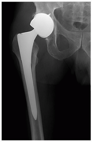
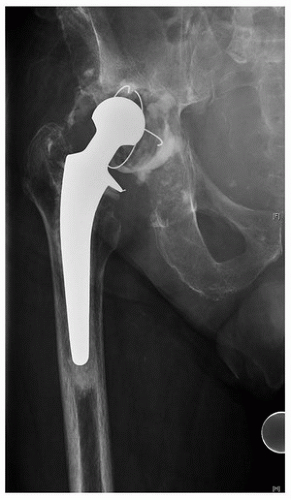
On the AP pelvis, look at the acetabular component first. Note the type of fixation used for the acetabular component and try to identify the model of the acetabular component based on its radiographic appearance. Evaluate the acetabular component abduction angle and get a sense of the acetabular anteversion (to be confirmed later on the cross-table lateral). Figure 21-1 shows the presenting radiograph of a patient who had undergone a metal on metal total hip arthroplasty and developed metal-related soft adverse local tissue reaction and hip instability due to an overabducted acetabular component. Look at the location of the hip center and note whether it is proximal or distal and medial or lateral compared to normal hip anatomy. Look at the quality of the bone supporting the acetabulum and look for osteolytic defects. If there is substantial osteolysis and the implant has migrated, the patient may have had a periprosthetic insufficiency fracture of the bone supporting the acetabular component. If concerned about pelvic discontinuity, order Judet views of the pelvis and a lateral view of the pelvis or a false-profile view of the hip, as these last views often provide a view of the posterior column without interference from the hip hardware (8,9). The lateral view of the pelvis done in the standing position can also give a sense of the natural pelvic tilt for that particular patient. Look for radiographic signs of loosening, including migration and radiolucent lines, and look for signs of fixation such as spot welds on an uncemented component. Figures 21-2 and 21-3 show dramatic examples of cemented and uncemented acetabular component migration. Figure 21-4 shows a more subtle case of acetabular component loosening associated with osteolysis and bead shedding.
FIGURE 21-1 A large head metal on metal total hip arthroplasty is shown. This radiograph demonstrates an acetabular component that was placed in excessive abduction. This patient had a large metal-related adverse local soft tissue reaction and hip instability.
FIGURE 21-2 An example of a grossly loose and migrated cemented acetabular component is shown. Note the broken cement fragments and the loss of superior bone stock. Reconstruction in this case would have been complicated by the acetabular bone loss, by the general osteopenia, and by the existing well-fixed cemented monoblock femoral component. The patient, however, was over 90 years old, had significant comorbidities, and had a reasonable quality of life. He elected not to have revision surgery.
Look at the femoral component on the low AP pelvis. Note the type of fixation used and try to identify the model of the femoral component. Evaluate the position of the femoral component in relation to the femur. This involves evaluating the center of the femoral head in relation to the tip of the trochanter. Locate the collar, or the proximal extent of the porous coating in a collarless implant, and note its relation to the calcar and lesser trochanter of the femur. Look at the quality of the bone supporting the femoral component, and look for osteolysis or a periprosthetic fracture—particularly in the area of the greater trochanter. Then, look for radiographic signs of loosening (Tables 21-5
Only gold members can continue reading. Log In or Register to continue