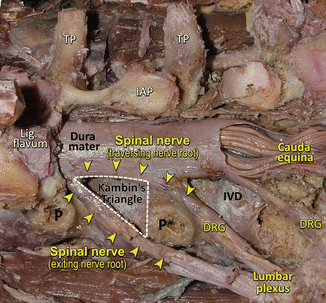
Fig. 47.1
Lumbar spine anatomy and lumbar plexus branches related to the bony anatomy. Spinal nerve and its anterior and posterior branches are demonstrated. Medial branch of the posterior ramus innervating the facet joint. FJ facet joint, SP spinous process, TP transverse process,SAP superior articular process, IAP inferior articular process
Pathologies related with lumbar and cervical regions occur more frequently, and therefore, the required surgical interventions are more frequent. During flexion or extension, changes in size and shape of the vertebral canal occur, and particularly those caused by the ligamenta flava essentially involve the central area of the spinal canal, and thus the dural sac undergoes constrictions at the intervertebral level [2].
It is possible to reach all these structures with anterior or posterior approaches. Although anterior approaches are preferred in procedures regarding infections, fractures, and tumors, posterior approaches are used in pathologies related to the spinal cord and intervertebral discs.
Clinical Relevance
Anatomical relations of the intervertebral disc are important in the planning of interventional investigative and therapeutic procedures ranging from diskography to open disc surgery.
Posterior Approaches to the Spine
Posterior approaches allow us to reach facet joint, lamina, pedicle, spinous process, cauda equina, and spinal cord. Posterior approaches were frequently used in procedures like spinal disc herniations, spinal nerve exploration, and tumor resections.
Additionally, spinous processes were used in deciding the localization of incision line. The line connects the iliac crests, which can be easily palpated, and passes between L4 and L5. Besides this rough method, the precise level can be determined by using needle-guided radiographic evaluation. Planned incision was made longitudinally over the spinous processes after the identification of the line. The length of the incision varies according to the severity of that particular pathology.
Related Anatomical Structures During Dissection via Posterior Approach
In this stage, the skin and the subcutaneous tissue were removed. Then, paraspinal muscles were reached using a dissector, and en block removal of these muscles was performed to the sides. If necessary, the incision can be extended laterally. This maneuver makes the facet joint, spinous, transverse, and the mammillary processes visible.
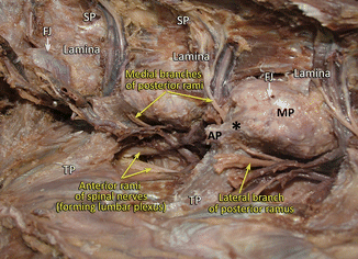
It should be taken into consideration that the nerves and vessels supplying the paraspinal muscles are between the transverse processes and the facet joints (Fig. 47.2). The blood vessels supplying this area aroused directly from the aorta. Therefore, it is essential to protect these branches of the lumbar vessels which may cause severe bleeding. Additionally, the posterior branches of these nerves [3] are located in this area to innervate the local muscles (Fig. 47.2). Damage to these nerves causes denervation and atrophy of paraspinal muscles.


Fig. 47.2
Posterolateral view to the left side of the lumbar spine. Facet joints, laminae spinous processes are demonstrated. Anterior and posterior rami of the spinal nerve and medial and lateral branches of posterior rami are seen. TP transverse process, MP mamillary process, TP transverse process, AP accessory process, FJ facet joint, * mamilloaccessory ligament
All parts of the yellow ligament (ligamentum flavum) were elevated in the profound dissection in order to reach the epidural space and the dura mater. Meanwhile, blunt dissection must be performed and extra caution should be taken.
Each nerve root must be identified individually and should be protected. In general, spinal nerves can be identified and retracted easily from its distal approach. The venous structures surrounding the peripheral nerves may cause gross bleeding throughout the dissection; therefore, they should be handled with great care. The dissections should be done gently in the inferior lumbar region due to the fact that the iliac vessels located and lying on the anterior aspect of the vertebral bodies and nerve roots may be injured.
To gain better exposure, laminas and facet joints need to be removed. Enlarging the approach provides better access for the fusion of the facet joint and the transverse process.
All these approaches can be applied for all levels from C1 to the sacrum.
Surgical Anatomical Relations
The superficial layer consists of the latissimus dorsi, which contributes a part of the posterior wall of the axilla and extends until the humerus.
Deep layers contain paraspinal muscles. During this approach, these muscles are divided into two layers in which erector spinae muscles are located superficially, and rotatores and multifidus muscles are located deeply. Although this arrangement is not obvious in surgical procedures as the approach involves detaching all these muscles together, it should be kept in mind.
Posterior superior iliac spine (PSIS) and iliac crest make a 45° angle toward the midline. This area can be easily palpated as it is the bonding surface of the muscles to the iliac crest. PSIS passes through the second segment of the sacrum. Additionally, the top of the iliac crests marks the level of disc found between L4 and L5.
Superficial Anatomical Structures: Related with Surgical Dissection
Thoracolumbar fascia and supraspinous ligament were found immediately underneath the skin. This fascia is a broad, relatively thick, wide sheath of tissue and attaches to the spinous processes. Supraspinous ligament extends to the cervical region and continues as a nuchal ligament toward the neck. Thoracolumbar fascia, medially, was attached to the iliac crest. Laterally, it continues on the latissimus dorsi and erector spinae muscles. Supraspinous ligament merges and continues with the thoracolumbar fascia. It was anatomically reported that paraspinal muscle contraction leads to posterior displacement of the posterior layer of the thoracolumbar fascia and that dysfunctional paraspinal muscles would reduce posterior displacement [4].
Arterial supply of the muscles found in this area is also crucial. These segmental lumbar arteries arise directly from the aorta and ascend close to the pedicle, where they divide into two branches. The first branch supplies the spinal cord and the other supplies the muscles around. These arteries pass between the transverse processes and are visible around facet joints. Therefore, dissections close to the midline can be performed without the risk of cutting the arteries. Injury to the lumbar arteries during anterior spinal approaches is often encountered mostly during anterior approaches, and an anatomic relationship of the lumbar segmental arteries is important [5].
Profound Anatomical Structures Related with Surgical Dissection
Yellow ligament (ligamentum flavum in all levels) is the most important structure in the profound layer. These ligaments, consisting of yellow elastic tissue, posteriorly covers the spinal canal between the laminas. This arrangement allows yellow ligament to be attached to the anterior surface of the superior lamina and the posterior surface of the inferior lamina [6]. The two yellow ligaments from each side meet in the midline but do not fuse.
It is important to remove yellow ligament with caution due to its proximity to the dura mater. The endangered major structure in the profound dissections involves dura mater. Hence, in this stage, to protect the dura mater, a thin spatula should be placed beneath the yellow ligament. Additionally, perforations of dura mater can occur easily. Finally, it could be so difficult to distinguish spinal nerves when epidural veins start bleeding.
Anatomy of the Intervertebral Foramen and Its Clinical Relevance
Intervertebral foramen (IF) is a pass between the spinal canal and the periphery and serves as a doorway [7]. The spinal roots, nerves, and ganglia may be damaged here. This large osseous hole or canal is a unique pass in comparison to other foramens of the body due to the presence of two movable joints that attach to its boundaries, where anteriorly lies the ventral intervertebral joint and dorsally lies the zygapophysial joint. There is a small notch in the upper edge of the pedicle and a larger notch in the lower edge. Close distance of these joints increased the susceptibility of narrowing from arthritic pathological and structural alterations [8]. Additionally, IF gains a dynamic structure due to the joints and attached ligaments, and it is not limited by bony structures. Its mobility in the main part changes its diameter and structure. Narrowing of the foramen and all these dynamic changes in morphology can be tolerated easily by passing the neurovascular structures through this canal [7]. One of the functions of the ligaments is to serve as protection elements in preventing injury to the passing neurovascular structures.
Despite numerous studies about IF, there is not a consensus on its anatomical description, and still, agreement has not been made on which classification best describes this area. Some studies describe IF as the exit or the lateral side of the spinal canal, whereas others describe it differently as a complete anatomical canal with real borders. Lee et al. examined IF or intervertebral canal in 1988 by dividing this canal into three sections, as lateral recess zone, midzone, and exit zone [9]. Previously, Crock et al. described IF in 1981 differently, as the narrowest part of the spinal canal [10].
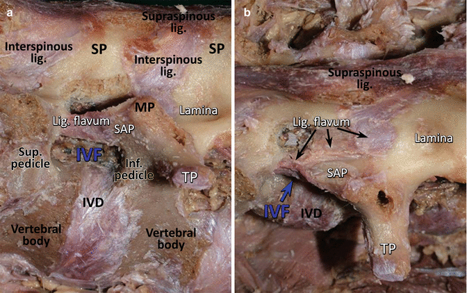
In vitro IF was observed oval and round, and was described to have an inverted teardrop-shaped window when examined medially through the spinal canal and looking outward through the intervertebral foramen [11]. The superior limit of the IF is the inferior aspect of the upper pedicle and the inferior limit of that is the superior aspect of the lower pedicle. As shown in Fig. 47.3, the floor of IF (namely, the anterior border) from above downward is formed by the posteroinferior margin of the superior vertebral body [10], the intervertebral disc, and the posterosuperior margin of the inferior vertebral body. The roof of the intervertebral foramen is bounded by the inferior, removed from this area, and superior articular processes of the adjacent vertebrae [7] at the same level as the foramen, and the lateral prolongation of the ligamentum flavum [10]. Multiple structures bound the anterior aspect of the foramen. These are posterior aspect of the adjacent vertebral bodies, the intervertebral disc, and lateral expansion of the posterior longitudinal ligament; here, additionally located structure is the anterior longitudinal venous sinus. The superior base (posterior border) is formed by the superior and inferior articular processes of the facet joint (zygapophysialjoint) at the same level as the foramen, and by the free lateral side prolongation of the ligamentum flavum. The medial canal border contains the dural sleeve. Fascial layer and the neighboring structure is psoas muscle, and it forms its lateral boundary [7]. In addition to these structures, the superior and the inferior borders are defined by the pedicles of the superior and the inferior vertebrae, respectively. The medial border is defined by the dural sac, and the lateral border is defined by the psoas muscle and the medial fascial sheath of this muscle [7, 13].


Fig. 47.3
The anterior and posterior boundaries of intervertebral foramen. (a) The floor (namely anterior border) of the intervertebral foramen is formed by the posteroinferior margin of the superior vertebral body, the intervertebral disc, and the posterosuperior margin of the inferior vertebral body. The roof of the intervertebral foramen is bounded by the inferior and superior articular processes of the adjacent vertebrae and the lateral prolongation of the ligamentum flavum. (b) The inferior articular process was removed from this area in this specimen. Lig. flavum ligamentum flavum, IVF intervertebral foramen, Inf. pedicle inferior pedicle, Sup. pedicle superior pedicle, SAP superior articular process, IVD intervertebral disc, Corpus body of vertebra, SP spinous process, Lig. flavum yellow ligament, Lamina lamina arcus vertebra, Supraspinous lig. supraspinous ligament, Interspinous lig. interspinous ligament, MP mamillary process, TP transverse process
The most popular and accepted opinion about true anatomic nerve root canal is that it initially arises from the lateral aspect of the dural sac and travels through the intervertebral canal or neural foramen. At each level, two to six small anterior and posterior nerve rootlets (fila radicularia) converge in the thecal sac to form anterior and posterior roots [12]. Clinically, it is important to know that the nerve roots regularly exit the thecal sac approximately one segmental level above their respective foraminal canals in the lumbar spine region [25]. The anatomic relation between the nerve roots and their corresponding discs in the inervertebral foramina depends on the spinal level. Therefore, for the cervical region of the spine, IF’s relative structures are different from other regions. Tanaka et al. found that the shape of the intervertebral foramen is like a funnel. The entrance zone is a narrow part, and the shape of the radicular sheath is conical, with its takeoff points from the central dural sac being the largest part [15]. Consequently, nerve root compression occurs mostly in the entrance zone of the intervertebral foramina. One reason is the equal depth of its superior and inferior vertebral notches. Also, these notches face anterolaterally, which is the same as the pedicles. Finally, the transverse foramen makes up its unique structure [14]. The cervical dorsal root ganglions almost fill the cervical intervertebral foramens. For these anatomic reasons, it seems reasonable to suggest that cervical radiculopathy is strongly correlated with both the cross-sectional area and shape of the intervertebral foramen. Additionally, the general trend of the foraminal height and width increased from the cephalad to caudal except at C2–C3. In addition, in the cervical region, the anterior border of the IF is also defined by the uncovertebral joint [15]. During the procedure just lateral to the uncinate process, the vertebral artery is defined. The vertebral artery pulsation is often visible lateral to the uncinate process. Anteriorly, compression of the nerve roots is caused by protruding discs and osteophytes of the uncovertebral region, whereas the superior articular process, the ligamentum flavum, and the periradicular fibrous tissues affect the nerve posteriorly. IF usually faces the lateral side, and the transverse processes are positioned more posteriorly in the thoracic and lumbar regions. In the thoracic region (T1–T10), the anteroinferior border of IF is surrounded by the synovial space and with intraarticular ligament (intraarticular ligament of the head of rib), which connects the crest of the head of rib and the intervertebral disc. Approaches as endoscopic posterolateral thoracic microdiscectomy under fluoroscopic guidance is between the pedicle and rib head line to be into the safety zone for discectomy. After the removal of the intercostal muscles in this area, we can find the intercostal vein, artery, and nerve. Parietal pleura is located under these structures, and the route passes through this area. Surgeon must avoid causing trauma to the lung, the rib heads, the pedicles, the intercostal nerves and arteries, and the spinal cord. Throughout this route, a probe placed too close to the midline may cause neurologic injury. If a probe is placed too laterally, pneumothorax may occur.
Many studies about spinal ligaments indicated an increased presence of ligaments in the fifth lumbar foramen. The outer opening of intervertebral foramens usually is covered with ligaments, which are formed by the contribution of the medial fascia of psoas major muscle in the lumbar region. The passages found between these ligaments allow the passage of spinal nerve branches and segmental vascular structures [13].
In a study conducted on bones, the foraminal height was determined to be between 19.14 mm (L5–S2 foramen) and 21.06 mm (L3–L4 foramen) [11]. In this study, the sagittal diameter was determined to be changing between 8.02 mm (L3–L4 foramen) and 8.41 mm (L1–L2 foramen) along the sagittal line (the area which meets the spinal nerve or the spinal ganglion), which passes through the inferior border of the superior pedicle and which limits the IF. The narrowest sagittal diameter of IF’s minimum distance is between 5.13 mm (L4–L5 foramen) and 6.58 mm (L1–L2 foramen), and it has been shown that this diameter is narrower at the lower lumbar levels compared to the upper lumbar levels. Experimentally, it has been shown that the removal of the intervertebral disc or the reduction of intervertebral disc space decreases the vertical diameter of IF significantly and does not change the sagittal diameter [16]. There is a direct relationship between the height of the intervertebral foramen (vertical diameter) and the height of the intervertebral disc. Age-related disc degeneration and reduction in disc height cause reduction in the height of this pass, thus narrowing this area [15, 16]. Disc space is the major and the determining factor in vertical diameter and has no effect on the sagittal diameter. The sagittal diameter of IF is directly related to the width of the spinal canal and the length of the pedicle [17]. In cadaver studies, it has been shown that the height of IF, which has an inverted drop shape, in a lumbar region was between 11 and 12 mm, and a cross-sectional area was measured between 40 and 160 mm2 [18]. In healthy subjects with MR imaging, this distance was determined to be 17.1 ± 2 mm in L1–L2, 18. 4 ± 1.7 mm in L2–L3 level, 18.1 ± 1.5 mm in L3–L4 level, and 17.1 ± 3.6 mm in L4–L5 level [17]. The shortest distance between the disc and nerve root was L1–L2 (by mean, 8.2 mm), and the greatest distance was L3–L4 (mean, 10.5 mm). In the midline, the disc–root distance decreased from L1–2 to L5–S1 [19]. It was found that the distance consistently increased from L1–L2 to L5–S1 for distances between an intersection point between the medial edge of the nerve root and the superior edge of the disc and lateral line of the foramen. At point at which the nerve root crosses the shortest distance from nerve root to the lateral border of the foramen, where disc was at level L1–L2 (mean, 2.6 mm) and the biggest distance, L5-S1 (mean, 8.8 mm). The width of the foramina progressively increased in a craniocaudal direction as in L1–2 was 8.3 to L5-S1 was reported 17.8 mm [19]. Again, the mean height of the foramina was about 19.3–21.5 mm for all disc levels [19].
Spinal Nerve Canal
The true anatomic nerve root canal initially begins from the lateral aspect of the dural sac and continues toward IF (Fig. 47.4). The actual anatomical spinal never canal is a tube that starts from the lateral surface of the dural sac. At each level, anywhere from two to six anterior and posterior rootlets converge in the dural sac to form anterior and posterior roots. These roots merge to form the spinal nerves [12]. Damage of neural structures occurs in the vertebral and “root” canals and at the intervertebral foramina. The spinal nerve is wrapped with a sheath made out of dura mater when it exits the dural sac. For lumbar segments level, the spinal nerve is wrapped with a sheath. Then, it passes through the IF and extends downward and then outward on an oblique route. This route changes angle slightly depending on the lumbar level. This angle becomes more right-angled in the upper lumbar segments and becomes narrower and oblique in the lower segments of the lumbar spine. The intraspinal section of the spinal nerve is slightly shorter as it exits the dural sac with a more perpendicular angle at the upper segments. In fact, the spinal nerve root directly enters IF as it immediately exits the dural sac in the upper lumbar segments due to the medial position of the thecal sac to the pedicle [9]. The diameter of the dural sac narrows as it goes distally from L3, and the spinal nerve exiting the sac gains a more oblique angle as it reaches IF. In MR studies, spinal nerve root at L1 level was shown to have a larger angle while exiting the thecal sac and a smaller angle while exiting the thecal sac at S1 level [20].
 < div class='tao-gold-member'>


Only gold members can continue reading. Log In or Register to continue
Stay updated, free articles. Join our Telegram channel

Full access? Get Clinical Tree


