Physiotherapy in thoracic surgery


Physiotherapy in thoracic surgery


Anne Dyson and Kelly L. Youd


Anatomy of the thorax


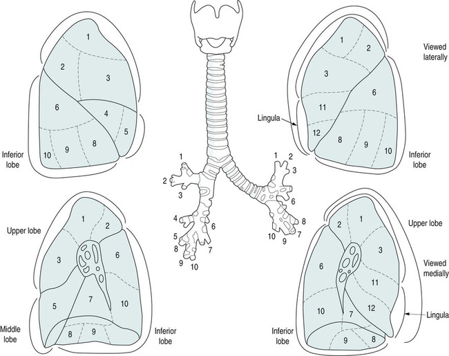
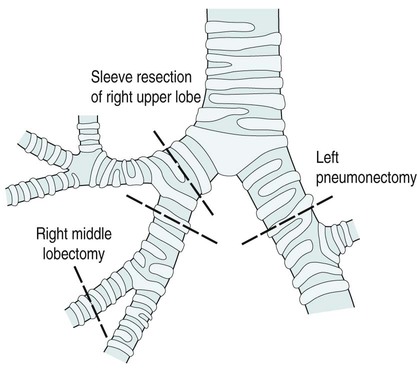
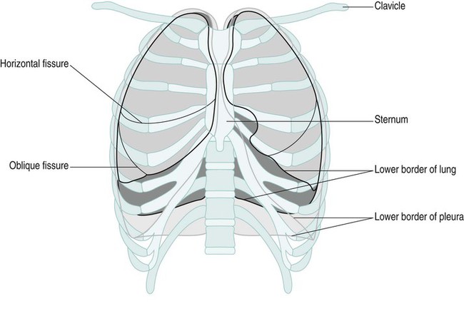
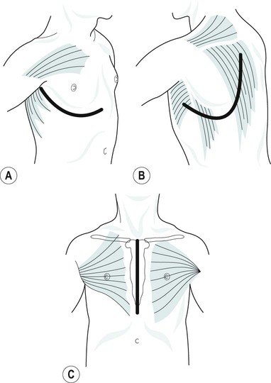
The skeleton of the thorax is an osteocartilagenous framework within which lie the principal organs of respiration, the heart, major blood vessels, and the oesophagus. It is conical in shape, narrow apically, broad at its base and longer posteriorly. The bony structure consists of 12 thoracic vertebrae, 12 pairs of ribs and the sternum (Figure 9.1).


The musculature of the thoracic cage is in two layers. The outer layer consists of latissimus dorsi and trapezius, the inner layer of the rhomboids and serratus anterior muscles. Anteriorly, the chest wall is covered by pectoralis major and minor. The intercostal muscles run obliquely between the ribs. The diaphragm forms the lower border of the thorax. It is convex upwards showing two cupolae, the right being slightly higher than the left. It is made up of muscle fibres peripherally and is tendinous centrally.


The lungs


The two lungs are basically very similar (Figure 9.2). The right lung is made up of three lobes and the left of two lobes. The lingular segment of the left lung corresponds to the middle lobe on the right. Each lobe is divided into segments.


The thoracic cage is lined by the pleura. There are two layers, the parietal and visceral, which are continuous with each other and enclose the pleural space. The parietal pleura is the outer layer and lines the thoracic cavity. The visceral pleura covers the surface of the lung, entering into the fissures and covering the interlobar surfaces. The two layers are lubricated by a thin layer of pleural fluid lying within the pleural space, which, in healthy individuals, contains no other structure.


Thoracic surgery


Indications for surgery


Tumour


The most common reason for pulmonary and oesophageal resection is a malignant tumour (carcinoma). A small percentage of tumours can be benign.


Lung cancers are classified into two main categories (NICE 2005):


Non-small-cell tumours are treated by resection if possible, if the tumour can be safely removed with clear margins and if metastatic disease is not in evidence. Small-cell cancer is virtually always widespread at diagnosis, so surgery is usually not an option.


Malignant tumours of the oesophagus are generally adenocarcinoma, especially in the lower end. They may have arisen in the cardia of the stomach and spread proximally. In the middle and upper oesophagus, squamous carcinomas predominate.


Benign tumours of the oesophagus and lungs are rare.


Pneumothorax


This is a collection of air in the pleural cavity. It usually occurs spontaneously and is caused by rupture of the visceral pleura of an otherwise healthy lung. This is more common in men than women and more usual in those under 40 years of age.


Patients with chronic obstructive pulmonary disease (COPD) can rupture a bulla resulting in a pneumothorax. Other, much rarer, causes include tumour, abscess and tuberculosis (TB). Traumatic pneumothoraces can occur with blunt trauma to the chest wall, such as following a car accident or heavy fall, or from a penetrating chest wound, i.e. a stab or gunshot wound. Iatrogenic (medical in origin) pneumothoraces can occur following intravenous line insertion, after pacemaker insertion or in ventilated patients on high levels of positive end expiratory pressure (PEEP).


Pre-operative investigations


Patients are assessed pre-operatively in order to establish the nature of the lesion and whether they are fit for surgery. The following investigations are commonly done.


Types of thoracic incision


Posterolateral thoracotomy


This incision is most commonly used for operations on the lung (Figure 9.3). It is a curved incision that starts at the level of the third thoracic vertebra and follows the vertebral border of the scapula and the line of the rib extending forward to the anterior angle or costal margin. An incision through the bed of the fifth or sixth rib is used for pneumonectomy or lobectomy.


The muscles involved are the trapezius, latissimus dorsi, rhomboids, serratus anterior and the corresponding intercostal. A small piece of rib, approximately 1 cm, may be removed to allow easier retraction and avoid a painful fracture.


Operations on the lung


Figure 9.5 shows resection margins in lung surgery.


Complications of pulmonary surgery


The major complications of pulmonary surgery are listed in Table 9.1.


Stay updated, free articles. Join our Telegram channel

Jan 7, 2017 | Posted by in PHYSICAL MEDICINE & REHABILITATION | Comments Off on Physiotherapy in thoracic surgery

Full access? Get Clinical Tree

Get Clinical Tree app for offline access