Chen et al. investigated the clinical value of physical therapy for postoperative outcome after TKR [8]. They assigned patients to one of three physical therapy groups:
1.
Start within 2 weeks after surgery
2.
Start after 2 weeks after surgery
3.
No-rehabilitation group
The patients in the no-rehabilitation group and group 2 had higher incidences of periprosthetic infection and deep venous thrombosis than those in group 1. It appears that the timing of rehabilitation may be a factor affecting TKR complications and promoting high levels of medical service utilisation. These findings could be useful for clinicians and health policymakers attempting to improve TKR services.
The question of how much physical therapy represents the optimum for each patient, however, remains unanswered. According to the theory of “the envelope of function”, tissue homeostasis needs to be restored or maintained to turn the knee joint into a “happy joint” [11].
In the case of TKR, this means that the knee needs sufficient time to heal from surgery as well as from daily exercises. Only then does it allow adequate soft and bony tissue remodeling. In a perfect sense, physical therapy should represent a positive trigger. The physical therapy needs to be individually tailored to the soft tissue condition of each patient.
It is the responsibility of both the orthopaedic surgeon and the physical therapist to identify problems with extension or flexion or persistent pain early after TKR. Only then can these be addressed in the early phase to prevent chronic problems like pain or range-of-motion deficits.
It is the responsibility of both the orthopaedic surgeon and the physical therapist to identify problems with extension or flexion or persistent pain early after TKR. Only then can these be addressed in the early phase to prevent chronic problems like pain or range-of-motion deficits.
For example, the patient should have more than 90° of flexion and full extension at discharge from the hospital. If there is a lack of normally expected improvement in ROM, the reason needs to first be identified and then either modification of physical therapy or pain management is required. Sometimes manipulation under anaesthesia might be considered at the early stage.
39.4.1 Treatment Recommendations for the Painful Knee After TKR
Identification of the cause(s) for pain after TKR is of paramount importance. This is critical before an effective treatment plan can be employed. In patients with clear mechanical problems or infection after TKR, the treatment is surgical. In these patients with clear causes of the pain after TKR, physical therapy will not permanently change the symptoms. No miracles can be expected. There is, however, a group of patients with more functional problems, with no clear cause of their problems or a low probability of improvement by revision surgery who should be treated nonsurgically. Physical therapy remains a cornerstone for the treatment here. It also represents the primary treatment in most of the patients with problems after TKR for at least two different reasons. Firstly, it is not rare that the physical therapist helps to establish the cause of the problem while performing a detailed functional assessment. Secondly, it is a less invasive treatment modality and includes a variety of different methods such as manual therapy, electrotherapy, cryotherapy and muscle strengthening exercises.
Identification of the cause(s) for pain after TKR is of paramount importance. This is critical before an effective treatment plan can be employed. In patients with clear mechanical problems or infection after TKR, the treatment is surgical. In these patients with clear causes of the pain after TKR, physical therapy will not permanently change the symptoms. There is, however, a group of patients with more functional problems, with no clear cause of their problems or a low probability of improvement by revision surgery who should be treated nonsurgically.
39.4.2 The Burning Question Remains: What Are Good Indications for Physical Therapy in Unhappy Patients After TKR?
The physical therapist should make a choice of the right interventions with the goal of preventing complications related to “over treatment”. Up to five percent of patients undergoing TKR will develop severe postoperative motion loss that does not respond to the normal standard of care [5, 13]. In most of the cases, early signs are already present during the hospital stay. Range of motion should normally be 0–110° by weeks 6–8 postoperatively. Physical therapists should contact the orthopaedic surgeon if there is no further weekly improvement in range of motion. A “yellow flag” situation is present when knee flexion is less than 90° after 4–6 weeks. Then a more aggressive physical therapy in combination with sufficient pain management is necessary. This is the case when the patient lacks motivation or shows low self-efficacy. In the situation when the patient’s ROM decreases due to swelling and pain, a too aggressive therapy might have been administered. In these cases, physical therapy should be changed from more stressing range-of-motion exercises to more relaxing and calming physical therapy options. Diabetes mellitus and the type of implant are negative predictors for limitation in range of motion after manipulation under anaesthesia MUA [2].
If physical therapy fails to improve knee motion, MUA might be considered. MUA can be performed before or after 30 days after surgery [27]. The entire spectrum of physiotherapy needs to be tried before MUA is considered.
However, the interval between TKR and MUA seems to correlate inversely with the ROM after MUA. Therefore, manipulation should be performed within the first 4–6 weeks after index surgery.
In our opinion, physical therapy in patients with pain and problems after TKR should consist of the following.
Training dosage is critical to achieve an optimal balance between improving knee function and preventing a further increase in pain that subsequently will lead to even further reduced knee function. As a general rule, patients should be informed about the following guidelines in regard to training dosage. “Exercise smart but not hard” should be the guideline to be adopted and followed [30]:
Pain should never be visual analog scale (VAS) >5 during and after exercise.
Pain should subside <2 h after cessation of an exercise session.
Swelling of the leg at the end of the day should return to minimal levels by the next morning.
Exercise should be performed three times a day and gradually increased to an hourly exercise regimen.
These guidelines need to be tailored to each patient on an individual basis. Never use these guidelines as a “cookbook”; every patient’s recipe needs a different mixture of spices.
39.4.3 Manual Lymph Drainage Therapy
Manual lymph drainage therapy (MLD) is helpful to treat postsurgical oedema of the lower leg. The inclusion of MLD as part of the postoperative rehabilitation pathway following TKR has immediate benefits to swelling and knee motion, arguably the two most important factors that affect early recovery. It has a sympathicolytic, analgetic and muscle tonus decreasing effect. MLD should be performed in the early postoperative phase and has been shown to be effective in improving flexion up to 6 weeks after TKR when compared to conventional care [12] (Figs. 39.1 and 39.2).
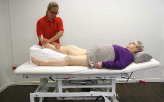
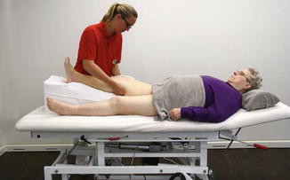
Fig. 39.1
Manual lymph drainage therapy. Elevation of the leg supports lymph drainage
Fig. 39.2
Manual therapy and relaxation of the popliteus region
39.4.4 Continuous Passive Motion (CPM)
CPM combined with physical therapy may offer beneficial results compared to physical therapy alone in the short-term rehabilitation following TKR (Fig. 39.3) [25]. Although there is a lack of strong evidence, a prolonged use of CPM slightly improved short-term ROM in patients with limited ROM at the time of discharge after TKR when added to a semi-standard PT program [20]. CPM may be most effective in a subset of patients with very stiff or painful knees. The frequency and duration of ROM exercises should be tailored to the individual tolerance of the patient, keeping in mind that we want to move forward in terms of ROM and reduction of pain [11]. The Cochrane analysis showed that CPM in combination with PT significantly improves active knee flexion and decreases the length of hospital stay and also the risk of post-surgical MUA [15].
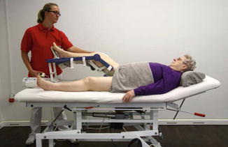
Fig. 39.3
Continuous passive motion. The patient’s position needs to be checked and the splint needs to be adjusted in order to make sure that the joint of the splint and the knee is at the same level
39.4.5 Cycling Exercises
Cycling is an excellent activity to help regain muscle strength and knee mobility (Fig. 39.4). The seat height should be adjusted so that at the bottom of the pedal stroke, the patient’s foot barely touches the pedal. Initially, backward pedaling is easier compared to forward pedaling. Forward pedaling should only be started if a comfortable backward cycling motion is achieved. At the beginning, cycling should last no longer than 5–10 min twice a day, which then can be gradually increased to 20–30 min on a daily basis. Patients should start cycling with an adjusted seat height and short crank to allow for repetitive motion without exceeding the pain level VAS pain >5. If tolerated, the patient can progress to cycling with a normal crank setting.
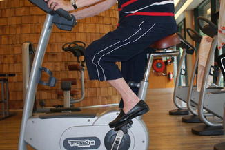
Fig. 39.4
Cycling with adapted sitting position as training for the knee-stabilising muscles
39.4.5.1 Joint Mobilisation
Joint mobilisation should be frequently performed and based on the concept “train smart, not hard”. Exercises should be done with low intensity but with high frequency (up to every hour).
The patient should begin patellar mobilisation techniques as early as possible after wound healing. Patella mobilisation is performed in a mediolateral (Fig. 39.5) and craniocaudal direction (Fig. 39.6).
Fig. 39.5
Right knee. Patella mobilization in the mediolateral direction
Fig. 39.6
Right knee. Patella mobilization in the cranial caudal direction.
Knee flexion is improved actively and passively while the patient is either supine (Figs. 39.7 and 39.8) or in a sitting position (Figs. 39.9, 39.10 and 39.11).
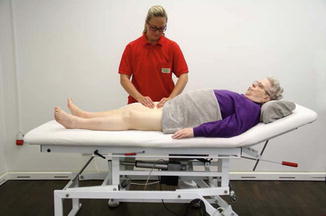
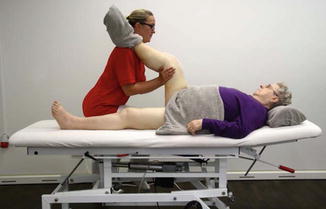
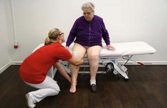
Fig. 39.7
Manual mobilisation using transverse friction technique to the rectus muscle
Fig. 39.8
Manual stretching aiming for improved extension
Fig. 39.9
 Diagnosis of Periprosthetic Joint Infection After Total Knee Replacement
How Can Preoperative Planning Prevent Occurrence of a Painful Total Knee Replacement?
Causes and Diagnosis of Aseptic Loosening After Total Knee Replacement
Discussion to Chap. 16: Mid-Flexion Instability After TKR Due to Femoral Malrotation
Specific Orthopaedic Imaging Analysis Software: Clinical Benefit for TKR Revision Surgeon
Constrained Condylar Total Knee Replacement
Diagnosis of Periprosthetic Joint Infection After Total Knee Replacement
How Can Preoperative Planning Prevent Occurrence of a Painful Total Knee Replacement?
Causes and Diagnosis of Aseptic Loosening After Total Knee Replacement
Discussion to Chap. 16: Mid-Flexion Instability After TKR Due to Femoral Malrotation
Specific Orthopaedic Imaging Analysis Software: Clinical Benefit for TKR Revision Surgeon
Constrained Condylar Total Knee Replacement


Training of knee flexion in sitting position
Related posts:
Diagnosis of Periprosthetic Joint Infection After Total Knee Replacement
How Can Preoperative Planning Prevent Occurrence of a Painful Total Knee Replacement?
Causes and Diagnosis of Aseptic Loosening After Total Knee Replacement
Discussion to Chap. 16: Mid-Flexion Instability After TKR Due to Femoral Malrotation
Specific Orthopaedic Imaging Analysis Software: Clinical Benefit for TKR Revision Surgeon
Constrained Condylar Total Knee Replacement
Stay updated, free articles. Join our Telegram channel
Full access? Get Clinical Tree
