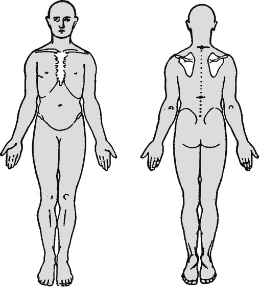
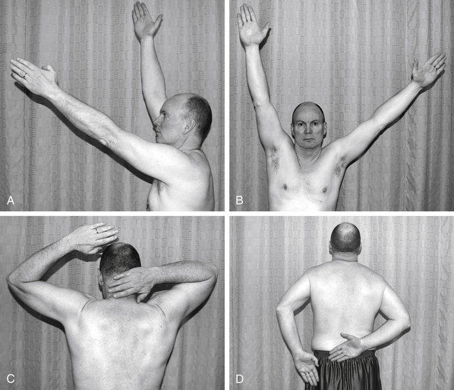
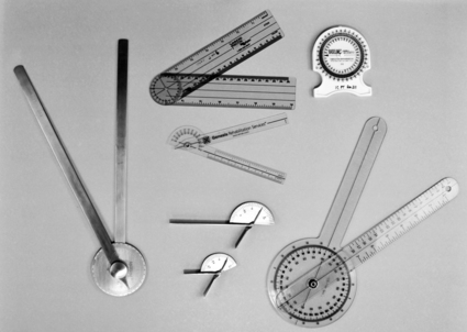
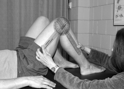
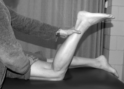
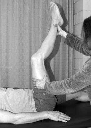
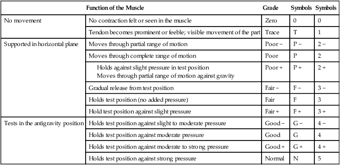
Physical Therapy for Musculoskeletal Conditions
After reading this chapter, the reader will be able to:
List common conditions seen in musculoskeletal physical therapy
Identify the components of an initial musculoskeletal examination
Define and give examples of the various tests and measures used in physical therapy for musculoskeletal conditions
List the general goals of a therapeutic exercise program
Describe the various types of therapeutic exercises
Describe various physical agents used to address musculoskeletal problems
Conditions that affect the musculoskeletal system are the primary domain of physical therapists who specialize in orthopedic physical therapy. One of the largest clinical specialties within the physical therapy profession, orthopedic physical therapy encompasses a wide array of therapeutic techniques and philosophies of treatment. Physical therapists (PTs) and physical therapist assistants (PTAs) in the field of orthopaedics work in a variety of clinical settings and treat patients of diverse ages, focusing on the prevention and rehabilitation of musculoskeletal injuries. This chapter describes the types of patients with whom an orthopaedic PT would work and presents some commonly used examination tests and measures and interventions.
General Description
Although the clinical interests or approaches to patient care may be diverse, the common thread throughout physical therapy for musculoskeletal conditions is the focus on a patient’s function. By examining the patient’s functional abilities, the PT determines the cause and extent of any limitations and restrictions in desired activities, and works with the patient/client to return the individual to his or her preinjury level of function in the shortest time possible.1 A person’s function can be affected when a disruption occurs in the musculoskeletal system. This disruption may be the result of traumatic or repeated stress to tissue, structural imbalances of muscle or bone, congenital conditions, surgery, or degenerative changes in the body. Dysfunctions of the musculoskeletal system often result in symptoms of pain, stiffness, edema (swelling), muscle weakness or fatigue, or loss of range of motion (ROM; movement at a joint).
Because these symptoms overlap with conditions managed by other clinicians, patients often arrive unsure of who they should be seeing in the first place. A quick read on chiropractor vs physical therapist roles can clarify that chiropractors typically focus on spinal adjustments for alignment-related complaints, while physical therapists address broader musculoskeletal dysfunction through exercise, manual therapy, and functional retraining, which is the framework the rest of this chapter builds on.
To conduct a comprehensive examination, generate an accurate diagnosis, and develop an appropriate plan of care, therapists must have an extensive understanding of anatomy, biomechanics, pathokinesiology, and exercise physiology. They must also be knowledgeable in the application of a variety of interventions and be able to make clinical decisions using the best available evidence in order to establish a plan of care that is appropriate for each individual patient situation,. Effective communication skills are also critical for PTs and PTAs so they can establish good rapport with patients and provide the necessary education to gain the patient’s adherence to the plan of care.
Development
Several factors contribute to the continued growth of musculoskeletal physical therapy. New contributions to the scientific literature provide therapists with evidence supporting the effectiveness of various procedural interventions that allow better evidence-based clinical decision making. The development of sophisticated technology and new intervention techniques has also provided new treatment options for PTs who evaluate and treat this patient population. Changes in lifestyle have contributed to the growth of orthopaedic physical therapy. Increasing interest and participation in physical fitness by the general population have resulted in an increase in musculoskeletal disorders caused by overuse or traumatic injuries. The increased use of computers and other technical machinery requiring repeated motions has also had an impact on the incidence of overuse injuries in the upper extremity. Individuals who must sustain postures at a computer or operate machinery while performing repeated motions with their hands may be at risk for the development of muscle injury or nerve entrapment requiring intervention by a PT. An increase in life span has also resulted in the growth of this area of physical therapy, as people are living longer, more active lives and experiencing symptoms related to degenerative changes in their bodies.
A great deal of similarity exists between orthopaedic physical therapy and sports physical therapy. In both areas the focus of rehabilitation is to regain optimum function and return the patient to the previous level of activity. A sports PT must therefore incorporate sport-specific activities into the treatment program to make sure that the patient can meet the physical demands of the sport with respect to strength, endurance, balance, speed, and coordination. An orthopaedic PT may work with athletes but may also treat a variety of musculoskeletal conditions that are not related to sports activities.
Common Conditions
Within the broad scope of musculoskeletal physical therapy, a variety of patient problems may be treated. These problems include injuries sustained through athletic participation, work-related injuries, conditions resulting from orthopaedic surgical procedures, and degenerative changes that accompany the aging process. As previously stated, patients with musculoskeletal conditions may report pain, swelling, weakness, or loss of motion resulting from stress to the musculoskeletal system. This stress may include damage to bones or soft tissue such as muscles, tendons, joint capsules, ligaments, bursae, cartilage, and fascia in the extremities or spine.
Overuse Injuries
Repeated stress to the musculoskeletal system can cause overuse injuries that may result in pain, inflammation, and dysfunction. The following examples are some common conditions caused by overuse.
Bursitis
Bursitis is an inflammation of bursae, which are fluid-filled sacs located throughout the body that decrease friction between structures. Bursae become irritated and painful when they are repeatedly pinched between two structures. A common example of this mechanism of injury occurs at the shoulder; the subacromial bursa may be pinched during repeated movements when the shoulder is in an overhead position, such as when painting, reaching, or throwing.
Tendinopathy
Tendons are the structures that connect muscle to bone. Repeated use of or rapid overstretching of muscles can overload and injure the tendons. Disorders of tendons (tendinopathies) can be the result of inflammation (tendinitis) or degenerative changes caused by overuse (tendinosis). Tendinopathies usually result in painful movements and are frequently seen in the patellar tendon at the knee in people who perform repeated jumping (e.g., dancers, basketball players) and at the elbow in people who do repeated or sustained gripping activities (e.g., carpenters, tennis players). Excessive overload of a tendon can also result in a partial or complete tear of the tendon, which is commonly seen in the ankle (Achilles rupture) or elbow (biceps rupture) and may need to be surgically repaired.
Nerve Entrapment
Pressure on a nerve, causing nerve entrapment, may result from a variety of sources and usually causes symptoms of tingling, pain, weakness, or any combination of these. A common condition of nerve compression at the wrist is referred to as carpal tunnel syndrome. Patients with carpal tunnel syndrome usually complain of numbness and pain in the hand and fingers, which commonly results from repeated activities with the wrist in a flexed position (e.g., musicians, computer keyboard operators).
Traumatic Injuries
Musculoskeletal injuries may also occur as a result of direct trauma. Bones, muscles, ligaments, and other soft tissues may be injured when they sustain a direct blow or when they are placed under excessive stretch. The following are just a few of the common conditions that can result from direct trauma to the musculoskeletal system.
Ligament Sprain
Ligaments are supporting structures at joints that serve to stabilize the joint and prevent excess movement. When ligaments are overstretched, their fibers can tear and cause pain and instability at the joint. A common site of sprain is at the ankle when the lateral (outside) ligaments are overstretched. This injury occurs when a person lands on the foot in a turned-in position. Another common site of ligament sprain is the anterior cruciate ligament (ACL) at the knee. Injuries to this ligament are usually the result of a cutting or twisting movement of the knee when the foot is planted, commonly occurring in sports that require jumping or quick changes in direction, such as soccer or volleyball.
Fracture
Direct trauma to bone can result in a break, or fracture, of the bone. Fractures can occur in any bone in the body but are commonly seen at the wrist or the hip after falls. Older adults are particularly prone to fractures because of changes in the structure of their bones resulting from inactivity, inadequate nutrition, and degenerative conditions. Fractures are best diagnosed through the use of radiographs.
Muscle Strain
A sudden contraction of a muscle or excessive stretch on a muscle can cause tearing of the muscle fibers, known as a strain. Muscle strains can occur in any area of the body and can range in severity. A strain of the rotator cuff muscles at the shoulder can result in shoulder pain and weakness when lifting the arm, and cervical strains may be the result of a sudden trauma to the neck, as with a whiplash injury.
Surgical Conditions
Individuals who have had surgery are another group of patients commonly seen by an orthopaedic PT. Injuries resulting from repeated stress, acute trauma, or disease processes may require surgical intervention for appropriate healing. The following are examples of orthopaedic surgery in which patients can benefit from physical therapy intervention to reduce pain and regain motion and strength, which will allow optimal movement and function.
Total Joint Arthroplasty
Painful movement caused by degenerative changes at joint surfaces can be alleviated through surgical replacement of the joint surfaces. Joints most commonly replaced are weight-bearing joints, primarily the hips and knees. A variety of plastic and stainless steel implants are used to effectively replace degenerated joint surfaces. Physical therapy intervention is necessary postoperatively for the patient to regain maximum strength and function and to receive education to prevent complications such as dislocation.
Amputation
Surgical amputation is the removal of a portion of an extremity because of trauma, inadequate blood flow, or the presence of a malignant growth. Inadequate circulation can be a result of disease processes such as diabetes mellitus or peripheral vascular disease, whereas a growth may indicate the presence of cancer. Postoperative physical therapy often includes exercises to maintain strength in the remaining portion of the limb, functional training with a prosthesis, and activities to improve overall fitness and well-being.
Medical Conditions
Numerous medical conditions may also affect the musculoskeletal system, resulting in pain, weakness, or loss of function. Systemic diseases such as rheumatoid arthritis, obesity, or cancer may cause impairments that disrupt the musculoskeletal system and result in functional challenges that can be addressed by the orthopaedic PT.
Principles of Examination
Working with a patient with a musculoskeletal condition requires the PT to have a comprehensive understanding of anatomy, pathology, biomechanics, and pathokinesiology. The first step in understanding the needs of a patient is to perform a thorough examination. Reexaminations are performed throughout the rehabilitative process to monitor patient progress toward established functional goals.
This section describes the following components of an initial examination: patient history, systems review, and tests and measures performed by the PT. The history is part of the subjective examination, whereas the remaining parts constitute the objective examination.
Patient History
The history involves gathering information about the current and past health status of the patient related to why the patient/client is seeking the services of a PT.2 The information may be obtained by interviewing the patient or the patient’s family, by accessing the patient’s medical record, and by consulting with other members of the patient’s health care team. The history is qualitative information based on the patient’s perception of the problem and is therefore included in the “S” portion of the “SOAP” note (see Chapter 2).
The role of the therapist during the interview is to guide the patient through pertinent questions about the patient’s musculoskeletal condition. This interaction allows the therapist to develop a rapport with the patient and to understand the patient’s insight into and opinion of the problem. The interview also assists the therapist in appropriately directing the remainder of the examination. Often, the patient interview will give the therapist ample information to make a preliminary physical therapy diagnosis. Questions asked during the interview include information about the onset of the condition, current symptoms, previous physical therapy treatments, past medical history, and lifestyle and health habits pertaining to work and recreation. Box 8-1 lists typical questions asked during the patient interview.
In addition to these questions specific to the patient’s reason for seeking physical therapy, the therapist should perform a review of symptoms (ROS) in order to identify symptoms that may have been overlooked in the history and to screen for medical conditions that may require referral to other health care providers.3 The ROS is usually performed by using checklists of common symptoms typically associated with various systems of the body (e.g., cardiovascular system, gastrointestinal system).
For more specific information about the location of symptoms, the patient is often asked to draw the location of the symptoms on a body chart (Figure 8-1). Pain scales may also be used to gauge the amount of pain the patient is experiencing (Figure 8-2). On completion of the history taking, the therapist should have gained information regarding the description and location of symptoms, nature of the disorder (acute versus chronic condition), behavior of the symptoms (what activities make the symptoms either better or worse), health risk factors that may be present, and limitations in activities the patient may be experiencing.
Systems Review
The objective portion of the examination refers to quantitative or qualitative measurements that are taken by the PT. This portion of the examination begins with a systems review and is included in the “O” section of the SOAP note (see Chapter 2). The systems review includes a brief examination of the other systems of the body related to physical therapy (e.g., cardiovascular, neuromuscular, integumentary) and information about the patient’s cognition, communication, and preferred learning style.2 The information gathered during the systems review assists the therapist in developing an appropriate, individualized plan of care and may further identify health problems that may require consultation with or referral to another health care provider. In a patient with a musculoskeletal condition, common systems reviews may include monitoring of heart rate and blood pressure, assessment of skin integrity, and a gross assessment of joint ROM, strength, and coordinated movements.
Tests and Measures
During the tests and measures portion of the examination, specific numbers or grades may be assigned (quantitative measurement), as is the case with ROM or strength measurements. At other times, parts of the examination are performed by observing and describing patterns of movement, deformities, or both (qualitative measurement). The purpose of the tests and measures is to establish baseline values and observations that can be used for comparison after a single treatment or a series of treatments. The PT can then make appropriate changes in the plan of care based on the amount of progress or lack of progress found with repeated tests and measures.
This section briefly describes some of the tests and measures performed in an orthopaedic physical therapy setting. The purpose is to familiarize introductory level students with common terms used when working with a patient who has a musculoskeletal problem.
Observation
Observation is the “looking” phase of the examination. It may begin in the waiting room, where the therapist can observe the patient’s general attitude, posture, and willingness to move. A perfunctory gait assessment may be made as the patient enters the examination area. Once the patient is appropriately undressed, a more detailed inspection can be made, including observation of obvious deformities such as an abnormal curvature of the spine, joint subluxations (a condition in which a joint partially dislocates), asymmetrical body contours, swelling, and color and texture of the skin. Many musculoskeletal injuries are a result of or are exacerbated by poor sitting and standing postures. Therefore particular attention is paid to the standing and sitting postures of the patient.
Active Range of Motion
Active range of motion (AROM) refers to the ability of the patient to voluntarily move a limb through an arc of movement. AROM provides the therapist with information regarding the quality of the movement (smooth versus rigid movement), the willingness of the patient to move the limb, any pain produced during movement, and whether the patient has any limitations in the motion as compared with the unaffected side. An example of AROM of the shoulder in multiple planes is provided in Figure 8-3.
Passive Range of Motion
Passive range of motion (PROM) refers to the amount of movement at a joint that is obtained by the therapist moving the segment without assistance from the patient. In some instances, because of injury or prolonged immobilization, a joint may have less motion than is considered functional. A joint in this condition is referred to as a hypomobile joint. In other cases, such as a joint subluxation, the joint may have excessive motion and is referred to as a hypermobile joint. PROM also gives the therapist an indication of the degree and pattern of pain, as well as the “feel” of the movement.
Many methods may be used to measure and document AROM and PROM. The most common measurement technique, goniometry, is performed with a goniometer and measures joint angles. Examples of goniometers are shown in Figure 8-4. The amount of motion available at any joint depends on the structure of the joint. In addition, norm values for joint ROM depend on several factors, including the patient’s age and gender.2 Typically the therapist compares ROM values of the affected joint with those on the unaffected side. Figure 8-5 shows a PT conducting a PROM measurement of a patient’s knee flexion.
Strength
Strength can be defined as the amount of force produced during a voluntary muscular contraction. This contraction may be performed statically (no motion) or dynamically (through an assessment of ROM). When one is assessing the status of the muscles and tendons, a quick resisted test is used. This test allows the therapist to determine the general strength of a muscle group and assess whether the muscle contraction produces pain. If the resisted test shows that a muscle or muscle group is weak or painful, further testing may be performed to isolate the specific muscle. To isolate and test specific muscles, manual muscle testing (MMT) is performed (Figure 8-6). MMT allows the therapist to assign a specific grade to a muscle. This grade is based on whether the patient can hold the limb against gravity, how much manual resistance can be tolerated, and whether the joint has full ROM. Several systems of grading are widely used. One of the most common grading systems was initially described by Robert Lovett, MD, and later modified by Henry Kendall, PT, and Florence Kendall, PT.4 This key to muscle grading is outlined in Table 8-1.
Table 8-1
Key to Manual Muscle Testing Grades
| Function of the Muscle | Grade | Symbols | Symbols | |
| No movement | No contraction felt or seen in the muscle | Zero | 0 | 0 |
| Tendon becomes prominent or feeble; visible movement of the part | Trace | T | 1 | |
| Supported in horizontal plane | Moves through partial range of motion | Poor − | P − | 2 − |
| Moves through complete range of motion | Poor | P | 2 | |
| Poor + | P + | 2 + | ||
| Gradual release from test position | Fair − | F − | 3 − | |
| Holds test position (no added pressure) | Fair | F | 3 | |
| Hold test position against slight pressure | Fair + | F + | 3 + | |
| Tests in the antigravity position | Holds test position against slight to moderate pressure | Good − | G − | 4 − |
| Holds test position against moderate pressure | Good | G | 4 | |
| Holds test position against moderate to strong pressure | Good + | G + | 4 + | |
| Holds test position against strong pressure | Normal | N | 5 |

Modified from Kendall FP, McCreary EK, Provance PG, et al: Muscles: testing and function, ed 5, Baltimore, 2005, Lippincott Williams and Wilkins.

With the development of sophisticated technical equipment, many other methods are now available to measure strength, including hand-held dynamometers and computerized instruments such as isokinetic devices. These devices allow the therapist to obtain strength curves of isolated muscles, as well as specific force values.
Flexibility
Flexibility refers to the ability to move a limb segment through a specific ROM. The amount of flexibility at a given joint depends on two factors. First, the soft tissue surrounding the joint must be pliable to allow movement between the joint surfaces. This feature is referred to as accessory motion of the joint. Accessory motion is the ability of the joint surfaces to glide, roll, and spin on each other. Second, the muscle or muscles crossing the joint must be at an appropriate length to allow motion to occur. For example, the ability to stand up and touch the toes while keeping the knees straight depends on the flexibility of the back and posterior hip muscles, as well as the ability of the spinal vertebrae to move.
Appropriate flexibility or balance of muscles is a key component of proper posture and body mechanics. Many musculoskeletal problems seen in the physical therapy clinic can be linked to muscle imbalances that have caused movement dysfunctions.5 For example, if the muscles surrounding the shoulder did not act synergistically (because of lack of flexibility), compensation might occur at joints distal and proximal to the shoulder, such as the elbow and cervical spine.
A PT may perform a number of tests to determine flexibility. One common test for the lower extremity is the 90/90 straight leg raise (Figure 8-7). This test objectively measures flexibility of the hamstring muscles, located on the posterior aspect of the thigh.
Functional Tests
The ultimate goal of therapy is to return the patient to the previous level of activity, which may include anything from the ability to go grocery shopping independently to returning to athletic competition. With some types of injuries a return to the previous level of activity is not feasible. In these cases the ultimate goal would be to return the individual to the highest level of function achievable.
Traditionally, functional assessment has referred to such activities as the patient’s bed mobility, transfers between a variety of surfaces (e.g., moving from a sitting position in a wheelchair to a standing position), and ability to perform activities of daily living (ADLs), such as hair combing, dressing, and bathing. PTs may spend a large percentage of their time during the initial examination assessing the patient’s ability to perform these ADLs. Box 8-2 lists examples of ADLs.
Individuals who wish to return to activities other than ADLs require more aggressive types of functional testing. Examples of these types of functional tests are hop tests, jump tests, lunge tests, excursion tests, and balance tests. Gary Gray and others have described these and other types of functional tests in detail.6,7
Outcome Measures
Outcome measures are standardized instruments that measure an individual’s actual or perceived activity limitations and participation restrictions, and/or an individual’s quality of life or health status. These instruments are typically closed-ended questions completed by the patient and scored by the PT. Scores can then be used to assess the initial degree of limitation as well as changes in ability or health status before and after treatment.8 Examples of common outcome measures include the Oswestry Low Back Disability Questionnaire, the Medical Outcomes Study 36-Item Short Form (SF-36), the Patient Specific Functional Scale (PSFS; Box 8-3), and the Lower Extremity Functional Scale (Table 8-2).
Table 8-2
Lower Extremity Function Scale
| We are interested in knowing whether you are having any difficulty at all with the activities listed below because of your lower limb problem. (Circle one number on each line.) | |||||
| Activities | Extreme Difficulty or Unable to Perform Activity | Quite a bit of Difficulty | Moderate Difficulty | A Little Bit of Difficulty | No Difficulty |
| Any of your usual work, housework, or school activities | 0 | 1 | 2 | 3 | 4 |
| Your usual hobbies, recreational or sporting activities | 0 | 1 | 2 | 3 | 4 |
| Getting into or out of the bath | 0 | 1 | 2 | 3 | 4 |
| Walking between rooms | 0 | 1 | 2 | 3 | 4 |
| Putting on your shoes or socks | 0 | 1 | 2 | 3 | 4 |
| Squatting | 0 | 1 | 2 | 3 | 4 |
| Lifting an object, such as a bag of groceries from the floor | 0 | 1 | 2 | 3 | 4 |
| Performing light activities around your home | 0 | 1 | 2 | 3 | 4 |
| Performing heavy activities around your home | 0 | 1 | 2 | 3 | 4 |
| Getting into or out of a car | 0 | 1 | 2 | 3 | 4 |
| Walking two blocks | 0 | 1 | 2 | 3 | 4 |
| Walking a mile | 0 | 1 | 2 | 3 | 4 |
| Going up or down one flight of stairs | 0 | 1 | 2 | 3 | 4 |
| Standing for 1 hour | 0 | 1 | 2 | 3 | 4 |
| Running on even ground | 0 | 1 | 2 | 3 | 4 |
| Running on uneven ground | 0 | 1 | 2 | 3 | 4 |
| Making sharp turns while running fast | 0 | 1 | 2 | 3 | 4 |
| Hopping | 0 | 1 | 2 | 3 | 4 |
| Rolling over in bed | 0 | 1 | 2 | 3 | 4 |
| Column totals: | |||||
| Score: ____/80 | |||||

Modified from Stratford PW, Binkley JM, Watson J, Heath-Jones T: Validation of the LEFS on patients with total joint arthroplasty, Physiother Can 52:97, 2000.
Special Tests
Special tests are used to examine specific joints to indicate the presence or absence of a particular problem. The purpose of these tests is to confirm or reinforce a physical therapy diagnosis. Because so many special tests are available for each joint, only those that appear to be indicated based on the results of tests and measures are performed. Examples of special tests are those that examine nerve compression (Phalen’s test; Figure 8-8, A), shoulder impingement (Hawkins test; Figure 8-8, B), and ligamentous knee injuries (Lachman test; Figure 8-8, C).
Palpation
A comprehensive understanding of anatomy is essential for any PT. In the clinical situation the therapist uses the sense of touch, known as palpation, to assess what is occurring below the skin and what musculoskeletal structures are involved in an injury. When palpating an area of the body, the therapist is feeling for areas of pain and tenderness, areas of restriction, swelling, and proper orientation of structures.
Other Diagnostic Procedures
Depending on the patient’s injury or complaint, other examination procedures may be performed to provide a more complete assessment of the patient. The patient may be referred to other personnel for these procedures. Such complementary procedures may include a variety of imaging techniques, such as plain film radiographs (“x-rays”), computed tomography (CT) scans, and magnetic resonance imaging (MRI). If the patient has had neurologic damage, full sensory testing may be indicated. Additional tests for patients with cardiopulmonary conditions may include an assessment of lung capacity. Some of these tests are presented in more detail in Chapters 9 and 10.
Principles of Evaluation, Diagnosis, and Prognosis
Based on evaluation of the findings from the comprehensive examination, the PT identifies the patient’s impairments, activity limitations, and participation restrictions and then determines the diagnosis and prognosis. Once the problems have been identified, the therapist and patient develop goals to address each problem. Common short-term goals are to decrease pain and edema and increase strength and motion. The ultimate long-term goal for patients with musculoskeletal dysfunctions is to achieve an optimal level of function, whether that means returning to work, resuming athletics, or performing daily activities independently. Therapeutic goals should include how each goal is to be measured and the expected time frame to achieve each goal. Once goals have been established, the therapist develops a plan of care designed to achieve these outcomes.
The plan of care is based on determining which interventions will most effectively improve a patient’s function. The process begins by identifying the relevant evidence in the literature, combined with the therapist’s clinical expertise and preferences of the patient. Numerous intervention options and rehabilitation approaches are available to the orthopaedic PT. Some therapists focus their treatment approaches on exercises, whereas others incorporate physical agents or manual techniques. In most instances a combination of techniques is appropriate when designing a comprehensive plan to address the needs of a patient with a musculoskeletal dysfunction. When selecting a plan of care, the therapist must consider the goals of the patient and the desired outcome of therapy.
Principles of Procedural Intervention
The following discussion of intervention options introduces the reader to the typical indications and uses of various techniques. The techniques described include physical agents, manual techniques (including soft tissue and joint mobilization), and therapeutic exercise. The reading list at the end of the chapter gives sources for in-depth information regarding the application of these and other interventions used in musculoskeletal physical therapy.
Physical Agents
Many physical agents are available for PTs to incorporate into the plan of care when treating patients with musculoskeletal problems. Physical agents may be classified by the tissue’s response to treatment (e.g., thermal changes caused by heating or cooling agents) or classified based on the type of energy used by the agent (e.g., ultrasound, electrotherapy). Physical agents can be used to address a variety of impairments; their therapeutic benefits include pain management, increased flexibility, improved ROM, increased muscle strength, and wound healing. The decision to include physical agents as a direct intervention is based on a thorough examination of the patient’s symptoms, the desired outcomes of therapy, and the therapist’s knowledge of the physiologic and clinical effects of each physical agent. Table 8-3 lists common physical agents used in physical therapy according to their physical effects and includes their physiologic effects and clinical indications.
Table 8-3
Summary of Common Physical Agents Used in Physical Therapy
| Physical Effect | Physical Agents | Physiologic Effects | Clinical Indications |
| Superficial heat | |||
| Deep heat | |||
| Cold | |||
| Electrical stimulation |

Thermal Agents
When human tissue sustains an injury, an automatic response is initiated in an attempt to heal the tissue and return it to its preinjured state. These naturally occurring processes are referred to as inflammation and repair.8 The inflammation and repair stages of tissue healing can be altered through the use of thermal agents or electrical stimulation. Thermal agents are used to modify the temperature of surrounding tissue and result in a change in the amount of blood flow to the injured area. Besides vascular changes, temperature changes affect the metabolism of the surrounding tissue, in addition to altering neuromuscular and connective tissue. Through the use of therapeutic changes in temperature, the healing process can be accelerated and the injured tissue restored to optimal strength and integrity.
The extent of the therapeutic changes caused by an alteration in tissue temperature depends on the intensity of the thermal agent applied, the length of time the tissue is exposed to the agent, and characteristics of the tissue being treated. The therapist must continually monitor and reexamine the patient to ensure that the thermal agent selected is appropriate and that the treatment outcomes are being achieved.
Thermal agents can be classified as those that provide superficial heat, deep heat, or cold. Superficial heat modalities create an increase in blood flow to cutaneous tissue close to the surface of the skin and are effective in reducing pain and stiffness, increasing ROM, and promoting healing.9 Examples of superficial heat agents include hot packs, paraffin, fluidotherapy, and whirlpools.
A hot pack is a pouch available in various shapes that is filled with silica gel and soaked in thermostatically controlled water (Figure 8-9). Hot packs are applied to the affected body part with layers of towels to prevent overheating (see Figure 2-11). Paraffin treatment involves dipping a patient’s involved extremity (usually hands or feet) into a mixture of melted paraffin wax and mineral oil that is maintained at a temperature of approximately 135° F (Figure 8-10). The heat from the paraffin produces the relaxing and pain-reducing effects of other superficial heat treatments and leaves the skin feeling warm and soft, which increases comfort when ROM exercises are performed.

Fluidotherapy is the use of a self-contained unit filled with corncobs finely chopped into a sawdust-type substance. The particles are heated to the desired temperature and circulated by air pressure around the involved body part. In addition to receiving the effects of heating, the patient can exercise while the treatment is in progress. Use of the therapeutic effects of water is known as hydrotherapy. A whirlpool can be used when the body part or entire body is immersed in a tank of water. Various sizes of tanks are available, ranging from a small tank for the distal ends of extremities to a full-body tank known as a Hubbard tank. In addition to its heating effects, hydrotherapy can assist with wound healing.
Deep heat modalities produce physiologic effects similar to those of superficial heat agents, but at a greater tissue depth. Therefore patients with deep muscle or joint dysfunction may receive more therapeutic benefit from the application of deep heat than from a superficial heating agent. Deep heat modalities include ultrasound and short-wave diathermy. Thermal ultrasound is the therapeutic application of high-frequency sound waves that penetrate tissue and increase tissue temperature to promote healing and reduce pain (Figure 8-11). Similar results are achieved with short-wave diathermy, which is the use of electromagnetic energy to produce deep therapeutic heating effects.

In contrast to heating agents, therapeutic cold (cryotherapy) may be applied to decrease tissue temperature. Temperature differences produced by the application of cold agents cause a decrease in blood flow and metabolism, which results in a decrease in swelling and pain. Cryotherapy is the physical agent of choice for patients who have acute injuries with clinical symptoms of swelling or pain or both (see Figure 2-12). Cold may also be incorporated into a treatment protocol after exercise to help reduce postexercise soreness. Cryotherapy may take the form of commercial cold packs, ice massage, cold whirlpool, or cold used in conjunction with compression.
Electrical Stimulation
PTs and PTAs may use electrical stimulation as part of their plan of care to achieve therapeutic results. With the use of electrical stimulation units, electrodes are placed on the skin at specified locations to stimulate nerves, muscles, and other soft tissues in an attempt to reduce pain and swelling, increase strength and ROM, and facilitate wound healing (Figure 8-12).10 The therapist must have a clear understanding of the desired effects of the electrical stimulation intervention and have knowledge of the appropriate parameters to use with regard to treatment intensity, voltage, and current type. Common electrotherapy applications are listed in Table 8-3.
Other Physical Agents
Additional physical agents that may be used in the treatment of patients with musculoskeletal dysfunction include mechanical traction, hyperbaric oxygen, biofeedback, LASER therapy, and ultraviolet treatment. These modalities achieve therapeutic benefit through mechanisms different from those of thermal or electrical agents, but they may also be used to decrease a patient’s pain, promote healing, or improve strength or motion in an attempt to maximize function.
Manual Therapy Techniques
PTs working with patients who have musculoskeletal dysfunction always have two tools at their ready disposal—their hands. Whether palpating a structure during an examination, providing manual force for a patient to resist against when performing strengthening exercises, or performing a mobilization to increase ROM, a therapist’s hands are important therapeutic instruments. A variety of manual techniques are frequently used by orthopedic PTs, and many of these techniques are the subject of current clinical research to validate and clarify their purpose and clinical efficacy.
For the purpose of this text, manual techniques are divided into two categories: soft tissue mobilization and joint mobilization. A discussion of specific procedures and the schools of thought behind the various techniques is beyond the scope of this text. The reading list at the end of this chapter provides further information regarding this topic.
Soft Tissue Mobilization
Soft tissue mobilization includes a variety of “hands-on” techniques designed to improve movement and function by restoring extensibility to tight tissue. The techniques are designed to decrease pain or swelling and relax muscle or fascia tension to create proper postural alignment and optimal muscle function.
Two common forms of soft tissue mobilization are massage and myofascial release. Massage involves the systematic use of various manual strokes to produce certain physiologic, mechanical, and psychological effects. Swedish massage strokes promote relaxation by decreasing pain or swelling, relieving tension, and improving the metabolism of surrounding tissue. More vigorous massage strokes may be used before physical activity to stimulate and prepare the muscles for exertion. Another soft tissue technique, known as transverse friction massage, is useful for promoting proper healing and improving the flexibility and function of soft tissues such as muscles, ligaments, and tendons.11
Myofascial release involves manual stretching of the layers of the body’s fascia, which is connective tissue that surrounds muscle and other soft tissue in the body (see Figure 2-9).12 Myofascial release techniques are reported to soften and reduce restrictions in muscles and fascia that are limiting normal movement.
Joint Mobilization
In contrast to soft tissue mobilization, which focuses on stretching or relaxing soft tissue, joint mobilization techniques are used when a patient’s dysfunction is the result of joint stiffness or hypomobility (limited motion). Based on knowledge of the anatomy of joint surfaces and the findings from joint examination, the therapist applies specific passive movements of varying amplitude to a joint, in either an oscillatory (rhythmic, repeated movements) or a sustained manner. Joint mobilization techniques are used to reduce pain and stiffness affecting movement and restore normal joint motion.
Therapeutic Exercise
Therapeutic exercise forms the core of most rehabilitation programs.2 This foundation is based on scientific principles and the knowledge that the human body has the ability to react and respond to physical stresses placed on it. In particular, the muscular and cardiovascular systems are adaptable, depending on the stresses and forces placed on them. When these systems are stressed with a program of progressive exercise, positive changes such as improvement in strength and endurance occur. Similarly, the effects of abnormal stresses, such as prolonged bed rest, can lead to detrimental changes, including osteoporosis and muscle atrophy.2
The goals of therapeutic exercise are not only to facilitate and restore normal function in an individual, but also to prevent an initial injury, educate the patient on how to prevent recurrence of an injury, and help maintain normal function. These goals are based on the results of the patient’s examination and assessment of needs.
The level of sophistication of an exercise program should not be determined by the type of equipment the clinic has. Some of the most sophisticated exercises can be performed with inexpensive equipment. With creativity, various pieces of equipment can be adapted to incorporate many of the goals of therapeutic exercise. This section describes a variety of therapeutic exercise techniques that may be used with a patient who has a musculoskeletal dysfunction. These techniques include exercises to improve ROM, strength, flexibility, balance and coordination, cardiovascular endurance, and function.
Range-of-Motion Exercise
As mentioned earlier in the chapter, range of motion exercise can be categorized into two types: passive and active. PROM may be provided manually by the therapist or mechanically by a machine. This type of exercise might be used with (but is not limited to) patients who are restricted to bed rest or have paralysis of one or more limbs. It may also be used when AROM is contraindicated. AROM can be subdivided into active assisted movement, active free movement, and active resisted movement. When performing active assisted range of motion, the patient may be assisted either manually or mechanically if the prime muscle mover is weak (Figure 8-13, A). Pendulum exercises in which the patient does not receive any support or resistance are an example of active free exercises (Figure 8-13, B). In active resisted exercises an external force resists the movement. The last category includes a variety of techniques, several of which are described in the next section.
Resistance Exercise
Resistance exercise is a form of active movement in which some form of resistance is provided. The goals of a resistance exercise program are to increase muscular strength, endurance, and power. Muscular strength is the maximal amount of tension an individual can produce in one repetition. Muscle endurance is the ability to produce and sustain tension over a prolonged period. Muscle power is the amount of work produced by the muscle in a given amount of time. If the goal is to increase strength, the program would concentrate on low repetitions with heavy resistance. If the goal is to increase endurance, the exercise program would concentrate on using low resistance for high repetitions. When the goal is to increase power, the exercise program would consist of explosive high-intensity muscle activity such as jumping. The type of exercise performed depends on the types of activities to which the patient is planning to return. When designing a program, the therapist must consider the type or types of resistance exercise on which the patient should concentrate. Resistance exercise can be categorized into three types: isometric, isotonic, and isokinetic. Definitions and examples of these types are outlined in Table 8-4. Typically, a combination of all three types of exercise is necessary to perform any type of functional activity.
Table 8-4
Classification of Resisted Exercises
| Type of Exercise | Definition | Example |
| Isometric | Muscle contraction without visible joint movement | Pushing against a wall |
| Isotonic concentric | Muscle contraction that produces or controls joint motion, resulting in muscle shortening | Flexing elbow with dumbbell in hand (biceps brachii muscle) |
| Isotonic eccentric | Muscle contraction that produces or controls joint motion, resulting in muscle lengthening | Extending elbow with dumbbell in hand (biceps brachii muscle) |
| Isokinetic | A concentric or eccentric muscle contraction that occurs at a constant speed | Knee extensions using an isokinetic device |
Resistance can be applied either manually by the therapist or mechanically by the use of equipment. Manual resistance can be applied to isolated muscle groups (as is the case with MMT positions) or to patterns of movement that involve several muscle groups. An example of the latter is a technique called proprioceptive neuromuscular facilitation, which is described in Chapter 9. The use of manual resistance offers many advantages, the primary one being that the therapist can control the amount of resistance provided. This advantage is particularly useful when working with patients in the early stages of rehabilitation when ROM may need to be limited or the patient can tolerate only mild to moderate resistance. The disadvantage of manual resistance is the difficulty in quantifying the amount of resistance provided. Inability to accurately document the resistance makes it difficult for another therapist to replicate the same force on that patient.
Many pieces of equipment can be used when applying mechanical resistance, from an inexpensive strip of elastic tubing to expensive and highly technologic isokinetic equipment. Other common and frequently used equipment in the clinic includes free weights exercise machines, and pulley systems (Figure 8-14).
Core Strengthening
Core strengthening refers to the strengthening of the deep postural muscles of the lumbar and cervical spine. The premise behind core strengthening is that it helps provide stability to proximal structures, allowing for appropriate movement patterns in the distal extremity. A number of core conditioning exercise classes such as Pilates and tai chi emphasize core strengthening during functional movement. Examples of core strengthening for the lumbar spine are shown in Figure 8-15.
Flexibility Exercise
Patients recovering from a musculoskeletal injury frequently have decreased flexibility in the muscles crossing the involved joint. Conditions that may produce decreased flexibility include prolonged immobilization and tissue trauma. Many times, prior limitations in flexibility contributed to or were the primary cause of the injury. Therefore flexibility exercise is an important component to address with the patient.
Soft tissue, such as muscle, has the ability to change length or adapt over time with stress. Although a variety of techniques can be used to increase flexibility, no consensus has been reached on the most effective way to stretch. Furthermore, a stretching technique that works well for one patient may be ineffective for another.
Stretching techniques can be performed passively with an external force applied either manually or mechanically. Stretching can also be performed by actively inhibiting the shortened muscle. This technique, called contract-relax, requires that the shortened muscle actively contract before a stretching force is applied.
Balance and Coordination Exercise
Loss of balance is an impairment that PTs frequently address in a rehabilitation program. Balance refers to the ability to maintain body position in equilibrium.13 Proper balance requires the nervous and musculoskeletal systems to interact with the environment to control body position. After injury, if these systems are not properly integrating information, loss of balance and coordination may result. Several clinical tests are used to assess a patient’s balance, and numerous exercises and equipment can be used to facilitate proper balance. One popular piece of equipment seen in the clinic is a BOSU balance trainer (Figure 8-16). The patient may be progressed from double-leg stance to single-leg stance. The exercise can be made more challenging by having patients close their eyes and by incorporating upper extremity movement with and without weights.
Cardiovascular Endurance Training
Cardiovascular or aerobic training is exercise performed over a long period at low intensity. Aerobic exercise typically involves large muscle groups used in a rhythmic type of activity. Many modes of exercise are available to improve cardiovascular endurance, including walking, running, stair climbing, cycling, cross-country skiing, and swimming. The PT will choose the most appropriate exercise modality for the patient. For example, a patient who is recovering from a low back injury and has difficulty sitting may participate in a walking program rather than a cycling program. See Chapter 10 for a more detailed description of cardiovascular exercise.
Functional Exercises
As mentioned earlier in this chapter, the ultimate goal in physical therapy is to allow the patient to return to the previous level of function or highest level of function achievable. Therefore exercises mimicking functional movements and activities must be incorporated into the rehabilitation program. A functional exercise incorporates strength, flexibility, balance, and coordination. Incorporating all these factors allows patients to return to function with confidence because they know that they performed the same or similar exercises in the clinic.
The use of closed kinetic chain exercises allows the patient to incorporate these functional movements. A closed kinetic chain exercise is an exercise in which movement at one joint affects movement at other joints. For example, when a person squats, movement occurs simultaneously at the ankle, knee, and hip joints. This is in contrast to an open kinetic chain exercise in which the end limb segment is free. An example of an open chain exercise is the seated leg extension machine frequently seen in fitness centers. Because the lower extremity typically functions with the foot on the ground, closed chain exercises are particularly important in the rehabilitation of the lower extremity. Therefore exercises involving the movement of joints while the foot is on the ground facilitate movement that mimics function (Figure 8-17). Closed kinetic chain exercises are also used with patients with upper extremity injuries, particularly those with shoulder dysfunctions.
Aquatic Therapy
The use of water for therapeutic benefit dates back to the ancient Greeks and Romans, who used therapeutic baths for relaxation and pain reduction.14 “Pool therapy” developed in the 1920s in the United States as part of the rehabilitation program for children with poliomyelitis (see Figure 1-4). As polio declined with the introduction of vaccines, so did the therapeutic use of pools. Recently, however, aquatic physical therapy has been shown to be beneficial for a variety of orthopaedic dysfunctions. The popularity of aquatic therapy has grown significantly, and in 1992, the American Physical Therapy Association (APTA) established the Aquatic Physical Therapy Section.
The Aquatic Physical Therapy Section offers a comprehensive description of this form of therapy (Box 8-4).15 The description indicates that this form of rehabilitation is effective for a variety of conditions, in addition to maintaining health and fitness in well individuals.
The beneficial effects of aquatic therapy depend largely on the fundamental principles of physics, such as the buoyancy, viscosity, and hydrostatic pressure of water. Both physiologic and psychological benefits are derived from aquatic physical therapy. The physiologic benefits include improved cardiovascular status, increased muscle strength and flexibility, decreased pain, and improved balance without the impact that occurs with exercises on land. Psychological benefits include general relaxation from the warmth of the water, the socialization process that may be associated with group sessions in a pool, and the increased patient confidence and level of satisfaction that accompany patient performance.
Certain contraindications and precautions must be considered when planning and implementing any aquatic therapy session (Box 8-5). These contraindications and precautions must be carefully considered to ensure the safety of the individual.
Home Exercise Programs
The use of therapeutic exercise in a rehabilitation program is an important component of the physical therapy plan of care. Aside from the physical benefits derived from exercise, it also encourages active participation in the rehabilitative process and allows patients to assume responsibility for their care. Home exercise programs are an important aspect of patient care. Performing exercises in the clinic two or three times per week is not usually adequate to achieve the desired long-lasting effects of rehabilitation, so home exercises given by the PT become an important component of physical therapy.
Patient Education
As mentioned earlier in the chapter, communication is a critical component of the orthopaedic physical therapy experience. The therapist’s depth of knowledge and effectiveness in performing and interpreting the examination and the variety of treatment options available are of little value if the therapist cannot share this information effectively with patients and include them in the rehabilitation process. The patient and therapist must work as a team and focus on common goals and sharing of information to achieve optimal results.
The PT and PTA are responsible for educating the patient about his or her diagnosis and about exercises to perform at home, postures or positions to avoid during daily activities at work or home, and strategies to prevent the dysfunction from recurring (Figure 8-18). To communicate effectively, the therapist must create a treatment atmosphere that takes into account the patient’s values and goals and must also provide the necessary information in a clear, easily understood manner.

It is important for the PT and PTA, when working with a patient, to treat the whole person rather than just an injured joint. Each patient comes to physical therapy with a different set of values, different expectations, and a different cultural background. All these factors must be considered if the patient is to be successfully and effectively treated as an individual.
Summary
This chapter has presented the role that PTs and PTAs play in physical therapy for musculoskeletal conditions. Common conditions described were overuse and traumatic injuries and surgical and medical conditions. Components of the patient examination were presented. Interventions focused on physical agents, manual techniques, therapeutic exercise, home programs, and patient education. The emphasis in physical therapy for musculoskeletal conditions is on evaluating a patient’s function and developing a plan of care that will assist the patient to return to optimal function in the environment, whether that be the athletic field, work site, community, or home.
Stay updated, free articles. Join our Telegram channel
Full access? Get Clinical Tree


