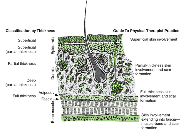
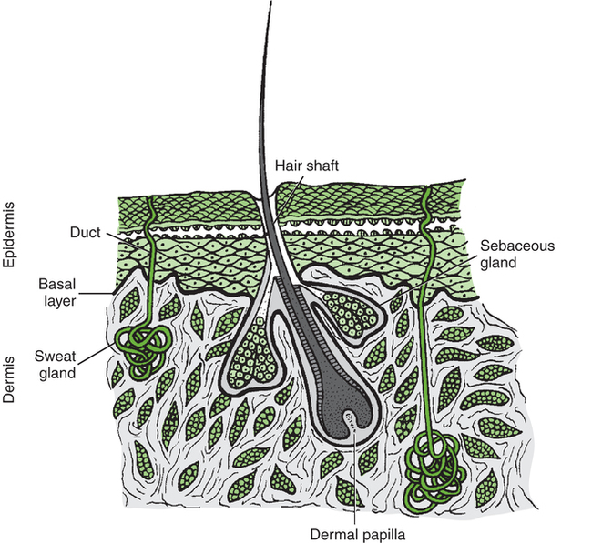
Physical Therapy for Integumentary Conditions


Physical Therapy for Integumentary Conditions


R. Scott Ward


Various types of integumentary (skin) wounds or impairments in skin integrity, the consequences of these wounds, and any associated effects of the wound such as inflammation, pain, edema, and scar formation can lead to significant functional limitations and disability. Physical therapists (PTs) and physical therapist assistants (PTAs) must be aware of the importance of the integumentary system in normal human function. They should be able to provide programs or interventions to prevent loss of skin integrity. Appropriate management of patients with various impairments of the skin is a critical part of physical therapy practice.


General Description


Integument


The integument, the largest organ of the body, ranges from about 1 to 4 mm in thickness and consists of two layers—the epidermis and the dermis. Beneath the dermis lies a layer of subcutaneous tissue. The integument is basically a protective organ, but it also plays a role in temperature control and provides important sensory information regarding the environment. Figure 11-1 illustrates the structure of the skin and its appendages.


Epidermis


The epidermis is very thin in comparison with the overall thickness of the skin. The thickness of the epidermis generally ranges from about 0.06 to 0.1 mm. It is thicker only on the soles of the feet and the palms of the hands, where the most superficial layer of the epidermis, the stratum corneum, may increase the thickness to 0.6 mm. This thicker stratum corneum is often referred to as callus. The preponderant cells in the epidermis are keratinocytes produced in the basal cell layer. The basal cell layer is also where the epidermis is anchored to the dermis. Keratinocytes take a minimum of 28 days to differentiate through their epidermal phases until they are finally sloughed off the most external surface of the stratum corneum. It is the stratum corneum that restricts the loss of fluids from internal tissue and separates this same internal tissue from the external environment.


Other cells that make up the epithelium are Langerhans cells, Merkel cells, and melanocytes. Langerhans cells play a role in the immune response in skin. Merkel cells are acknowledged as sensory receptor cells that provide information about tactile stimuli. Melanocytes (located in the basal cell layer) synthesize melanin, which is a pigment that principally serves as primary protection against harmful ultraviolet radiation. Once produced, melanin is transferred from melanocytes to keratinocytes. Melanocytes are also present in the dermis and hair follicles (as well as other sites, such as the retina).


Other components of the epidermis that penetrate into the dermis are hair follicles, sebaceous glands, apocrine glands, and sweat (eccrine) glands. The basal cell layer surrounds each of these structures because of their connection with the epidermis. Hair is formed at the follicle by a process of keratinization that produces three layers of cells. The hair follicle is an invagination of the epidermis. Hair type and amount depend on several factors, including hormonal influence, age, and heredity. Sebaceous glands produce a fatty secretion and are found in association with every hair follicle (pilosebaceous glands). Some sebaceous glands not associated with hair follicles are also found in a general distribution over the body with the exception of the soles of the feet, the palms of the hands, and the lower lip. The main function of the sebaceous glands is to keep the skin “moisturized” and pliant and to prevent it from drying and cracking. The apocrine glands begin to secrete a commonly colorless and odorless oily sweat at the onset of puberty. These glands are localized in the anogenital and axillary areas. The odor associated with perspiration in these areas results from bacterial decomposition of the secretions. Sweat is a hypotonic solution that is delivered to the skin surface by sweat glands. Normal function of the sweat glands is critical in temperature regulation.


Dermis


The dermis consists of fibrous and elastic connective tissue encompassed by a ground substance. The dermis varies from 1 to 4 mm in thickness and has two subdivisions—the papillary dermis and the reticular dermis. The papillary dermis, which is composed of a loosely organized collagen matrix and is highly vascular, forms in reflection to the basal cell layer of the epidermis. The junction between these two layers of skin is far from flat. The ridges formed at the dermal-epidermal junction (dermal papillae and epidermal ridges, respectively) provide protection against potentially damaging perturbations such as shearing and deepen the dispersion of the epidermal basal cell layer. The reticular dermis is composed of more densely bundled collagen fibers and less ground substance than the papillary dermis. The ground substance of the dermis is made up of various proteoglycans, glycoproteins, hyaluronic acid, and water. This “gel” forms the interstitial environment that accommodates the composite of dermal elements—fibroblastic collagen, blood vessels, and nerves—along with the epidermal appendages. The fibrous collagen supplies fortification against mechanical stresses on the skin while still allowing the deformation necessary for movement. The elastic connective tissue restores the collagen network to its “resting” arrangement, and the ground substance acts as a “cushion” to protect against many detrimental compression forces.


Blood vessels and nerves are also found within the dermis. The vascular structure in the dermis is vast and allows typically efficient diffusion of gases and nutrients to promote healthy cell function. The vascular system of the dermis also participates in the inflammatory response, an important component of wound healing. Along with the sweat glands, the capillaries in the skin also contribute to human thermal regulation. An equally expansive and efficient lymphatic system is associated with the vascular system in the dermis. The dermal nervous network provides the central nervous system with essential sensory information about temperature, pain, and various tactile stimuli (light touch, deep touch, and vibration) singly or in combination to allow for recognition of objects and textures. Efferent nerves innervate the vessels, sweat glands, and arrector pili muscles of the hair follicles.


Wound Healing


Wound healing is commonly described in three phases: the inflammatory phase, the proliferative phase, and the remodeling phase.1 Each of the phases, along with applicable interventions for each phase, are discussed briefly in this section. It is important that all the phases of wound repair occur simultaneously to some extent. For example, inflammation can occur while the proliferative process is in progress.


Inflammatory Phase


With any injury comes an inflammatory phase during which repair of the damaged tissue is initiated.2 Local cellular and vascular reactions are included in this wound-healing phase.3 Initial blood loss is decreased by the immediate vasoconstriction of vessels. The vasoconstrictive response may last about 5 to 10 minutes. This time frame also allows the accumulation of platelets and the formation of temporary “platelet clots” along the damaged endothelial lining of the vessels. Activation of the clotting cascade leading to the eventual formation of fibrin clots begins at this time.


The period of vasoconstriction is followed by an episode of vasodilatation and increased capillary permeability. Leukocytes, which are chemotactically recruited to the wound site, are delivered by the increased flow of blood with vasodilatation. Early battles against infection are waged at this point by neutrophils.47 Macrophages also migrate to the wound site to phagocytose wound debris and spent cells. Macrophages release factors important in wound repair, such as cytokines, growth factors, and collagenases.8 Lymphocytes also follow neutrophils into the wound site. They play an important role in the immune response because they release factors that stimulate macrophages and fibroblasts.9 The increased capillary permeability during inflammation can lead to the formation of local edema. Edema hinders healing by reducing the local arterial, venous, and lymphatic circulation and increases the chance of infection for the same reasons. Edema may also restrict motion, which increases the possibility of tissue fibrosis.


Exposure of injured nerves and release of chemical mediators at the wound site can produce pain. Pain often causes a patient to restrict activity because the activity may increase the pain. Decreases in appropriate activity can lead to a reduction in motion and mobility.


The inflammatory phase of healing may normally last about 2 weeks. Longer periods of inflammation are referred to as chronic inflammation.


During this phase, appropriate physical therapy interventions might include wound care, edema management, positioning, splinting, cautious passive range-of-motion exercises, active range-of-motion exercises, ambulation, and functional activities such as activities of daily living.


Proliferative Phase


Fibroblasts start converging on the wound site during inflammation, and the proliferative phase of wound healing commences with the production of collagen by these cells. Fibroblasts produce a connective tissue scaffold made up of elastin, collagen, and glycosaminoglycans. This process contributes to one of the major events during the proliferative phase of healing—rebuilding and strengthening of the wound site. Elastin is an elastic fibrous protein that provides flexibility to the wound, but it makes up only a small percentage in comparison with collagen.


Collagen is the chief protein produced by fibroblasts.10 Collagen fibrils formed by fibroblasts combine and form collagen fibers. Collagen fibers supply the preponderance of strength to the wound. The strength lies in the collagen fiber, not in the amount of collagen at the wound site,11 so a patient does not need a big scar to have a strong and well-healed wound.


Ground substance (glycosaminoglycans, water, and salts) occupies the space among the elastin, collagen, vascular structures, and other cells in the healing wound.12 The ground substance allows cell proliferation and migration and provides some cushion for the healing tissue.


Angiogenesis (the formation of new blood vessels) begins during the inflammatory phase of healing, but the majority of regrowth occurs during the proliferative healing phase.13 Vascular genesis is important for the distribution of nutrients and oxygen to cells at the site of healing.


Wounds that are not deep enough to destroy the epidermal basal cell layer can heal through real epidermal regeneration. In epidermal regeneration, proliferation of both epithelial cells at the margin of a wound and epidermal cells from any existing basal cell (such as those in the dermis that encompass hair follicles or sweat glands) ultimately leads to wound coverage. Deeper wounds that do not have basal cells available may still achieve wound closure with epithelium that migrates from adjacent uninjured skin. This process generally occurs only in smaller wounds.


One other concern associated with the proliferative phase of healing is wound contraction. Wounds begin to contract slightly during inflammation; however, aggressive contraction at the wound commences during the proliferative phase. Fibroblasts, particularly myofibroblasts, have contractile capability.1416 It appears that the physiologic function of wound contraction is to decrease the surface area of the wound, but contraction takes place in wounds of all sizes. Although potentially beneficial in small wounds, contraction is more frequently the cause of decreased mobility and cosmetic change, particularly in wounds associated with joints.


Physical therapy interventions for the proliferative phase of healing may include wound care, edema management, positioning, splinting, cautious passive range-of-motion exercises, active range-of-motion exercises, ambulation, and functional activities such as activities of daily living, similar to interventions during the inflammatory phase. In addition, active assisted range-of-motion exercises, stretching, strengthening exercises, and endurance exercises may be appropriate. During this phase wounds must be handled carefully because a wound will not be as strong as normal skin.17


Maturation Phase


The maturation phase of healing is also often referred to as the remodeling phase. During the maturation phase, collagen continues to be actively deposited while it is also going through active lysis. The balance between the amount of collagen deposition by fibroblasts and the magnitude of collagen lysis influences the ultimate appearance of the scar (if scar formation occurs). If deposition exceeds lysis, either a hypertrophic scar or a keloid scar forms.18,19 Keloid scars differ from hypertrophic scars in that they extend beyond the original boundaries of the wound, they take longer to mature, and they are not associated with contracture, and there may be some biologic distinction between these types of scars.2022


During the maturation phase, collagen fibers are deposited in an unorganized fashion. The arrangement of these fibers, however, is influenced by stresses placed on them. For example, stretching an actively forming scar will cause the collagen fibers to align themselves along the length of the stretch and therefore become oriented in an alignment that favors mobility over restriction of movement.


The maturation phase of wound healing may last for several months. While the phase is active—that is, while collagen is being produced—the wound continues to contract with varying degrees of vitality. As the phase nears its end, wound contraction tends to diminish. Contraction during this phase is often referred to as scar contraction. If scar contraction leads to either a permanent or a semifixed positional fault at a joint, it is referred to as a scar contracture. Race, family history, depth of the wound, size of the wound, patient age, and location of the wound all appear to be factors affecting scar formation.2225


All therapeutic interventions listed for the previous two phases may be applied to the maturation phase of healing. However, the PT or PTA can generally be more aggressive with manipulation of the wound site. Depending on circumstances, the maturation phase may also be the phase when work-hardening and work-conditioning exercises are energetically pursued. Moreover, depending on the size and location of the scar, techniques to control scar formation should be instituted.


Additional Considerations


The variables of repair and patient response to skin wounds include depth of the damage, location of the injury, size of the wound, healing time, and cause of the disruption. The depth of injury probably has the greatest impact on repair and eventual healing of a wound. For example, superficial wounds that leave a majority of the epidermal basal cells intact often heal without complication. Deeper skin damage that destroys much if not all the epidermal basal cell layer may take weeks to heal or require surgical intervention to hasten repair. Generally, the deeper the wound, the longer it takes to heal. Figure 11-2 illustrates depths of wounds and the integumentary structures involved at the varying depths.


The location of the injury can affect rehabilitation in many ways. For example, wounds on the feet can affect gait, wounds on hands can affect activities that require hand function, and wounds over any joint can lead to impairment in motion and therefore also lead to changes in strength and performance of activities of daily living. Furthermore, wounds at cosmetic sites such as the face and hands may offer psychological challenges for a patient to overcome.


The size of a wound, often measured as the percentage of total body surface area (TBSA) affected, has an effect on the extent of the physiologic response. A few of the physiologic responses that should be considered include the local inflammatory response, the basal metabolic rate, temperature control, cardiopulmonary stresses, hematopoietic reactions, and pain. For example, a large skin wound (such as an extensive burn injury) may dangerously decrease the ability of a patient to control body temperature. Also, a big wound will lead to an increased basal metabolic rate and impose extra nutritional demands on a patient that must be met to avert the protein catabolism that could lead to muscle loss. Infection is a potential problem with any open wound, and increased wound size may increase the risk of infection. As wound size increases, so does the magnitude of the physiologic response.


Wounds that require a long time to heal are associated with two primary problems. First, the risk of infection increases the longer that the wound is open. Second, a wound that takes longer than 2 to 3 weeks to heal is more likely to scar.


Common Conditions


Damage to the integument most commonly results from vascular compromise or trauma. Vascular deficiencies such as local tissue ischemia (pressure ulcers and arterial disease) and venous insufficiency can create an unhealthy tissue environment that leads to skin breakdown. Wounds from various types of trauma may include cuts, abrasions, and burns.


The cause of a wound can provide insight into its prognosis. For example, patients with wounds caused by arterial disease usually require surgical intervention to improve arterial function and eventually improve healing of the associated skin wound. Electrical injuries should lead to the suspicion that tissue deeper than the skin has been damaged.


Vascular Compromise


Wounds caused by arterial insufficiency are most commonly situated on the foot or ankle, but they also occur at other locations. These wounds are caused by primary loss of vascular flow to an anatomic site, which leads to tissue death.26 Venous insufficiency (venous stasis) can also lead to ulceration of the skin and generally occurs on the lower part of the legs.27 Venous stasis may result from venous hypertension, venous thrombosis, varicose (dilated) veins, or obstruction of a portion of the venous system. The precise cause of ulcers caused by venous stasis has not been determined. Various theories forwarded to explain venous stasis ulceration include the notion of “fibrin cuff formation” that occurs as a result of an increase in capillary leakage of fibrinogen (as well as other large molecules) secondary to venous hypertension.4 Fibrin then accumulates in the interstitial space and around capillaries and produces an obstacle to the transportation of oxygen and nutrients to tissue. Another theory regarding venous stasis ulcers is referred to as “white cell trapping.” Venous hypertension decreases capillary flow and the subsequent removal of leukocytes. The trapped cells then occlude capillaries, which leads to ischemic damage and may also release substances that bring about direct local tissue damage. 2831


Pressure on tissue causes ischemia, producing damage, tissue hypoxia and death, and a wound referred to as a pressure ulcer.32 Only a few hours of pressure can cause severe tissue injury.33,34 Pressure occurs most commonly over areas of bony prominence, such as the sacral or coccygeal area, ischial tuberosity, heel, lateral malleolus, and greater trochanter. Pressure may increase or decrease, depending on the patient’s position.35 Table 11-1 lists sites at risk for pressure ulcers by position. For example, simply being positioned incorrectly in bed can damage the skin.


Stay updated, free articles. Join our Telegram channel

Mar 13, 2017 | Posted by in PHYSICAL MEDICINE & REHABILITATION | Comments Off on Physical Therapy for Integumentary Conditions

Full access? Get Clinical Tree

Get Clinical Tree app for offline access