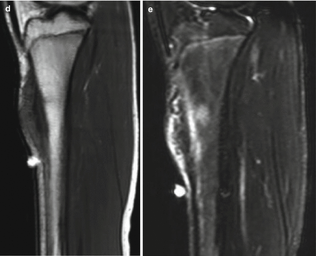
Fig. 14.1
AP radiograph (a) of a 15-year-old male shows a region of cortical thickening, aggressive periosteal reaction, and subtle matrix production along the medial aspect of the proximal tibial diaphysis with the suggestion of an associated unmineralized soft tissue mass. The axial and sagittal T1 (b, d) and axial and sagittal fat-suppressed T2 weighted (c, e) MR images show that the mass involves the surface of the bone without involvement of the medullary canal. The mass is heterogeneous with a region of predominantly low T2 signal intensity near the attachment to the cortex that correlates with the periosteal reaction and matrix production on the radiograph. In addition, there is an unmineralized component to the soft tissue mass with nonspecific intermediate T1 and hyperintense T2 signal
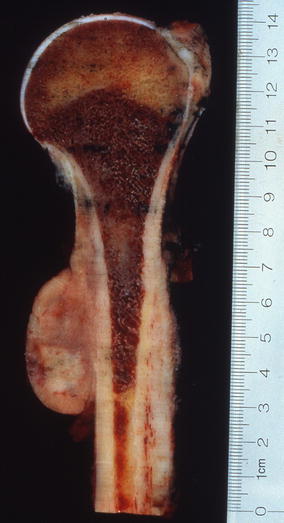
Fig. 14.2
Periosteal osteosarcoma forming a surface-based mass involving the diaphyseal portion of the underlying bone. The tumor contains zones of gray-white tissue reflecting areas of cartilaginous differentiation
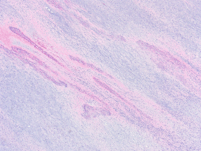
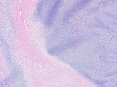
Fig. 14.3
Periosteal osteosarcoma showing histological features of a chondroblastic osteosarcoma. The malignant osteoid forms spicules of bone arranged perpendicular to the surface of the underlying cortical bone
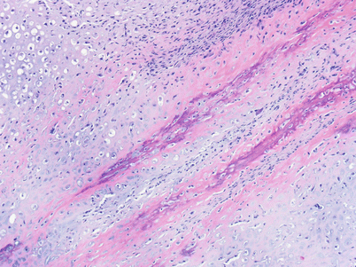
Fig. 14.4
Periosteal osteosarcoma composed of an intermediate-grade chondroblastic osteosarcoma