Fracture type
Internal fixation performed (%)
Type A
55
3.9
Type B
25
37.3
Type C
20
54.3
Complex trauma
12
57.5
+ Acetabular fracture
15
Concomitant injuries
Head
40
Thorax
36
Abdomen
25
Spine
15
Peripheral skeleton
69
Urogenital
5 (50 in complex trauma)
Table 17.2
Epidemiology of isolated acetabular fracture (study group pelvis of AO, n = 704; and according to Letournel and Judet, n = 940)
Fracture type | Frequency (%) | |
|---|---|---|
Letournel & Judet (%) | Study group pelvis (AO) (%) | |
Posterior wall | 24 | 14 |
Posterior column | 3 | 11 |
Anterior wall | 2 | 2 |
Anterior column | 4 | 13 |
Transverse | 7 | 17 |
Posterior column + posterior wall | 3 | 5 |
Transverse + posterior wall | 20 | 7 |
T-type | 7 | 8 |
Anterior column + post. hemitransverse | 7 | 3 |
Both columns | 23 | 20 |
The rate of neurological concomitant lesions was 11 % in complex trauma.
In fractures of type C, 50–70 % of the patients were polytraumatized.
In the above-mentioned study, isolated acetabular fractures are seen in 21 % of all pelvic injuries. In a large series by Judet and Letournel (940 patients), the most frequent fracture types (for classification, see below) were the posterior wall fracture, the transverse and the posterior wall fracture, and the two columns fracture, each with a frequency of 20–25 % (Table 17.2). In 15 % of the acetabular fractures, concomitant injuries of the pelvic ring were found, and in 45 % there were additional fractures of the peripheral skeletal system. According to Judet and Letournel, neurological damages were seen in 12 %; in two-thirds of the cases there were fractures of the posterior column or of the posterior wall.
Pediatric pelvic ring and acetabular injuries are extremely rare. Because of the increased elasticity and therefore associated high restoring forces of the pediatric pelvic ring, only radiographically minimally displaced fractures can be found, despite high-energy impact, whereby those fractures are often undervalued. The frequency of complex pelvic ring injuries in children is about 20 %, twice as much as in adolescents. Acetabular fractures in children are also extremely rare; the percentage of all pediatric fractures accounts for about 0.01 %. Mostly, disruptions of the Y-shaped epiphyseal plate are found.
17.3 Classification of Injuries
Generally, injuries of the pelvis can be divided into pelvic ring injuries and acetabular fractures.
The pelvic ring injuries can be classified as isolated osseous or ligamentous injuries, with or without instability of the pelvic ring, and as complex pelvic trauma.
Patients presenting with a combination of pelvic fracture and concomitant soft tissue injuries have a fourfold higher lethality compared with those suffering from an isolated, “uncomplicated” pelvic fracture. Therefore, the term complex pelvic injury has been established. Complex pelvic injuries are characterized by soft tissue injuries, including extensive subcutaneous décollements (Morel-Lavallée syndrome), urogenital injuries, and anorectal impalements. The stability of the pelvic girdle is mainly dependent upon the integrity of the posterior sacroiliac ring segment, consisting of the posterior parts of the iliac bones, the sacrum, and their ligamentous connections. The extent and localization of the interruption of the pelvic girdle determine the classification, whereas smooth transitions between the different directions of instability are possible.
Acetabular fractures are classified according to Letournel, who established a consideration of an “anterior” and “posterior” column into the morphological analysis of acetabular fractures. The classification of Letournel is the basis of all further analyses and classifications. Based on this classification, there are five elementary fractures, where it is about column, transverse, and wall fractures, and five combined fracture types.
17.4 Pelvic Ring Injuries
17.4.1 Injury Mechanism
Pelvic ring injuries are mostly the result of high-energy trauma with direct or, transferred via the femur, indirect energy impact. In about 52 % of cases pelvic ring injuries are caused by car accidents, in 37 % by falls from a great height, and in 11 % by other injuries. Severe open pelvic trauma (crush injury) is mostly caused by a run over by a vehicle.
In principle, three vectors of energy impact to the pelvic girdle can be defined (Fig.17.1):
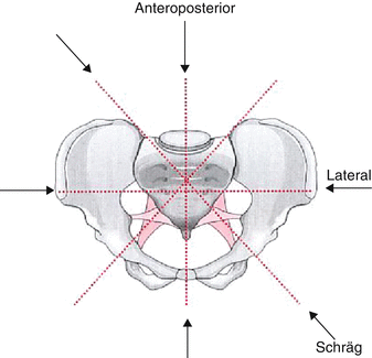

Fig. 17.1
Vectors of energy impact to the pelvic ring
The anterior–posterior compression with an anterior or posterior impact to the pelvic girdle results in an external rotation movement of the hemipelvis presenting the so-called “open-book” mechanism. In this type of injury, the posterior ligamentous structures are partially intact.
The lateral compression is the most common energy impact to the pelvic girdle (75 %). Posterior-lateral impact will result in a rupture of the symphysis or in pubic rim fracture, whereas anterior-lateral impact will cause an internal rotation movement of the hemipelvis with partial disruption of the posterior ligamentous structures.
The vertical shear mechanism consists of a vertical energy impact to the posteriorly stabilizing structures of the pelvic ring segment. Translational movements will result in a complete disruption of the pelvic girdle posteriorly and anteriorly. The posterior injuries can be presented as disruptions of ligamentous structures or as osseous lesions of the sacrum or the iliac bone. Because “directions” in the various positions of the pelvis often cannot be determined sufficiently, the vertical shear mechanism is increasingly constituted as a translation injury because the direction of movements are irrelevant for the complete disruption of the sacroiliac structures.
17.4.2 Classification
The classification is based on the direction of energy impact to the pelvic girdle. Thereby, the fracture course and the stability of the integrity of the pelvic girdle are determined. The A-B-C system according to Müller, Isler, and Ganz, which is assumed by the AO/OTA, has been established for the classification of pelvic ring injuries. The graduation proposed by Pennal considering the pure injury mechanism is combined with the classification according to Tile, who respects the degree of instability of the pelvic girdle.
In daily practice, the pelvic ring injury is divided into a stable and unstable type. Additionally, the direction of the instability is indicated, that is, in the anterior pelvic ring segment, transpubic and transsymphyseal instability, and in the posterior ring segment, transiliac, transsacroiliac, transsacral, and transacetabular instability. The AO/Ota classification is the first system that allows a complete alphanumeric graduation of all injuries by separated reflection of the anterior, posterior, right, and left pelvic ring segment.
17.4.2.1 Type A Injuries
Pelvic rim or pelvic ring injuries without loss of pelvic stability (Fig. 17.2).
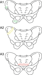

Fig. 17.2
Classification of type-A pelvic ring fractures
Type A fractures are stable fractures. Examples are avulsion fractures, iliac wing fractures, pubis or ischial rim fractures, as well as transverse sacral fractures below the sacroiliac joint.
Type A 1: Iliac wing fracture
Type A 2: Anterior pelvic ring fracture with minimal lesion of the posterior ring segment not compromising the stability of the pelvic ring
Type A 3: Transverse sacral or coccygeal fracture (below the sacroiliac joint)
17.4.2.2 Type B Injuries
Type B injuries are characterized by an anterior lesion of the pelvic girdle with partial disruption of the posterior ring segment (Fig. 17.3). In addition to instability of the anterior pelvic ring segment, the anterior ligamentous structures of the sacroiliac joint are damaged, mostly as a result of an anterior-posterior or a lateral energy impact. The posterior pelvic ring can be affected ipsilaterally, contralaterally, or bilaterally to the anterior lesion. External rotation injuries cause the so-called “open-book” mechanism, whereas internal rotation injuries are the result of a lateral compression with possible impression fracture of the ala of the sacrum or with partial rupture of the dorsal ligamentous structures of the sacroiliac joint.


Fig. 17.3
Classification of type-B pelvic ring fractures
The lateral compression can impact aslope, yielding a rotation of the affected hemipelvis in the vertical as well as in the sagittal pane. By disruption of additional ligamentous structures, this very unstable Type B injury is often difficult to differentiate from a type C injury. The one hemiplevis can be internally rotated like a handle of a bucket. Therefore, this injury type is termed by Pennal as a “bucket handle” injury.
Type B 1:External rotation injuries:
Anterior-posterior impact or indirect energy impact transferred via the femur will result in an external rotation of one or both hemipelvis (“open book” mechanism). Type B1.1 and type B1.2 represent the classical open book mechanism: a disruption of the symphysis of, respectively, less or more than 2.5 cm. In cases of type B 1.3 lesions, there are additional flexion or extension rotational components.
Type B 2:Internal rotation injuries:
Ipsilateral energy impact yields a lesion of the posterior ring segment with internal rotation of the affected hemipelvis. There are often compression fractures of the ala of the sacrum (type B 2.1). Type B 2.2 injury is characterized by partial dislocation of the sacroiliac joint, and type B 2.3 injury shows an incomplete posterior iliac fracture.
Type B 3:Bilateral posterior rotation injury:
Type B 3.1 is characterized by bilateral external rotation injury and type B 3.2 by an internal rotation injury of the one side and, contralaterally, external rotation injury. Type B 3.3 represents a bilateral fracture of the sacrum caused by internal rotation injury.
17.4.2.3 Type C Injuries
The type C injuries are caused by translation or shearing movements of the involved hemipelvis resulting in a complete disruption of the anterior as well as the posterior pelvic ring segment (Fig. 17.4).


Fig. 17.4
Classification of type-C pelvic ring fractures
Type C 1:Unilateral type C injury–the posterior ring segment is completely interrupted at the iliac bone, the sacroiliac joint, or at the sacrum.
Type C 2:Unilateral type C injury with additionally contralateral type B injury–in cases of increased vertical shear energy impact, the contralateral hemipelvis can be involved by a type B injury of the dorsal ring segment. Depending on the direction of force impact, internal or external rotation injury will result.
Type C 3:Bilateral type C injury–the type C 3.1 injury represents the bilateral extrasacral instability, the type C 3.2 injury is characterized by a combined transsacral and contralateral extrasacral lesion, and the type C 3.3 injury shows a bilateral transsacral instability.
Depending on the potency of the energy impact, smooth transitions are possible between solitary rotational and additional vertical instabilities.
There exist different classifications for sacral fractures, which can appear with type A 3, type B, and type C fractures. For clinical daily practice, the classification according to Dennis (Fig. 17.5) has been proved.
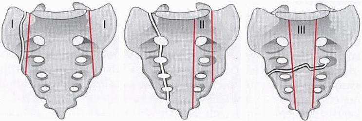

Fig. 17.5
Classification of sacral fractures according to Dennis
This classification is based on three zones in relation to the sacral foramina. It is easy to remember and allows an estimation of the expected rate of neurological damage of the sacral neural plexus. Zone I is located laterally and zone III medially of the sacral foramina; zone II is bordered by the sacral foramina. Furthermore, longitudinal fractures, which can proceed from one of the above-called zones, are differentiated from transverse fractures.
17.4.3 Diagnostic Procedures
17.4.3.1 Medical History and Clinical Examination
Because of potentially extensive extravasation of blood volume and concomitant intraabdominal injuries, patients with pelvic fracture may face life-threatening injuries. In principle, the life-threatening situation (polytrauma, complex trauma, crush injury) must be distinguished from the hemodynamically stable situation in solitary pelvic ring injuries without danger to life. The diagnostic procedures as well as the emergency treatment must proceed according to a graduated scheme (ATLS® concept) in polytraumatized patients (Fig. 17.6).
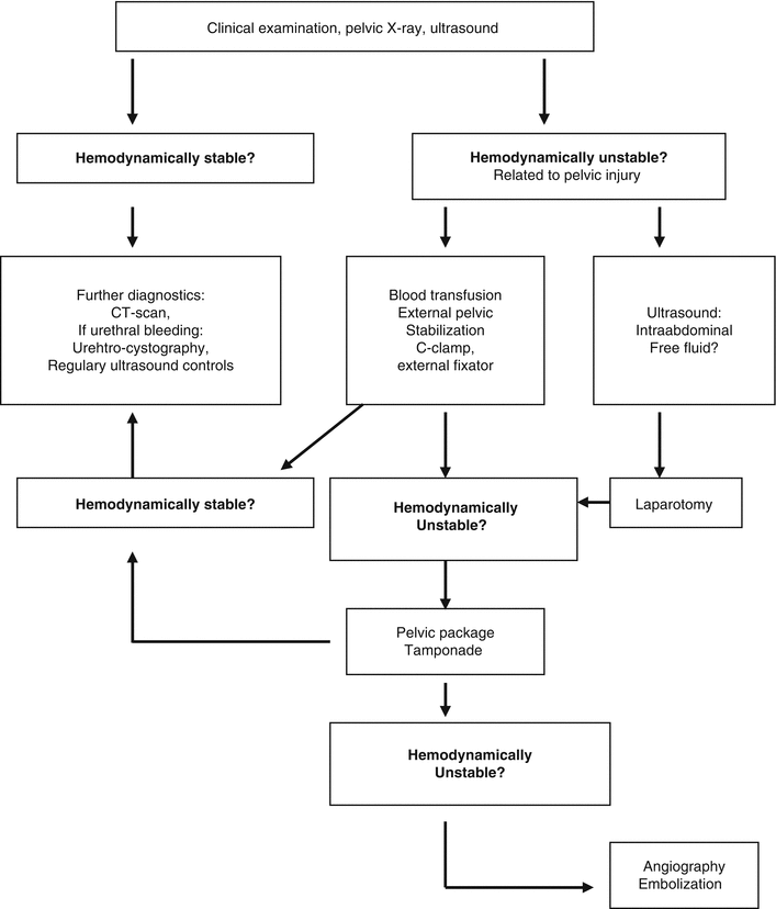

Fig. 17.6
Diagnostics in pelvic trauma
The clinical history, which should essentially consider the injury mechanism, gives primary information for an injury of the pelvic girdle. Clinical examination starts with the inspection of all orifices and concomitant soft tissue injuries (wounds, hematomas, contusions, impalements).
Pelvic ring instability should be tested by gentle but firm compression and distractive movements of the iliac wings. The insertion of a urethral catheter is part of the primary diagnostic procedure. Urethral hemorrhage, scrotal hematoma, and difficult or impossible insertion of the urethral catheter must raise suspicion of a urogenital injury.
When the patient is alert, a precise neurological examination must be performed to exclude neurological damage; its frequency accounts for 11 % in complex trauma and 20–50 % in unstable fractures of the sacrum.
17.4.3.2 Diagnostic Devices
Basic diagnostic procedures include abdominal ultrasound and conventional pelvic X-rays. CT scan and angiography are considered extended diagnostic procedures.
Ultrasound
Ultrasound should be performed immediately in the emergency room. Within several minutes, the whole abdomen can be examined in consideration of free fluid and lacerations of parenchymatous organs (liver, spleen, and kidney). In the pelvis, extended retroperitoneal hematoma as well as peri- or intravesical bleeding or a tamponade of the bladder can be recognized.
Conventional X-Rays
Subsequently, the standard a.p.-view pelvic X-ray is taken. When there is suspicion of involvement of the posterior pelvic ring, additional radiographs using the inlet and outlet technique are performed by tilting the X-ray tube 45°in the frontal plane cranially and caudally, respectively (Fig. 17.7). The inlet view can demonstrate horizontal dislocations, whereas the outlet view shows vertical dislocations. In daily clinical practice, these techniques are being increasingly replaced by the CT scan. However, they are still recommended so as to have a comparison using the intraoperative image to check the reduction of vertical dislocation in type C injuries.
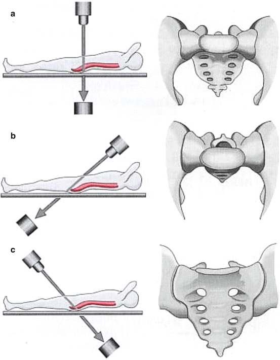

Fig. 17.7
Standard X-rays: (a) a.p., (b) inlet, (c) outlet view
CT Scan
Only the CT scan is able to visualize the whole dimension of the osseous and ligamentous lesions of the posterior pelvic girdle. The dislocation of fracture fragments and their three dimensional orientation are better visualized than with conventional X-rays. The CT scan enables the severity of the lesion of the posterior pelvic girdle and therefore its classification. Three dimensional CT scan reconstructions improve the spatial orientation of the fracture lines. By using contrast agents, additional urethral and bladder injuries can be recognized.
Angiography
Angiography is occasionally indicated in cases of persistent hemodynamic instability in pelvic trauma. Using digital subtraction angiography (DSA) technique, intrapelvic bleeding in the area of the internal iliac artery can be visualized if dynamics of the bleeding is greater than 3 ml/min. In the same procedure, the artery can be occluded using coils or gelofoam particles. The value of the angiography is differently discussed. Because the most hemodynamically relevant bleeding has a venous origin, angiography seems to not be indicated. In the own management we attach no importance to the angiography.
When a urethral or bladder injury is suspected, further investigation using contrast agents – retrograde urethrography and cystography – are performed. Rectal examination reveals a dislocation of the prostate (so-called “riding prostate”) in cases of posterior supradiaphragmal urethral laceration.
Following a high-energy impact to the perineum, rectal examination should be followed by a rectoscopy to exclude laceration of the rectum.
17.4.4 Treatment
17.4.4.1 Treatment Tactics
In addition to appraisal of the stability of the pelvic girdle, differentiation between pelvic fractures without or with life-threatening bleeding is made during the initial treatment phase. In principle, two initial situations can be defined in accordance with the type (direct, indirect trauma, crush injury) and severity of energy impact: the life-threatening emergency situation in cases of complex trauma or crush injury with hemodynamic instability and the solitary osseous or ligamentous injury without hemodynamic effects.
17.4.4.2 Treatment Concepts in Emergency Cases
Patients with pelvic ring injuries and life-threatening hemorrhages (hemoglobin < 8 mg/dl on arrival at the emergency room) should be managed according the “pelvic emergency algorithm” (Fig. 17.8). In cases of massive obvious hemorrhages following crush injuries or run-over, patients will be rushed to the operating room for emergency surgical procedures and, if necessary, emergency hemipelvectomy. In cases of life-threatening hemorrhages, where the source of bleeding is not obvious, it may be wise to initiate shock treatment and apply massive fluid challenge. When the patient is not responding to these procedures and is not in a hemodynamically stable condition within 10–15 min, self-tamponade of the bleeding into the retroperitoneal space cannot be assumed. These cases must be addressed by giving O negative packed red cells, and the pelvic girdle should be stabilized mechanically by external procedures to reduce the intrapelvic space. Bleeding caused by the cancellous bone and by the posterior sacral and anterior perivesical venous plexus will be compressed. The C-clamp (Fig. 17.15) is the appropriate external stabilizer for type C injuries. In cases of open-book injuries, the pelvic girdle can be closed using an anterior external fixator (Fig. 17.16).
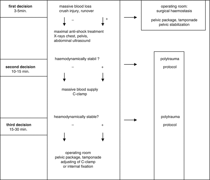

Fig. 17.8
Treatment algorithm in the emergency pelvic trauma
In continuous hemodynamic instability passing the next 15–30 min, the patient should be transferred to the operating room to perform a packing tamponade. The incision is performed from the symphysis to the umbilicus. Extraperitoneal inspection of the pelvis follows. Massive hematoma is evacuated and the true pelvis is packed with tamponades at both sides of the bladder to the posterior presacral region. Packing is only helpful when counterpressure can be established after external stabilization of the pelvic ring. If necessary, the C-clamp should be adjusted. The anterior pelvic ring is stabilized by a single internal fixation (plate or tension band wiring). The definitive stabilization and reconstruction of the posterior pelvic ring may be performed secondarily if applicable during a second-look procedure of the pelvic package.
Using military anti-shock trousers (MAST) for external compression of the pelvic girdle does not avoid dissemination of the hematomas and, furthermore, may increase the danger of a severe compartmental syndrome. The MAST suit makes also the artificial ventilation and the clinical handling of the patient difficult. Therefore, this equipment has no relevance in this situation.
17.4.4.3 Treatment Concept in Pelvic Injuries Without Hemodynamic Instability
Operation Time
All pelvic girdle injuries requiring stabilization will be operated on as soon as possible when control of hemorrhage and protection of the soft tissue are ensured.
Nonoperative Treatment
Stable pelvic ring fractures (type A injuries) can be treated nonoperatively. Fractures of the pubic rim, undisplaced iliac wing fractures, transverse fractures of the sacrum below the linea terminalis, and in zone I according to Dennis require only a short time of bed rest until relief of fracture pain is attained. Avulsion fractures of the iliac spine or of the ischial tuberosity can also be treated nonoperatively. The leg is carried in the corresponding unloaded position for 1–2 weeks. Avulsion of the inferior anterior iliac spine heals without problems because the alteration of the length of the muscle can be completely compensated. This can also be applied to the avulsion of the ischial tuberosity; the hip joint is carried in an extension position to relax the hamstrings. Internal fixation using lag screws, tension band wiring, or transosseous sutures is recommended only in active athletes.
In type B and C fractures, internal fixation is not often performed. Nonoperative treatment is possible but its result is a high percentage of deformities of the pelvic ring. The often painful instability of the ligamentous injury will lead to a poor clinical outcome. Therefore, internal fixation is considered as the standard and, therefore, pelvic ring injuries should be admitted to a trauma center.
Operative Treatment
Surgical treatment with internal fixation in cases of unstable type B and C fractures enables anatomical reconstruction of the pelvic girdle, resulting in possible weight bearing and in a reduction of the immobilization phase.
Indication for open reduction and internal fixation can also been seen in type A fractures, such as major dislocations of iliac wing or pubic rim fractures that are dangerous to vascular, neural, or bladder lesions (so-called “tilt-fracture”).
Fixation Tactic
The principle of internal fixation in type B fractures is the closure of the anterior pelvic ring. Transsymphyseal instabilities always require open reduction and internal fixation by plate. Transpubic instabilities can be fixed by an external fixator or by transpubic screws. In cases of additional external or flexional instability (type B 1.3, type B 2.3 injury) stabilization of the posterior ring may be necessary.
In type C injuries, the anterior as well as the posterior pelvic ring has to be reconstructed. Only in cases of minor instability of the anterior pelvic ring (in the context of an internal rotation injury) can the anterior stabilization be renounced (Fig. 17.9).
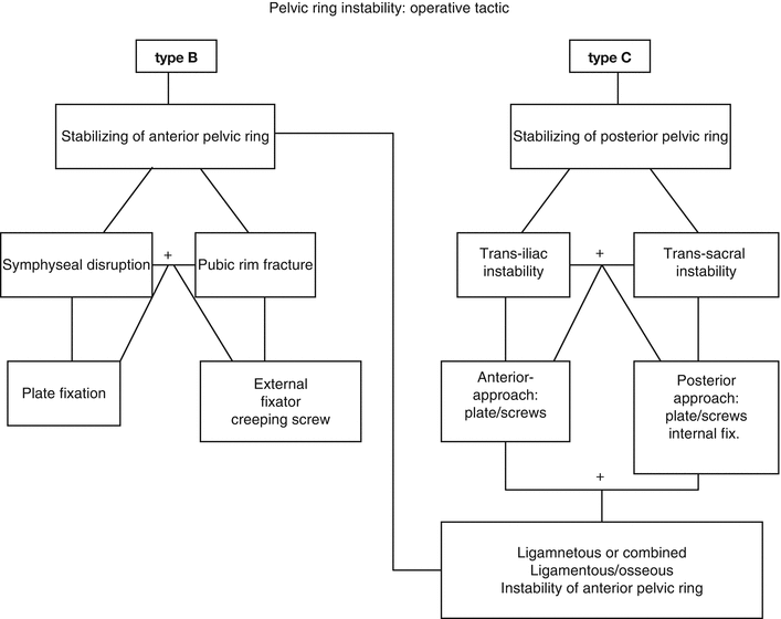

Fig. 17.9
Operative tactic in type-B injuries and in type-C injuries
Stabilization of the Anterior Pelvic Ring
The most common indication for stabilization of the anterior pelvic ring is the rupture of the symphysis.
Approach
In cases of isolated transsymphyseal instability, a Pfannenstiel approach is performed; in cases of laparotomy, via a median longitudinal laparotomy.
Required stabilization of transpubic instabilities is achieved via minimally invasive medial incisions. The complete exposure of the pubic rim is not necessary and increases the risk of damage of the femoral nerve and vessels. Both rectus abdominis muscles are engrailed at both sides only as much as necessary. The cavum retzii before the bladder becomes obvious. During wound closure, exact refixation of the abdominal muscles is mandatory to avoid a hernia.
Operation Technique
Small, 4.5-mm DC plates are normally used for fixation. A flush closure of the symphysis should be attempted. A sharp reduction clamp into the obturator foramen can be used for reduction. The plate is located at the cranial area of the pubic rims (Fig. 17.10). Normally, four-hole, 4.5-mm plate and cortical screws are used. The direction of drilling is 30° to the vertical plane dorsally and caudally. In children, tension band wiring over two screws near the symphysis can be performed alternatively.
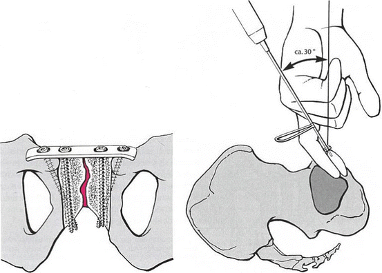

Fig. 17.10
Fixation of symphysis: localization of the plate and direction of drilling
In unstable fractures of the pubic rims, screw fixation can be carried out. Cortical screws are inserted via small medial incisions. Because only the medial cortex is drilled, the screws are creeping intramedullary (so-called “creeping screws”) and work as a lag screw when passing the fracture line.
Stabilization of the Posterior Pelvic Ring
In type C injuries, the approach is determined by the site of the instability. Depending on the site of instability, the soft tissue damage, and concomitant injuries, the stabilization is performed via an anterior-lateral or via a dorsal approach (open or closed). Transiliac instability is approached anterior-laterally (Fig.17.11). Stabilization can be performed using plates and screws (Fig. 17.12a). The anterior-lateral approach is also feasible for sacroiliac dislocation (Fig. 17.12b) and for transsacral instability with a small anterior fracture fragment of the sacrum.
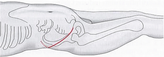


Fig. 17.11
Anterior-lateral approach to the posterior pelvic ring

Fig. 17.12


Stabilization by plate of iliac (a) and sacroiliac (b) instability
Stay updated, free articles. Join our Telegram channel

Full access? Get Clinical Tree


