Benign lesions
Malignant lesions
Enchondroma
Metastatic bone disease
Bone islands
Multiple myeloma
Paget’s disease
Lymphoma
Hyperparathyroidism
Chondrosarcoma
Malignant fibrous histiocytoma (undifferentiated sarcoma)
Very few benign lesions cause symptoms in adults. Paget’s disease occasionally causes symptoms. Patients with bone destruction secondary to hyperparathyroidism may also have symptoms. In general enchondromas and bone infarcts do not cause pain.
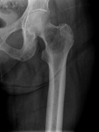
When a patient presents with a destructive bone lesion (Fig. 1), the clinician does an evaluation to try to find the cause of the destructive lesion. The evaluation scheme is quite simple:

Get Clinical Tree app for offline access


Fig. 1
Anteroposterior radiograph showing a destructive lesion in the proximal femur. The differential diagnosis of this lesion includes metastatic bone disease, multiple myeloma, lymphoma, chondrosarcoma, and malignant fibrous histiocytoma
Computerized tomography scan of the chest, abdomen, and pelvis: This test is rapid (generally less than 3 min) and can be done without contrast. This test is very sensitive for:
Lung cancer
Kidney cancer
Pulmonary metastases
Liver metastases
Lymph node enlargement
Technetium bone scan: This test is very sensitive for detecting bone metastases. If there is more than one destructive lesion present (increased uptake on the technetium bone scan and correlation with a radiograph), the differential diagnosis narrows as one can exclude monostotic processes (such as chondrosarcoma or malignant fibrous histiocytoma).
Laboratory tests
Complete blood count
Hemoglobin level – low levels of hemoglobin are often indicative of replacement of the bone marrow by a malignancy. Hemoglobin levels of less than 12 mg/dl are a presenting finding in two thirds of patients with muliple myleoma. Patients who have a hemoglobin less than 12 mg/dl and a sedimentation rate greater than 50 mm/h often have multiple myeloma.
Stay updated, free articles. Join our Telegram channel

Full access? Get Clinical Tree


