, Ratna Maheshwari2 and Shalin Maheshwari2
(2)
Pediatric Orthopedics, Childrens’ Orthopedic Centre, Mumbai, India
Take-Home Message
Ultrasound is a better test than plain radiographs in the first 4–6 months of life.
Routine ultrasound screening should be performed for infants with risk factors for the condition. Because of the poor specificity of ultrasonography in children younger than 1 month, hip ultrasonography should be deferred until after 1 month of life.
Pavlik harness treatment should be discontinued if the dislocated hip does not relocate within 3–4 weeks, to avoid Pavlik harness disease.
Excessive hip flexion in the Pavlik harness results in an increased risk of femoral nerve palsy. Excessive hip abduction in the Pavlik harness results in an increased risk of osteonecrosis of the femoral head.
Hip abduction does not become limited in DDH until approximately 6 months of age.
Definition
Developmental dysplasia of the hip (DDH) refers to the complete spectrum of pathologic conditions involving the developing hip, ranging from acetabular dysplasia to hip subluxation to irreducible hip dislocation.
Teratologic dislocation of the hip occurs in utero and is irreducible on neonatal examination. This condition always accompanies other congenital anomalies or neuromuscular conditions, most frequently arthrogryposis and myelomeningocele.
Aetiology
Multifactorial—genetic, hormonal and mechanical. Maternal hormone like relaxin may play a role.
It is more common in females with the left hip more commonly involved than the right hip.
There is an increased incidence in breech deliveries. It is associated with oligohydramnios, torticollis, talipes calcaneovalgus and metatarsus adductus (intrauterine packaging disorder).
Primary acetabular dysplasia may run in families.
Pathoanatomy
The femoral head and neck are anteverted. The head is pulled proximally and laterally by abductors.
Hip joint fills by fibrofatty tissue (pulvinar).
Acetabular labrum becomes inverted and enlarged. Limbus blocks reduction of the femoral head.
Acetabulum becomes dysplastic.
Ligamentum teres becomes lengthened and redundant. Transverse acetabular ligament is pulled superiorly.
Capsule becomes expanded.
Muscles crossing the hip joint—hamstring, hip adductors and psoas—become shortened and contracted and block reduction.
Evaluation
Clinical presentation—The clinical presentation varies with age.
In the neonatal period, instability of the hip is the key clinical finding.
Hip clicks are nonspecific physical findings.
Because asymmetric skin folds are common in children with normal hips, children with such asymmetry have a high rate of false positives.
In infants older than 6 months, limitation of motion and apparent limb shortening are common findings.
In toddlers, in addition to restricted motion and limb-length inequalities, a limp or waddling gait may be appreciated.
In adolescents, all the above findings may be present in addition to fatigue and pain in the hip, thigh or knee.
Physical examination—Accuracy of the physical examination requires that the child be relaxed.
The Galeazzi (or Allis) test is positive only in unilateral severe subluxations or dislocations. In the Galeazzi test, the hips are flexed to 90°; the test is positive if one knee (the involved side) is lower than the other.
The Barlow test is performed by applying a posterolateral force to the extremity with the hip in a flexed and adducted position. The test is positive if the hip subluxates or even dislocates.
The Ortolani test is performed by abducting and lifting the proximal femur anteriorly. The test is positive if the dislocated hip is reducible.
Range of motion (ROM) testing of the hip is important, with decrease in abduction as the most sensitive test for DDH. ROM however will be normal in children younger than 6 months because contractures will not yet have developed.
Diagnostic tests
Ultrasonography—In the first 4 months of life, plain radiographic evaluation is unreliable because the femoral epiphysis has not yet ossified. Ultrasonography of the infant hip (before the appearance of the proximal femoral ossific centre) is useful in confirming a diagnosis of DDH and also in documenting reducibility and stability of the hip in an infant undergoing treatment with a Pavlik harness or brace.
Graf classification (Table 1)—after taking a midcoronal section, alpha angle is measured between the line of the ilium and bony acetabulum. The beta angle is the angle between the line of the ilium and cartilaginous acetabular roof.
Table 1
Graf classification
Types
Alpha angle
Beta angle
Description
Treatment
1
>60
<55
Normal
None
2
43–59
55–77
Physiological in child <3 months, delayed ossification in child >3 months
?
3
<43
>77
Lateralization
Pavlik harness
4
Unmeasurable
Dislocated and labrum interposing between the head and acetabulum
Pavlik harness/closedor open reduction
Radiology
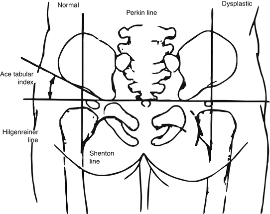
Radiographic lines in DDH (Fig. 1)

Fig. 1
Radiographic lines in DDH. Hilgenreiner line is drawn through the triradiate cartilages. The Perkin line is drawn perpendicular to the Hilgenreiner line at the margin of the bony acetabulum. The Shenton line curves along the femoral metaphysis and connects smoothly to the inner margin of the pubis. The acetabular index is the angle between a line drawn along the margin of the acetabulum and the Hilgenreiner line. It averages 27.5° in normal newborn and decreases with age
The Hilgenreiner line is a line drawn horizontally through each triradiate cartilage of the pelvis.
The Perkin line is drawn perpendicular to the Hilgenreiner line at the lateral edge of the acetabulum.
The Shenton line is a continuous arch drawn along the medial border of the femoral neck and superior border of the obturator foramen.
The acetabular index is the angle formed by an oblique line (through the outer edge of the acetabulum and triradiate cartilage) and the Hilgenreiner line.
In the newborn, a normal value averages 27.5°.
By 24 months of age, the acetabular index decreases to 21°.
The centre–edge angle of Wiberg is the angle formed by a vertical line through the centre of the femoral head and perpendicular to the Hilgenreiner line and an oblique line through the outer edge of the acetabulum and centre of the femoral head. The centre–edge angle is reliable only in patients older than 5 years. A centre–edge angle <20° is considered abnormal.
Arthrography of the hip—Useful in confirming the acceptability of a closed reduction and in diagnosing the blocks to reduction, such as capsular narrowing and labral hypertrophy.
CT—The standard for confirming acceptable reduction for a patient in a spica cast following closed or open procedures.
Magnetic resonance imaging:
MRI can also be used to confirm acceptable reduction of the hip following closed or open procedures.
MRI can also be useful in an older patient with suspected labral pathology.
Management
Based on the age of the child, stability of the hip (unstable versus dislocated hip) and severity of acetabular dysplasia. The aim of management is to obtain concentric, congruent and stable hips.
Dysplastic hip in neonate through 6 months of age
In a child with an abnormal α angle on ultrasound or with an unstable hip (subluxatable hip on examination), initial treatment usually includes a Pavlik harness.
Proper positioning of the Pavlik harness is critical.
The hips should be flexed to 100° with mild abduction (two to three finger breadths between knees when knees are flexed and adducted).
Excessive flexion should be avoided to lower the risk of femoral nerve palsy.
Excessive abduction should be avoided to lower the risk of osteonecrosis; osteonecrosis can occur in both the normal and dysplastic hip.
The child can be weaned from the Pavlik harness over a 3- to 4-week period when ultrasound parameters become normal.
Success rates for Pavlik harness treatment in this setting have been reported at >90 %.
The recurrence rate is 10 %; therefore, follow-up evaluation until maturity is necessary.
In a relatively large child or in a child older than age 6 months with a dysplastic hip or with hip subluxation, a fixed abduction orthosis or spica casting is an option. Pavlik harness treatment is ineffective in this setting because the child generally “overpowers” the brace.
Dislocated hip in neonate through 6 months of age
If the hip is Ortolani positive, Pavlik harness treatment is initiated.
Frequent (every 1–2 weeks) re-examination (clinical plus ultrasound) is necessary to ensure that the hip is reduced.
Once the hip becomes stable, treatment is the same as the protocol described above for treatment of dysplastic hip.
Success rate (i.e. the hip becomes reduced) is reported to be 85 %.
The risk of osteonecrosis is low (<5 %), especially if treatment is initiated early (i.e. in the first 3 months of life).
If the hip is Ortolani negative, initial treatment still includes a Pavlik harness. If reduction is not achieved by 3–4 weeks (confirmed by ultrasound), however, Pavlik harness treatment is abandoned, and closed reduction is necessary.
Pavlik harness treatment should be discontinued if the dislocated hip does not relocate within 3–4 weeks, to avoid Pavlik harness disease.
In Pavlik harness disease, the femoral head sits up against the edge of the acetabulum and worsens the acetabular dysplasia, particularly the posterolateral rim.
Dislocated hip in children 6–18 months of age
Closed reduction is the preferred method of treatment in children age 18 months or younger.
Secondary femoral or acetabular procedures are rarely necessary in this age group.
The evidence for preliminary traction is equivocal. Given this and the possible complications of skin slough and leg ischaemia, most centres have abandoned preliminary traction.
Closed reduction is performed under general anaesthesia.
Adductor tenotomy frequently is necessary.
Hip arthrography is used intraoperatively to confirm adequacy of reduction.
There should be <5 mm of medial contrast pooling between the femoral head and acetabulum.
The safe and stable zones for abduction/adduction, flexion/extension and internal/external rotation should be established.
A spica cast is applied with the hip maintained in the “human position” (hip flexion of 90–100° and abduction). Hip abduction should be <60° to minimize the risk of osteonecrosis.
The reduction of the hip in the cast must be confirmed.
Presently, CT is the standard.
MRI is used in some centres to confirm adequacy of reduction of the hip.
Cast immobilization is continued for 3–4 months.
Cast change is sometimes necessary.
Removable abduction brace is used afterwards until the acetabulum normalizes.
Dislocated hip in children >18 months of age
Open reduction is the preferred treatment.
Unilateral dislocation: Surgical treatment is generally indicated in children up to 8 years of age with a unilateral dislocation. After 8 years of age, the risks of surgery are felt to outweigh the benefits.
Bilateral dislocation: The upper age limit for surgical treatment in these children is typically 5–6 years.
Femoral shortening is indicated in children with significantly high-riding dislocations.
This is necessary in most, but not all, children ≥2 years of age.
Pelvic osteotomy is needed for significant acetabular dysplasia (typical in children >18–24 months old). If a pelvic osteotomy is performed in this age group at the time of initial surgery, the rate of reoperation is reduced significantly.
Open reduction
Open reduction is indicated if concentric closed reduction cannot be achieved or when excessive abduction (>60°) is required to maintain reduction.
The goal of open reduction is to remove the obstacles to reduction and/or safely increase its stability.
Impediments to congruent reduction are the iliopsoas, hip adductors, capsule, ligamentum teres, pulvinar and transverse acetabular ligament. An infolded labrum may be an impediment in some cases.
The most commonly used approaches are anterior, anteromedial and medial.
Secondary procedures
Secondary femoral and/or pelvic procedures are frequently necessary in children older than 2 years of age to achieve and maintain concentric reduction and minimize the risk of osteonecrosis.
Femoral osteotomy
Femoral osteotomy provides shortening (to decrease pressure on the femoral head, thereby minimizing the risk of osteonecrosis), derotation (external rotation to address the abnormally high femoral anteversion in DDH) and varus.
Avoid excessive varus, because the greater trochanter can impinge against the acetabulum and prevent concentric reduction.
Pelvic osteotomy
Indications for pelvic osteotomy include persistent acetabular dysplasia and hip instability.
There is considerable variability in clinical practice with regard to pelvic osteotomy in children >2 years of age.
The two general types of pelvic osteotomy are reconstructive and salvage:
Reconstructive osteotomies redirect or reshape the roof of the acetabulum with its normal hyaline cartilage into a more appropriate weight-bearing position. A prerequisite to a reconstructive pelvic osteotomy is a hip that can be reduced concentrically and congruently. The hip must also have near-normal ROM. Redirectional osteotomies include single innominate (Salter), triple innominate (Steel) and peri-acetabular (Ganz). Redirectional osteotomies involve complete cuts through various pelvic bones, and those that cut all three pelvic bones provide the ability to obtain greater coverage. Reshaping osteotomies involve incomplete cuts, and hinge on different aspects of triradiate cartilage can decrease volume. It must be done in patients with open triradiate cartilage. These include Dega and Pemberton osteotomies.
Salvage procedures are typically indicated in adolescents with severe dysplasia in whom acetabular deficiency precludes the use of a redirectional osteotomy. In salvage procedures, weight-bearing coverage is increased by using the joint capsule as an interposition between the femoral head and bone above it. The intent of these osteotomies is to reduce point loading at the edge of the acetabulum. These osteotomies rely on fibrocartilaginous metaplasia of the interposed joint capsule to provide an increased articulating surface. Salvage osteotomies include Chiari and shelf osteotomies.
Complications
Pavlik harness treatment should be discontinued if the dislocated hip does not relocate within 3–4 weeks, to avoid Pavlik harness disease.
Excessive hip flexion in the Pavlik harness results in an increased risk of femoral nerve palsy. Excessive hip abduction in the Pavlik harness results in an increased risk of osteonecrosis of the femoral head.
Bibliography
1.
Gillingham BL, Sanchez AA, Wenger DR. Pelvic osteotomies for the treatment of hip dysplasia in children and young adults. J Am Acad Orthop Surg. 1999;7:325–37.
2.
Staheli LT. Surgical management of acetabular dysplasia. Clin Orthop Relat Res. 1991;264:111–21.
3.
Vitale MG, Skaggs DL. Developmental dysplasia of the hip from six months to four years of age. J Am Acad Orthop Surg. 2001;9:401–11.
4.
Weinstein SL, Mubarak SJ, Wenger DR. Developmental hip dysplasia and dislocation: Part I. J Bone Joint Surg Am. 2003a;85:1824–32.
5.
Weinstein SL, Mubarak SJ, Wenger DR. Developmental hip dysplasia and dislocation: Part II. J Bone Joint Surg Am. 2003b;85:2024–35.
6.
Wientroub S, Grill F. Ultrasonography in developmental dysplasia of the hip. J Bone Joint Surg Am. 2000;82:1004–18.
2 Perthes Disease
Ashok Johari3 , Ratna Maheshwari3 and Shalin Maheshwari3
(3)
Pediatric Orthopedics, Childrens’ Orthopedic Centre, Mumbai, India
Take-Home Message
The disease more commonly affects boys than girls (4:1–5:1).
The hips are involved bilaterally in 10–12 % of cases.
The Herring classification is the most reliable classification scheme and is related to prognosis.
The older the child, the poorer is the prognosis.
The most important factor that influences the outcome in Perthes’ disease is extrusion of the epiphysis.
Definition
Perthes’ disease is a self-limiting form of osteochondrosis of the capital femoral epiphysis of unknown aetiology that develops in children commonly between the ages of 5 and 12 years.
Aetiology
The exact aetiology of LCPD is unknown.
Risk factors: Family history is positive in 1.6–20 % of cases.
LCPD is associated with attention-deficit/hyperactivity disorder (33 %).
Patients are commonly skeletally immature, with bone age delayed in 89 % of cases.
Pathophysiology
Current theories propose that a disruption of the vascularity of the capital femoral epiphysis occurs, resulting in necrosis and subsequent revascularization.
Deficient vascularity may be due to interruption of the blood supply to the femoral head.
The vascularity of the capital femoral epiphysis may also be threatened by thrombophilia and/or various coagulopathies (protein C and S deficiency, activated protein C resistance).
Thrombophilia has been reported to be a possible causative factor in 50 % of children with LCPD.
As many as 75 % of patients with LCPD will have a coagulopathy.
The capital epiphysis and physis are abnormal histologically, with disorganized cartilaginous areas of hypercellularity and fibrillation.
Evaluation
Onset is insidious, and children with LCPD will commonly have a limp and pain in the groin, hip, thigh or knee regions.
Examination of the child with LCPD may reveal an abnormal gait (antalgic and/or Trendelenburg).
ROM testing will often reveal decreased abduction and internal rotation. Hip flexion contractures are seen rarely.
Radiology
Standard AP and frog-leg lateral views of the pelvis are critical in making the initial diagnosis and assessing the subsequent clinical course.
LCPD typically proceeds through four radiographic stages.
Initial stage—Early radiographic findings are a sclerotic, smaller proximal femoral ossific nucleus (due to failure of the epiphysis to increase in size) and widened medial clear space (distance between the teardrop and femoral head).
Fragmentation stage—Segmental collapse (resorption) of the capital femoral epiphysis follows, with increased density of the epiphysis.
Reossification or reparative stage—Necrotic bone is resorbed with subsequent reossification of the capital femoral epiphysis.
Remodeling stage—Remodeling begins when the capital femoral epiphysis is completely reossified.
Classification
These stages are those of avascularity (stage I), fragmentation (stage II), regeneration (stage III) and complete healing (stage IV). The first three stages of the disease can be further subdivided into early (substage a) and late (substage b) parts of each respective stage:


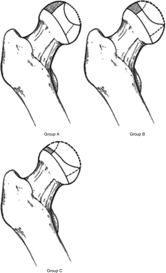
The Herring (lateral pillar) (Fig. 2) classification is based on the height of the lateral pillar of the capital epiphysis on AP view of the pelvis:

Fig. 2
Herring classification
Group A—No involvement of the lateral pillar, with no density changes and no loss of height of the lateral pillar.
Group B—More than 50 % of the lateral pillar height is maintained.
Group C—Less than 50 % of the lateral pillar height is maintained.
B/C border group—This group has been added more recently to increase the consistency of readings and to increase prognostic accuracy of the lateral pillar classification. In this group, the lateral pillar is narrow (2–3 mm wide) or poorly ossified, or exactly 50 % of lateral pillar height is maintained.
The Herring classification is the most reliable classification scheme and is related to prognosis. Its limitation is that the final classification cannot be determined at the time of initial presentation.
The Catterall classification is based on the amount of epiphyseal involvement. Although commonly used in the past, it has more recently been criticized for its poor interobserver reliability.
Group I—Involvement is limited to the anterior part of the capital epiphysis.
Group II—The anterior and central parts of the capital epiphysis are involved.
Group III—Most of the capital epiphysis is involved, with sparing of the medial and lateral parts of the epiphysis.
Group IV—The entire epiphysis is involved.
Catterall also described four at-risk signs, which indicate a more severe disease course.
Gage sign (radiolucency in the shape of a V in the lateral portion of the epiphysis)
Calcification lateral to the epiphysis
Lateral subluxation of the femoral head
A horizontal physis
Stay updated, free articles. Join our Telegram channel

Full access? Get Clinical Tree


