Prenatal
Perinatal
Postnatal (0–2 years)
Prematurity (gestational age less than 36 weeks)
Prolonged and difficult labor
CNS infection (encephalitis, meningitis)
Low birth weight (less than 2,500 g)
Premature rupture of membranes
Hypoxia
Maternal epilepsy
Presentation anomalies
Seizures
Hyperthyroidism
Vaginal bleeding at the time of admission for labor
Coagulopathies
Infections (TORCH)
Bradycardia
Neonatal hyperbilirubinemia
Bleeding in the third trimester
Hypoxia
Incompetent cervix
Severe toxemia, eclampsia
Hyperthyroidism
Drug abuse
Trauma
Multiple pregnancies
Placental insufficiency
Classification
Clinical classification
Tonus
Lesion site
Spastic
Cortex
Dyskinetic
Basal ganglia – extrapyramidal system
Hypotonic/ataxic
Cerebellum
Mixed
Diffuse
The importance of the classification is that, in general, surgical results are more reliable in patients with spasticity compared with patients with dystonia.
Anatomical classification
Location
Description
Hemiplegia
Upper and lower extremity on one side of the body
Diplegia
Four extremities, legs more affected than the arms
Quadriplegia
Four extremities plus the trunk, neck, and face
Triplegia
Both lower extremities and one upper extremity
Monoplegia
One extremity (rare)
Double hemiplegia
Four extremities, arms more affected than the legs
Functional classification – gross motor function classification system
Walks independently and speed, balance, and coordination reduced
Walks without assistive devices but limitations in community
Walks with assistive devices
Transported or uses powered mobility
Severely limited, dependent on wheelchair
Investigations and Clinical Assessment
Obtain baseline hip and spine radiographs. It is necessary to monitor hip instability. Reimer’s index which is the percentage of femoral head coverage by the acetabulum (Fig. 1).
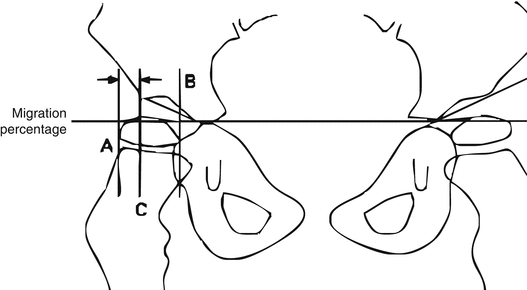
Fig. 1
Reimer’s migration index
In a normal hip, the entire femoral head is located medial to the lateral margin of the acetabulum. In a spastic hip, the lateral migration is measured as AC/ AB ×100.
Cranial magnetic resonance imaging (MRI) – useful for diagnosing lesions in the white matter.
Electroencephalogram – useful for diagnosis and follow-up of seizure disorders.
The typical clinical picture is established towards the age of 1 year. Primitive reflexes persist, and advanced postural reactions do not appear in the child with CP.
Musculoskeletal examination includes evaluation of joint range of motion (ROM), deformities, contractures, balance, posture, sitting, and gait.
Assessing Lower Extremities
Thomas test for hip flexion contracture.
Range of abduction with hip in flexion and extension. If abduction is limited when the hips are extended but better when they are flexed, then adduction contracture is caused by gracilis and medial hamstring spasticity.
The Ely test shows rectus femoris tightness.
Test hip rotation in prone position with the knee in flexion. Excessive internal rotation suggests persistent femoral anteversion.
Measure the popliteal angle to test for hamstring contracture.
Use the Silfverskiöld test to assess triceps surae tightness.
Examine tibial torsion with the patient in the prone position. Evaluate the thigh-foot angle with the knee flexed to 90°.
A spastic posterior tibialis muscle causes hindfoot varus.
A spastic peroneus or gastrocnemius muscle may cause a valgus deformity.
Assessing Upper Extremities
Most commonly these patients possess internal rotation contractures at the shoulder, flexion contractures about the elbow, pronation contractures of the forearm, flexion contractures at the wrist, and thumb-in-palm deformity.
A higher level of use and recognition of the affected upper extremity or extremities leads to more reliable surgical outcomes.
Commonly used assessment tools include the Manual Ability Classification System, the House functional classification system of upper extremity use (see Table 2), the Zancolli classification system of voluntary grasp and release patterns of the upper extremity (Table 3), and modified House classification for thumb deformities (Table 4).
Table 2
House functional classification for cerebral palsy
Grade
Designation
Activity level
0
Does not use
Does not use
1
Poor passive assist
Uses as stabilizing weight only
2
Fair passive assist
Can hold on to object placed in hand
3
Good passive assist
Can hold on to object and stabilize it for use by the other hand
4
Poor active assist
Can actively grasp object and hold it weakly
5
Fair active assist
Can actively grasp object and stabilize it well
6
Good active assist
Can actively grasp an object and then manipulate it against other hand
7
Spontaneous use, partial
Can perform bimanual activities easily and occasionally uses the hand spontaneously
8
Spontaneous use, complete
Uses hand completely independently without reference to the other hand
Table 3
Zancolli classification of active finger and wrist extension
Level
Designation
Description
1
Minimal flexion spasticityRelated posts:

Stay updated, free articles. Join our Telegram channel

Full access? Get Clinical Tree

 Get Clinical Tree app for offline access

